Advertisement

Main menu

  • Home
  • Content
    • Current Issue
    • Accepted Manuscripts
    • Article Preview
    • Past Issue Archive
    • Video Articles
    • AJNR Case Collection
    • Case of the Week Archive
    • Case of the Month Archive
    • Classic Case Archive
  • Special Collections
    • AJNR Awards
    • Low-Field MRI
    • Alzheimer Disease
    • ASNR Foundation Special Collection
    • Photon-Counting CT
    • View All
  • Multimedia
    • AJNR Podcasts
    • AJNR SCANtastic
    • Trainee Corner
    • MRI Safety Corner
    • Imaging Protocols
  • For Authors
    • Submit a Manuscript
    • Submit a Video Article
    • Submit an eLetter to the Editor/Response
    • Manuscript Submission Guidelines
    • Statistical Tips
    • Fast Publishing of Accepted Manuscripts
    • Graphical Abstract Preparation
    • Imaging Protocol Submission
    • Author Policies
  • About Us
    • About AJNR
    • Editorial Board
    • Editorial Board Alumni
  • More
    • Become a Reviewer/Academy of Reviewers
    • Subscribers
    • Permissions
    • Alerts
    • Feedback
    • Advertisers
    • ASNR Home

User menu

  • Alerts
  • Log in

Search

  • Advanced search
American Journal of Neuroradiology

American Journal of Neuroradiology

ASHNR American Society of Functional Neuroradiology ASHNR American Society of Pediatric Neuroradiology ASSR
  • Alerts
  • Log in

Advanced Search

  • Home
  • Content
    • Current Issue
    • Accepted Manuscripts
    • Article Preview
    • Past Issue Archive
    • Video Articles
    • AJNR Case Collection
    • Case of the Week Archive
    • Case of the Month Archive
    • Classic Case Archive
  • Special Collections
    • AJNR Awards
    • Low-Field MRI
    • Alzheimer Disease
    • ASNR Foundation Special Collection
    • Photon-Counting CT
    • View All
  • Multimedia
    • AJNR Podcasts
    • AJNR SCANtastic
    • Trainee Corner
    • MRI Safety Corner
    • Imaging Protocols
  • For Authors
    • Submit a Manuscript
    • Submit a Video Article
    • Submit an eLetter to the Editor/Response
    • Manuscript Submission Guidelines
    • Statistical Tips
    • Fast Publishing of Accepted Manuscripts
    • Graphical Abstract Preparation
    • Imaging Protocol Submission
    • Author Policies
  • About Us
    • About AJNR
    • Editorial Board
    • Editorial Board Alumni
  • More
    • Become a Reviewer/Academy of Reviewers
    • Subscribers
    • Permissions
    • Alerts
    • Feedback
    • Advertisers
    • ASNR Home
  • Follow AJNR on Twitter
  • Visit AJNR on Facebook
  • Follow AJNR on Instagram
  • Join AJNR on LinkedIn
  • RSS Feeds

AJNR Awards, New Junior Editors, and more. Read the latest AJNR updates

Research ArticleNeurointervention

Endovascular Treatment of High-Flow Carotid Cavernous Fistulas by Stent-Assisted Coil Placement

Fanny E. Morón, Richard P. Klucznik, Michel E. Mawad and Charles M. Strother
American Journal of Neuroradiology June 2005, 26 (6) 1399-1404;
Fanny E. Morón
  • Find this author on Google Scholar
  • Find this author on PubMed
  • Search for this author on this site
Richard P. Klucznik
  • Find this author on Google Scholar
  • Find this author on PubMed
  • Search for this author on this site
Michel E. Mawad
  • Find this author on Google Scholar
  • Find this author on PubMed
  • Search for this author on this site
Charles M. Strother
  • Find this author on Google Scholar
  • Find this author on PubMed
  • Search for this author on this site
  • Article
  • Figures & Data
  • Info & Metrics
  • Responses
  • References
  • PDF
Loading

Article Figures & Data

Figures

  • Tables
  • Fig 1.
    • Download figure
    • Open in new tab
    • Download powerpoint
    Fig 1.
    • Download figure
    • Open in new tab
    • Download powerpoint
    Fig 1.

    20-year-old woman (patient #3, Table 1) injured in an equestrian accident with multiple skull base fractures and bilateral carotid cavernous fistulas. A, Lateral projection of right common carotid artery arteriogram shows type A carotid cavernous fistula with no filling of the intradural segment of the internal carotid artery. Note prominent intracranial and orbital venous drainage. B, Lateral projection of right vertebral artery arteriogram demonstrates forward filling into right internal carotid artery. No demonstration of the communication between the proximal and distal internal carotid artery is seen. C, Lateral projection of right internal carotid artery angiogram following stent deployment with reconstruction of the vessel shows that there is now brisk antegrade filling of the internal carotid artery distal to the site of the fistula. D, Lateral projection of left internal carotid artery arteriogram shows type A carotid cavernous fistula. Although there is antegrade flow distal to the site of the fistula, the course and caliber of this segment of the internal carotid artery could not be determined. E, Lateral projection of left internal carotid artery arteriogram after stent deployment shows improved visualization of the cavernous segment of the internal carotid artery. Venous drainage to orbital veins persists. F, Lateral unsubtracted projection demonstrates bilateral stents in place (arrows). G, Lateral right internal carotid artery angiogram, unsubtracted, shows an inflated balloon inside the stent (arrow). This was used during coil deposition. H, Lateral projection of left internal carotid artery arteriogram demonstrates persistent flow after extensive coiling. I, Lateral projection of left internal carotid artery arteriogram shows a small aliquot of liquid adhesive (arrow) that was instilled in the proximal portion of the superior ophthalmic vein. This resulted in complete closing of the fistula. J-K, Frontal and lateral projections of right internal carotid artery (J) and left internal carotid artery (K) on 3-month follow-up arteriogram demonstrate patency of both internal carotid arteries with antegrade filling of their branches, dense coil packing, and complete closure of the fistulas.

  • Fig 2.
    • Download figure
    • Open in new tab
    • Download powerpoint
    Fig 2.

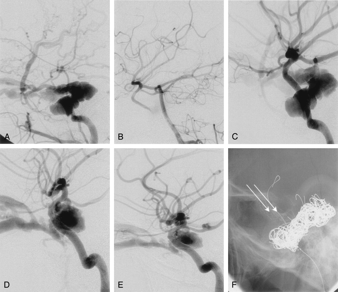
    62-year-old woman (patient 5, Table 1) with sudden left-sided ptosis. A, Frontal projection of left internal carotid artery arteriogram confirms type A carotid cavernous fistula with poor antegrade filling of the internal carotid artery branches. Note extensive venous drainage to superior ophthalmic vein, pterygopalatine plexus, and both inferior petrosal sinus and contralateral cavernous sinus. B, Lateral projection of left vertebral artery arteriogram does not demonstrate communication between the proximal and distal internal carotid artery segments. The right internal carotid artery arteriogram (not shown) also did not demonstrate a connection between these two segments. C, Lateral unsubtracted projection demonstrates the stent in place (arrow). D, Lateral road map image of left internal carotid artery arteriogram shows inflated balloon in stent (triple black arrows), used during coiling to prevent coils from coming into the arterial lumen, which could not be visualized. Note microcatheter in inferior petrosal sinus (white arrow) for transvenous embolization. E, Lateral projection of left internal carotid artery arteriogram after extensive coiling with almost complete obliteration of the aneurysm and carotid cavernous fistula. Note small residual aneurysm (arrow) and antegrade filling of the internal carotid artery branches. No additional coils could be placed. F, Lateral projection of left internal carotid artery on 6-month follow-up arteriogram demonstrates stable appearance of the aneurysm and carotid cavernous fistula with normal antegrade filling of the internal carotid artery. G, In lateral unsubtracted projection of left internal carotid artery on 6-month follow-up arteriogram, the stent demonstrates the lumen (arrow) of the internal carotid artery.

Tables

  • Figures
  • Summary of 5 patients with 6 type A CCF treated with a combination of metallic stent and platinum coils

    Patient No.Major SymptomsAgeSexMechanismVenous AccessArterial AccessEmbolic MaterialOutcomeClinical Outcome Last Follow-up
    1Headache Visual Loss55MTraumaSOV via IPSCoilsComplete occlusionNo CCF recurrence
    2Proptosis Visual loss19MGSW–ICACoilsComplete occlusionNo CCF recurrence
    3Proptopsis Chemosis Left CN palsy20FTrauma–ICACoils LAComplete occlusionNo CCF recurrence Traumatic CNP
    3Proptosis Chemosis Left CN palsy20FTrauma–SOV via facial veinICACoils LAComplete occlusion One coil stretchNo CCF recurrence Traumatic CNP
    4Proptosis83F?IPSCoilsComplete occlusionNo CCF recurrence
    5Ptosis Diplopia Headache62FCavernous ICA AneurysmIPSCoilsComplete occlusionNo CCF or aneurysm recurrence
    • Note—GSW indicates Gunshot wound, IPS, Inferior Petrosal Sinus; SOV, Superior Ophthalmic vein; LA, Liquid Adhesive; and CNP, Cranial nerve palsy.

PreviousNext
Back to top

In this issue

American Journal of Neuroradiology: 26 (6)
American Journal of Neuroradiology
Vol. 26, Issue 6
1 Jun 2005
  • Table of Contents
  • Index by author
Advertisement
Print
Download PDF
Email Article

Thank you for your interest in spreading the word on American Journal of Neuroradiology.

NOTE: We only request your email address so that the person you are recommending the page to knows that you wanted them to see it, and that it is not junk mail. We do not capture any email address.

Enter multiple addresses on separate lines or separate them with commas.
Endovascular Treatment of High-Flow Carotid Cavernous Fistulas by Stent-Assisted Coil Placement
(Your Name) has sent you a message from American Journal of Neuroradiology
(Your Name) thought you would like to see the American Journal of Neuroradiology web site.
CAPTCHA
This question is for testing whether or not you are a human visitor and to prevent automated spam submissions.
Cite this article
Fanny E. Morón, Richard P. Klucznik, Michel E. Mawad, Charles M. Strother
Endovascular Treatment of High-Flow Carotid Cavernous Fistulas by Stent-Assisted Coil Placement
American Journal of Neuroradiology Jun 2005, 26 (6) 1399-1404;

Citation Manager Formats

  • BibTeX
  • Bookends
  • EasyBib
  • EndNote (tagged)
  • EndNote 8 (xml)
  • Medlars
  • Mendeley
  • Papers
  • RefWorks Tagged
  • Ref Manager
  • RIS
  • Zotero
0 Responses
Respond to this article
Share
Bookmark this article
Endovascular Treatment of High-Flow Carotid Cavernous Fistulas by Stent-Assisted Coil Placement
Fanny E. Morón, Richard P. Klucznik, Michel E. Mawad, Charles M. Strother
American Journal of Neuroradiology Jun 2005, 26 (6) 1399-1404;
del.icio.us logo Twitter logo Facebook logo Mendeley logo
  • Tweet Widget
  • Facebook Like
  • Google Plus One

Jump to section

  • Article
    • Abstract
    • Methods
    • Results
    • Discussion
    • Conclusion
    • References
  • Figures & Data
  • Info & Metrics
  • Responses
  • References
  • PDF

Related Articles

  • No related articles found.
  • PubMed
  • Google Scholar

Cited By...

  • Combined use of Onyx and coils for transarterial balloon-assisted embolization of traumatic carotid-cavernous fistulas: a report of 16 cases with 17 fistulas
  • Complications of endovascular therapy for acute ischemic stroke and proposed management approach
  • Direct puncture of the highest cervical segment of the internal carotid artery for treatment of an iatrogenic carotid cavernous fistula in a patient with Ehlers-Danlos syndrome
  • Multimodal endovascular therapy of traumatic and spontaneous carotid cavernous fistula using coils, n-BCA, Onyx and stent graft
  • Comparison of the Risk of Oculomotor Nerve Deficits between Detachable Balloons and Coils in the Treatment of Direct Carotid Cavernous Fistulas
  • Crossref
  • Google Scholar

This article has not yet been cited by articles in journals that are participating in Crossref Cited-by Linking.

More in this TOC Section

  • A Retrospective Study in Tentorial DAVFs
  • Proximal Protection Devices for Carotid Stenting
  • Rescue Reentry in Carotid Near-Occlusion
Show more NEUROINTERVENTION

Similar Articles

Advertisement

Indexed Content

  • Current Issue
  • Accepted Manuscripts
  • Article Preview
  • Past Issues
  • Editorials
  • Editor's Choice
  • Fellows' Journal Club
  • Letters to the Editor
  • Video Articles

Cases

  • Case Collection
  • Archive - Case of the Week
  • Archive - Case of the Month
  • Archive - Classic Case

More from AJNR

  • Trainee Corner
  • Imaging Protocols
  • MRI Safety Corner
  • Book Reviews

Multimedia

  • AJNR Podcasts
  • AJNR Scantastics

Resources

  • Turnaround Time
  • Submit a Manuscript
  • Submit a Video Article
  • Submit an eLetter to the Editor/Response
  • Manuscript Submission Guidelines
  • Statistical Tips
  • Fast Publishing of Accepted Manuscripts
  • Graphical Abstract Preparation
  • Imaging Protocol Submission
  • Evidence-Based Medicine Level Guide
  • Publishing Checklists
  • Author Policies
  • Become a Reviewer/Academy of Reviewers
  • News and Updates

About Us

  • About AJNR
  • Editorial Board
  • Editorial Board Alumni
  • Alerts
  • Permissions
  • Not an AJNR Subscriber? Join Now
  • Advertise with Us
  • Librarian Resources
  • Feedback
  • Terms and Conditions
  • AJNR Editorial Board Alumni

American Society of Neuroradiology

  • Not an ASNR Member? Join Now

© 2025 by the American Society of Neuroradiology All rights, including for text and data mining, AI training, and similar technologies, are reserved.
Print ISSN: 0195-6108 Online ISSN: 1936-959X

Powered by HighWire