Advertisement

Main menu

  • Home
  • Content
    • Current Issue
    • Accepted Manuscripts
    • Article Preview
    • Past Issue Archive
    • Video Articles
    • AJNR Case Collection
    • Case of the Week Archive
    • Case of the Month Archive
    • Classic Case Archive
  • Special Collections
    • AJNR Awards
    • Low-Field MRI
    • Alzheimer Disease
    • ASNR Foundation Special Collection
    • Photon-Counting CT
    • View All
  • Multimedia
    • AJNR Podcasts
    • AJNR SCANtastic
    • Trainee Corner
    • MRI Safety Corner
    • Imaging Protocols
  • For Authors
    • Submit a Manuscript
    • Submit a Video Article
    • Submit an eLetter to the Editor/Response
    • Manuscript Submission Guidelines
    • Statistical Tips
    • Fast Publishing of Accepted Manuscripts
    • Graphical Abstract Preparation
    • Imaging Protocol Submission
    • Author Policies
  • About Us
    • About AJNR
    • Editorial Board
    • Editorial Board Alumni
  • More
    • Become a Reviewer/Academy of Reviewers
    • Subscribers
    • Permissions
    • Alerts
    • Feedback
    • Advertisers
    • ASNR Home

User menu

  • Alerts
  • Log in

Search

  • Advanced search
American Journal of Neuroradiology

American Journal of Neuroradiology

ASHNR American Society of Functional Neuroradiology ASHNR American Society of Pediatric Neuroradiology ASSR
  • Alerts
  • Log in

Advanced Search

  • Home
  • Content
    • Current Issue
    • Accepted Manuscripts
    • Article Preview
    • Past Issue Archive
    • Video Articles
    • AJNR Case Collection
    • Case of the Week Archive
    • Case of the Month Archive
    • Classic Case Archive
  • Special Collections
    • AJNR Awards
    • Low-Field MRI
    • Alzheimer Disease
    • ASNR Foundation Special Collection
    • Photon-Counting CT
    • View All
  • Multimedia
    • AJNR Podcasts
    • AJNR SCANtastic
    • Trainee Corner
    • MRI Safety Corner
    • Imaging Protocols
  • For Authors
    • Submit a Manuscript
    • Submit a Video Article
    • Submit an eLetter to the Editor/Response
    • Manuscript Submission Guidelines
    • Statistical Tips
    • Fast Publishing of Accepted Manuscripts
    • Graphical Abstract Preparation
    • Imaging Protocol Submission
    • Author Policies
  • About Us
    • About AJNR
    • Editorial Board
    • Editorial Board Alumni
  • More
    • Become a Reviewer/Academy of Reviewers
    • Subscribers
    • Permissions
    • Alerts
    • Feedback
    • Advertisers
    • ASNR Home
  • Follow AJNR on Twitter
  • Visit AJNR on Facebook
  • Follow AJNR on Instagram
  • Join AJNR on LinkedIn
  • RSS Feeds

AJNR Awards, New Junior Editors, and more. Read the latest AJNR updates

Getting new auth cookie, if you see this message a lot, tell someone!
Research ArticleSpine Imaging and Spine Image-Guided Interventions

MR Diffusion Tensor Imaging and Fiber Tracking in Spinal Cord Compression

David Facon, Augustin Ozanne, Pierre Fillard, Jean-François Lepeintre, Caroline Tournoux-Facon and Denis Ducreux
American Journal of Neuroradiology June 2005, 26 (6) 1587-1594;
David Facon
  • Find this author on Google Scholar
  • Find this author on PubMed
  • Search for this author on this site
Augustin Ozanne
  • Find this author on Google Scholar
  • Find this author on PubMed
  • Search for this author on this site
Pierre Fillard
  • Find this author on Google Scholar
  • Find this author on PubMed
  • Search for this author on this site
Jean-François Lepeintre
  • Find this author on Google Scholar
  • Find this author on PubMed
  • Search for this author on this site
Caroline Tournoux-Facon
  • Find this author on Google Scholar
  • Find this author on PubMed
  • Search for this author on this site
Denis Ducreux
  • Find this author on Google Scholar
  • Find this author on PubMed
  • Search for this author on this site
  • Article
  • Figures & Data
  • Info & Metrics
  • Responses
  • References
  • PDF
Loading

Article Figures & Data

Figures

  • Tables
  • Fig 1.
    • Download figure
    • Open in new tab
    • Download powerpoint
    Fig 1.

    A, MR imaging of a spinal cord compression due to a breast tumor metastasis in patient 1. Images are from C7–T10. Color-coded scale related to parameters values. Higher values are coded in red, medium values in green, and lower values in blue. T2-weighted imaging (first image) shows a high signal intensity (white arrow), ADC (second image) is slightly increased (blue-green areas; ADC = 1.1 10−3 mm2/s versus 0.84 10−3 mm2/s in normal cervicothoracic area) and FA (third image) is decreased (green areas; FA = 0.61 vs 0.75 in normal cervicothoracic area) after region of interest measurements. The region of interest in abnormal area was drawn by using the fiber tracking three-dimensional reconstruction.

    B, MR imaging of a spinal cord compression due to a breast tumor metastasis with epidural involvement in patient 1. Fiber tracking over b0 image shows a mass-effect on fibers tracts. The region of interest (green area) was drawn over the maximal level of compression (blue arrow) and then automatically reported on the coregistered ADC and FA maps to measure ADC and FA values in the compression site.

  • Fig 2.
    • Download figure
    • Open in new tab
    • Download powerpoint
    Fig 2.

    Time course from d1 to d30 of the averaged FA parameter estimated from the compression sites in 11 patients with abnormal FA values. FA values decreased from the 1st to the 21st days, then slightly increased, both related to the extracellular water diffusivity: restricted diffusivity in acute stage and increased diffusivity in chronic stage. Normal values range from 0.69 to 0.8.
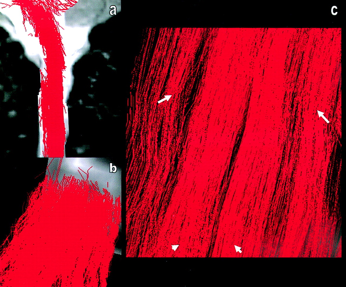
  • Fig 3.
    • Download figure
    • Open in new tab
    • Download powerpoint
    Fig 3.

    Fiber tracking performed on a volunteer’s cervical spinal cord. Sagittal (A), axial (B), and coronal (C) views show tracts reconstructed over the b0 sequence. Main white matter tracts are visible on axial (B) and coronal (C) views of the three-dimensional reconstructions: two individualized posterior lemniscal tracts (arrowheads), and posterolateral corticospinal tracts (arrows). Other tracts are visible, but have to be more correlated with known anatomy.

  • Fig 4.
    • Download figure
    • Open in new tab
    • Download powerpoint
    Fig 4.

    Three-dimensional reconstructions are mapped over the b0 images. Fiber tracking performed on the spinal cord volunteer shows some pitfalls of the FT method due to magnetic susceptibility effect of the DTI MR image: “hole” effect, gap in three-dimensional reconstruction (blue arrows).

Tables

  • Figures
    • View popup
    TABLE 1:

    Patients’ data with estimates of ADC and FA on compression level

    Patient (no.)Sex/AgeSymptomsIOTC/ICLT2 SIADCFADiagnoses
    1M/60MD + Thoracic Pain7T9Normal1.10.61Metastasis
    2F/80Thoracic Pain1T6High1.060.831Metastasis
    3M/59MD7T1–T3High0.820.78Metastasis
    4M/76MD + SD>30C1–C2High1.410.526Degenerative
    5M/43MD15C6–C7Normal1.170.73Degenerative
    6M/62MD>30C5–C7High1.260.642Degenerative
    7M/40SD>30T4–T5Normal1.030.736Degenerative
    8F/41Cervical Pain15C3–C4Normal0.850.6Spondylodiscitis
    9F/46SD21C5–C6Normal0.910.67Degenerative
    10F/73SD3T6High0.7750.64Metastasis
    11M/34SD + Lumbar Pain>30T12–L1Normal1.080.599Spondylodiscitis
    12M/77MD1T6–T7High1.190.8Spondylodiscitis
    13M/30MD + SD>30C6Normal0.970.676Spondylodiscitis
    14F/48MD>30C5–C7High0.880.614Spondylodiscitis
    15M/40MD21C4–C5Normal1.080.6Degenerative
    • Note.—Abnormal values are in red. Imaging compression level matched the clinical data.

      IOT = imaging/onset time: time between MR exam and onset of symptoms (in days); C/ICL = clinical/imaging compression level; SI = signal intensity; MD = motor deficit; SD = sensitive deficit; ADC = apparent diffusion coefficient (%10−3 mm2/s); FA = fractional anisotropy.

    • View popup
    TABLE 2:

    Statistical tests in volunteer

    AverageSDMedianMinimumMaximumMDP Value
    FA0.7480.0270.7430.7000.8007
        Cervical0.7480.0310.7470.7000.7801.86
        High thoracic0.7510.0270.7400.7200.8002.67
        Low thoracic0.7450.0270.740.7140.8004–
    ADC (%10−3)1.000.1301.000.771.257
        Cervical1.010.1571.020.771.251.36
        High thoracic0.960.1040.930.811.132–
        Low thoracic1.050.1111.060.891.194.15
    • Note.—Comparison of FA and ADC values according to medullar level on healthy volunteers doesn’t show a statistically significant difference. SD = standard deviation; MD = patient distribution (number of patients).

    • View popup
    TABLE 3:

    Statistical tests in volunteer

    AverageSDMedianMinimumMaximumMDP Value
    FA.42
        Healthy volunteers0.7480.0270.7430.7000.8007
        Patients0.7400.0340.7500.6900.7800
    ADC (%10−3).26
        Healthy volunteers1.000.1301.000.771.257
        Patients0.9490.1720.9280.7301.340
    • Note.—Comparison of FA and ADC of healthy level on healthy volunteers and patients doesn’t show a statistically significant difference. SD = standard deviation; MD = patient distribution (number of patients).

    • View popup
    TABLE 4:

    Statistical tests in volunteer

    AverageSDMedianMinimumMaximumMDP Value
    FA.012
        Healthy level0.7400.0340.7500.6900.7800
        Pathologic0.6700.0870.6420.5260.8310
    ADC (%10−3).13
        Healthy level0.9490.1720.9280.7301.340
        Pathologic1.030.1771.030.7751.410
    • Note.—Comparison of FA and ADC of pathologic and healthy levels in patients doesn’t show a statistically significant difference for ADC but shows one for FA (bold value). SD = standard deviation; MD = patient distribution (number of patients).

PreviousNext
Back to top

In this issue

American Journal of Neuroradiology: 26 (6)
American Journal of Neuroradiology
Vol. 26, Issue 6
1 Jun 2005
  • Table of Contents
  • Index by author
Advertisement
Print
Download PDF
Email Article

Thank you for your interest in spreading the word on American Journal of Neuroradiology.

NOTE: We only request your email address so that the person you are recommending the page to knows that you wanted them to see it, and that it is not junk mail. We do not capture any email address.

Enter multiple addresses on separate lines or separate them with commas.
MR Diffusion Tensor Imaging and Fiber Tracking in Spinal Cord Compression
(Your Name) has sent you a message from American Journal of Neuroradiology
(Your Name) thought you would like to see the American Journal of Neuroradiology web site.
CAPTCHA
This question is for testing whether or not you are a human visitor and to prevent automated spam submissions.
Cite this article
David Facon, Augustin Ozanne, Pierre Fillard, Jean-François Lepeintre, Caroline Tournoux-Facon, Denis Ducreux
MR Diffusion Tensor Imaging and Fiber Tracking in Spinal Cord Compression
American Journal of Neuroradiology Jun 2005, 26 (6) 1587-1594;

Citation Manager Formats

  • BibTeX
  • Bookends
  • EasyBib
  • EndNote (tagged)
  • EndNote 8 (xml)
  • Medlars
  • Mendeley
  • Papers
  • RefWorks Tagged
  • Ref Manager
  • RIS
  • Zotero
0 Responses
Respond to this article
Share
Bookmark this article
MR Diffusion Tensor Imaging and Fiber Tracking in Spinal Cord Compression
David Facon, Augustin Ozanne, Pierre Fillard, Jean-François Lepeintre, Caroline Tournoux-Facon, Denis Ducreux
American Journal of Neuroradiology Jun 2005, 26 (6) 1587-1594;
del.icio.us logo Twitter logo Facebook logo Mendeley logo
  • Tweet Widget
  • Facebook Like
  • Google Plus One

Jump to section

  • Article
    • Abstract
    • Methods
    • Results
    • Discussion
    • Conclusion
    • Acknowledgments
    • References
  • Figures & Data
  • Info & Metrics
  • Responses
  • References
  • PDF

Related Articles

  • No related articles found.
  • PubMed
  • Google Scholar

Cited By...

  • Cervical Spinal Cord DTI Is Improved by Reduced FOV with Specific Balance between the Number of Diffusion Gradient Directions and Averages
  • Diffusion Tensor Imaging Correlates with the Clinical Assessment of Disease Severity in Cervical Spondylotic Myelopathy and Predicts Outcome following Surgery
  • Diffusion tensor imaging of the spinal cord and its clinical applications
  • Reduced Field-of-View Diffusion Imaging of the Human Spinal Cord: Comparison with Conventional Single-Shot Echo-Planar Imaging
  • Quantification of Diffusivities of the Human Cervical Spinal Cord Using a 2D Single-Shot Interleaved Multisection Inner Volume Diffusion-Weighted Echo-Planar Imaging Technique
  • Assessment of spinal somatosensory systems with diffusion tensor imaging in syringomyelia
  • Propriospinal myoclonus revisited: Clinical, neurophysiologic, and neuroradiologic findings
  • In Vivo Tracing of Neural Tracts in the Intact and Injured Spinal Cord of Marmosets by Diffusion Tensor Tractography
  • Comparison Between Diffusion Tensor Imaging and Conventional MR Imaging Sequences in the Detection of Spinal Cord Abnormalities
  • Crossref
  • Google Scholar

This article has not yet been cited by articles in journals that are participating in Crossref Cited-by Linking.

More in this TOC Section

  • Management Outcomes For VO Spine Biopsy
  • Characteristics of SIH Type I Culprit Lesions
  • Advanced Imaging of Type 2 Spinal CSF Leaks
Show more Spine Imaging and Spine Image-Guided Interventions

Similar Articles

Advertisement

Indexed Content

  • Current Issue
  • Accepted Manuscripts
  • Article Preview
  • Past Issues
  • Editorials
  • Editor's Choice
  • Fellows' Journal Club
  • Letters to the Editor
  • Video Articles

Cases

  • Case Collection
  • Archive - Case of the Week
  • Archive - Case of the Month
  • Archive - Classic Case

More from AJNR

  • Trainee Corner
  • Imaging Protocols
  • MRI Safety Corner
  • Book Reviews

Multimedia

  • AJNR Podcasts
  • AJNR Scantastics

Resources

  • Turnaround Time
  • Submit a Manuscript
  • Submit a Video Article
  • Submit an eLetter to the Editor/Response
  • Manuscript Submission Guidelines
  • Statistical Tips
  • Fast Publishing of Accepted Manuscripts
  • Graphical Abstract Preparation
  • Imaging Protocol Submission
  • Evidence-Based Medicine Level Guide
  • Publishing Checklists
  • Author Policies
  • Become a Reviewer/Academy of Reviewers
  • News and Updates

About Us

  • About AJNR
  • Editorial Board
  • Editorial Board Alumni
  • Alerts
  • Permissions
  • Not an AJNR Subscriber? Join Now
  • Advertise with Us
  • Librarian Resources
  • Feedback
  • Terms and Conditions
  • AJNR Editorial Board Alumni

American Society of Neuroradiology

  • Not an ASNR Member? Join Now

© 2025 by the American Society of Neuroradiology All rights, including for text and data mining, AI training, and similar technologies, are reserved.
Print ISSN: 0195-6108 Online ISSN: 1936-959X

Powered by HighWire