Advertisement

Main menu

  • Home
  • Content
    • Current Issue
    • Accepted Manuscripts
    • Article Preview
    • Past Issue Archive
    • AJNR Case Collection
    • Case of the Week Archive
    • Classic Case Archive
    • Case of the Month Archive
  • Special Collections
    • Spinal CSF Leak Articles (Jan 2020-June 2024)
    • 2024 AJNR Journal Awards
    • Most Impactful AJNR Articles
  • Multimedia
    • AJNR Podcast
    • AJNR Scantastics
    • Video Articles
  • For Authors
    • Submit a Manuscript
    • Author Policies
    • Fast publishing of Accepted Manuscripts
    • Graphical Abstract Preparation
    • Manuscript Submission Guidelines
    • Imaging Protocol Submission
    • Submit a Case for the Case Collection
  • About Us
    • About AJNR
    • Editorial Board
  • More
    • Become a Reviewer/Academy of Reviewers
    • Subscribers
    • Permissions
    • Alerts
    • Feedback
    • Advertisers
    • ASNR Home
  • Other Publications
    • ajnr

User menu

  • Alerts
  • Log in

Search

  • Advanced search
American Journal of Neuroradiology

American Journal of Neuroradiology

ASHNR American Society of Functional Neuroradiology ASHNR American Society of Pediatric Neuroradiology ASSR
  • Alerts
  • Log in

Advanced Search

  • Home
  • Content
    • Current Issue
    • Accepted Manuscripts
    • Article Preview
    • Past Issue Archive
    • AJNR Case Collection
    • Case of the Week Archive
    • Classic Case Archive
    • Case of the Month Archive
  • Special Collections
    • Spinal CSF Leak Articles (Jan 2020-June 2024)
    • 2024 AJNR Journal Awards
    • Most Impactful AJNR Articles
  • Multimedia
    • AJNR Podcast
    • AJNR Scantastics
    • Video Articles
  • For Authors
    • Submit a Manuscript
    • Author Policies
    • Fast publishing of Accepted Manuscripts
    • Graphical Abstract Preparation
    • Manuscript Submission Guidelines
    • Imaging Protocol Submission
    • Submit a Case for the Case Collection
  • About Us
    • About AJNR
    • Editorial Board
  • More
    • Become a Reviewer/Academy of Reviewers
    • Subscribers
    • Permissions
    • Alerts
    • Feedback
    • Advertisers
    • ASNR Home
  • Follow AJNR on Twitter
  • Visit AJNR on Facebook
  • Follow AJNR on Instagram
  • Join AJNR on LinkedIn
  • RSS Feeds

Welcome to the new AJNR, Updated Hall of Fame, and more. Read the full announcements.


AJNR is seeking candidates for the position of Associate Section Editor, AJNR Case Collection. Read the full announcement.


Research ArticlePEDIATRICS

Disruption of Cerebellar Development: Potential Complication of Extreme Prematurity

Agnes Messerschmidt, Peter C. Brugger, Eugen Boltshauser, Gerlinde Zoder, Walter Sterniste, Robert Birnbacher and Daniela Prayer
American Journal of Neuroradiology August 2005, 26 (7) 1659-1667;
Agnes Messerschmidt
  • Find this author on Google Scholar
  • Find this author on PubMed
  • Search for this author on this site
Peter C. Brugger
  • Find this author on Google Scholar
  • Find this author on PubMed
  • Search for this author on this site
Eugen Boltshauser
  • Find this author on Google Scholar
  • Find this author on PubMed
  • Search for this author on this site
Gerlinde Zoder
  • Find this author on Google Scholar
  • Find this author on PubMed
  • Search for this author on this site
Walter Sterniste
  • Find this author on Google Scholar
  • Find this author on PubMed
  • Search for this author on this site
Robert Birnbacher
  • Find this author on Google Scholar
  • Find this author on PubMed
  • Search for this author on this site
Daniela Prayer
  • Find this author on Google Scholar
  • Find this author on PubMed
  • Search for this author on this site
  • Article
  • Figures & Data
  • Info & Metrics
  • Responses
  • References
  • PDF
Loading

Article Figures & Data

Figures

  • Tables
  • Fig 1.
    • Download figure
    • Open in new tab
    • Download powerpoint
    Fig 1.

    Coronal sonograms in a preterm neonate born at 27 + 5 gestational weeks.

    A, Scan on day 1 of life shows normal cerebellar hemispheres.

    B, Scan at 3 weeks shows reduced cerebellar volume.
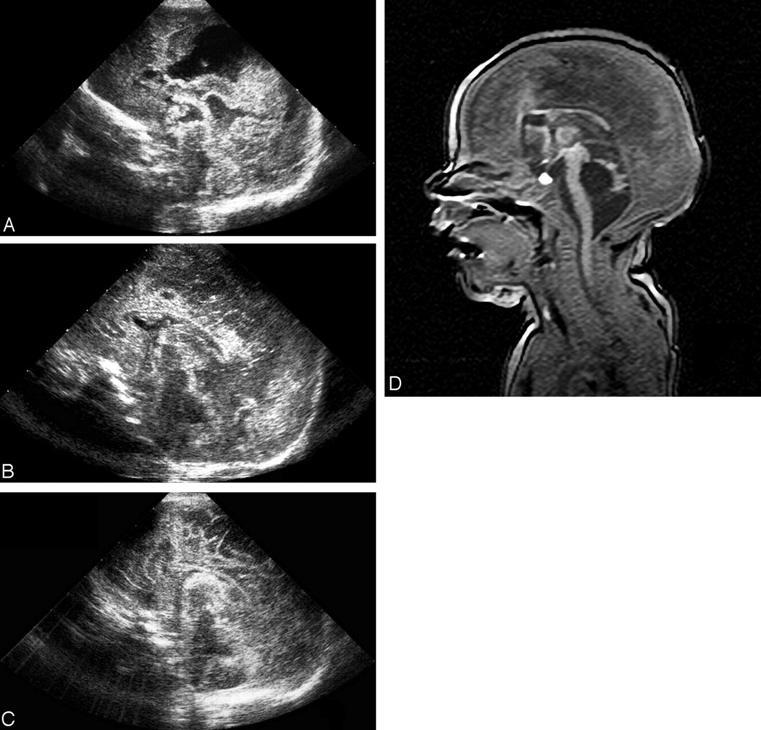
  • Fig 2.
    • Download figure
    • Open in new tab
    • Download powerpoint
    Fig 2.

    Type 2 disrupted cerebellar development in a preterm infant born at 26 + 3 gestational weeks.

    A–C, Sagittal follow-up sonograms obtained on days 57 (A), 86 (B), and 106 (C) of life show a vanishing cerebellar vermis with successive enlargement of the fourth ventricle.

    D, MR image obtained at 15 weeks.
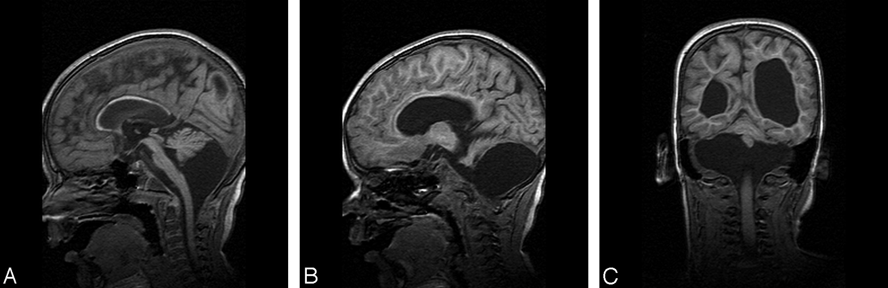
  • Fig 3.
    • Download figure
    • Open in new tab
    • Download powerpoint
    Fig 3.

    Type 1 disrupted cerebellar development in a preterm infant born at 29 gestational weeks. T1-weighted MR imaging was performed at 7 months.

    A, Sagittal image shows normal configuration of the fourth ventricle, a thin corpus callosum, and an inclined tentorium. The vermis is small but normally shaped. Dimensions of the brain stem are reduced.

    B and C, Parasagittal (B) and (C) frontal images demonstrate small cerebellar hemispheres immediately beneath the tentorium.
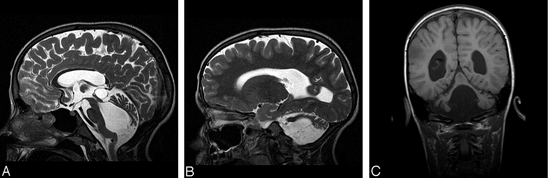
  • Fig 4.
    • Download figure
    • Open in new tab
    • Download powerpoint
    Fig 4.

    Type 2 disrupted cerebellar development.

    A, Sagittal T2-weighted image shows a balloon-shaped fourth ventricle; a longitudinal, small vermis; and remarkable kinking of the brainstem.

    B and C, Parasagittal T2-weighted (B) and frontal T1-weighted (C) images show small cerebellar hemispheres laterally located in the posterior fossa.
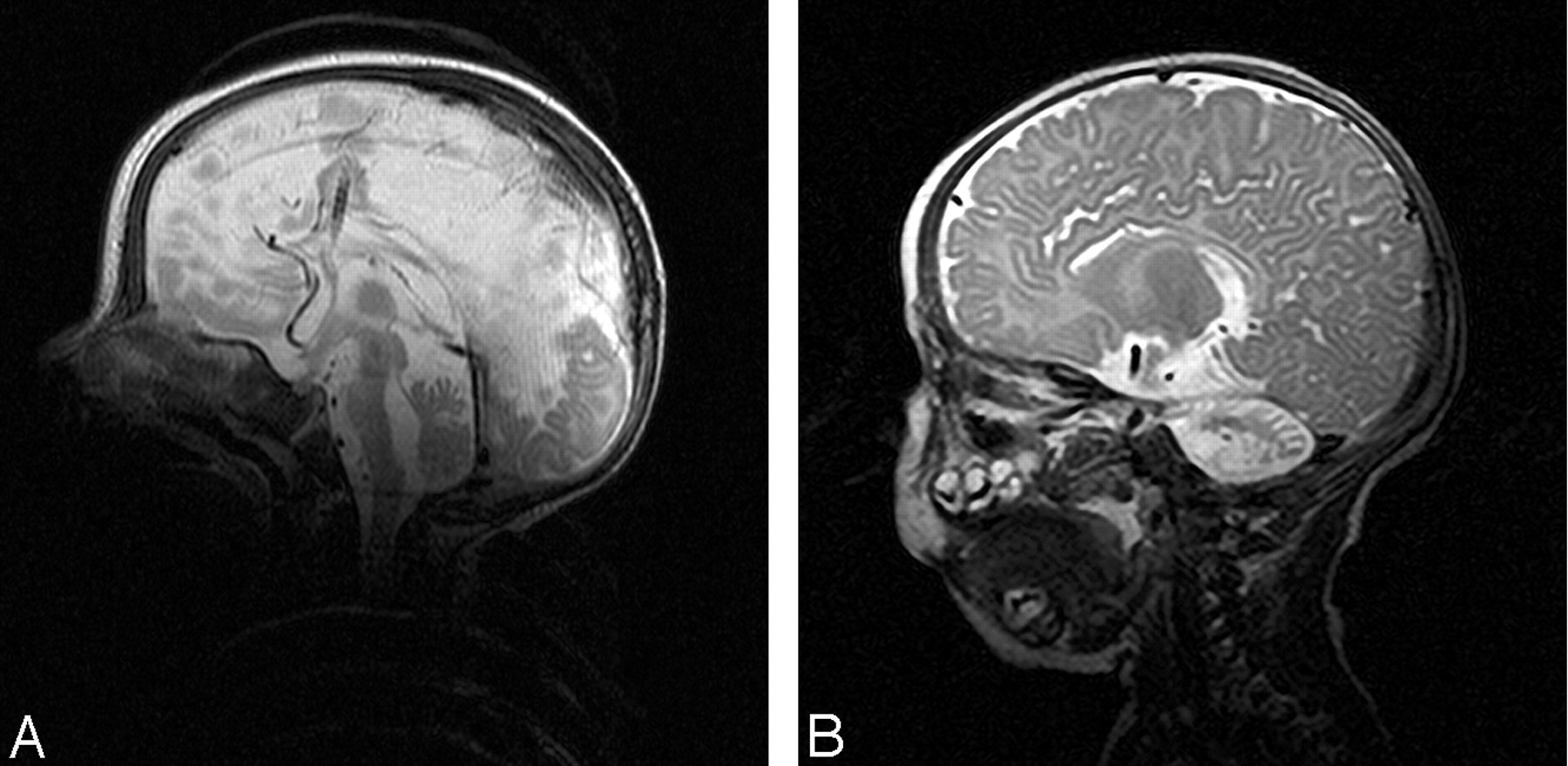
  • Fig 5.
    • Download figure
    • Open in new tab
    • Download powerpoint
    Fig 5.

    Type 3 disrupted cerebellar development in a preterm infant born at 26 + 4 gestational weeks. T2-weighted images show a skeletonized appearance of the cerebellum.

    A, Sagittal image.

    B, Parasagittal image.
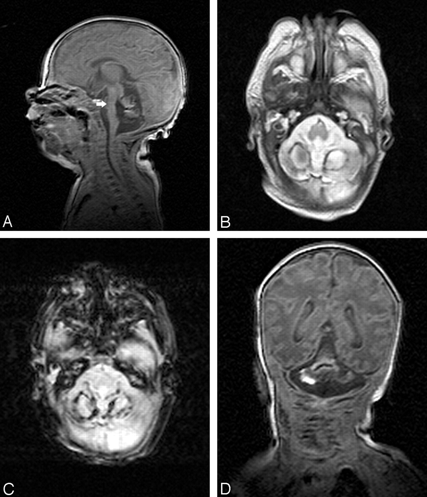
  • Fig 6.
    • Download figure
    • Open in new tab
    • Download powerpoint
    Fig 6.

    Unclassified pattern in a preterm infant (patient 26) born at 24 + 6 gestational weeks. Images show different blood breakdown products in the posterior fossa, shrunken cerebellar hemispheres, and cystic pontine (arrow) and vermian lesions.

    A, Sagittal T1-weighted image.

    B, Axial T2-weighted image.

    C, Axial T2*-weighted image.

    D, Coronal T1-weighted image.

Tables

  • Figures
    • View popup
    TABLE 1:

    Patients’ characteristics: intraventricular hemorrhage (IVH), hemosiderin infratentorial only estimated in hemosiderin-sensitive sequences

    No.TypeSexGestation WeekBirth Weight (g)IVH Grade RightIVH Grade LeftPosthemorrhagic Ventricular DilatationShuntCerebral White Matter LossHemosiderin Infratentorial
    11M27117622NoNoYes
    21F2796022YesNoYes
    31M2798523YesYesYesYes
    41M25101734YesYesYesYes
    51F2657402NoNoYesYes
    61M2687534YesYesYesNo
    71M2574022NoNoNo
    81M2664022NoNoNo
    91M29149033YesYesYes
    101M27113022NoNoYes
    111M2880011NoNoYes
    121F30131022NoNoYes
    132M2476840YesYesYesYes
    142F2682243YesYesYesYes
    152M2680033YesYesYesYes
    162M2471622YesYesYesYes
    172M2595032YesYesYes
    182M28130040YesYesYes
    192M27103033YesYesYes
    202F28133022YesYesYesYes
    212M26108022YesYesNo
    223M2697822YesYesYesYes
    233M2663733YesYesYesYes
    243M28122834YesYesYesNo
    253F27105822YesNoYesNo
    26M2486622YesYesNoYes
    27F3096022YesYesYesYes
    28M28108033YesYesYes
    • View popup
    TABLE 2:

    Common morphological features of disruptive cerebellar development

    Infratentorial FeaturesSupratentorial Features
    Vertical cerebellar diameter reducedWhite matter loss
    Tentorium steeply inclinedThin corpus callosum
    Small brain stem, flattened anterior curvature of the pons
    Dentate nuclei not detectable
    • View popup
    TABLE 3:

    Special morphological features of cerebellar disruptive development

    Cerebellar HemispheresVermisFourth Ventricle
    Type 1Marked volume reduction, immediately beneath the tentoriumSmall, shape preservedNormal width (8) widened (4)
    Type 2Volume reduction, position more lateralVery small, shape lostBalloon shaped
    Type 3Marked volume reduction, “Skeletonized,” shape preserved“Skeletonized,” shape preservedNormal width
PreviousNext
Back to top

In this issue

American Journal of Neuroradiology: 26 (7)
American Journal of Neuroradiology
Vol. 26, Issue 7
1 Aug 2005
  • Table of Contents
  • Index by author
Advertisement
Print
Download PDF
Email Article

Thank you for your interest in spreading the word on American Journal of Neuroradiology.

NOTE: We only request your email address so that the person you are recommending the page to knows that you wanted them to see it, and that it is not junk mail. We do not capture any email address.

Enter multiple addresses on separate lines or separate them with commas.
Disruption of Cerebellar Development: Potential Complication of Extreme Prematurity
(Your Name) has sent you a message from American Journal of Neuroradiology
(Your Name) thought you would like to see the American Journal of Neuroradiology web site.
CAPTCHA
This question is for testing whether or not you are a human visitor and to prevent automated spam submissions.
Cite this article
Agnes Messerschmidt, Peter C. Brugger, Eugen Boltshauser, Gerlinde Zoder, Walter Sterniste, Robert Birnbacher, Daniela Prayer
Disruption of Cerebellar Development: Potential Complication of Extreme Prematurity
American Journal of Neuroradiology Aug 2005, 26 (7) 1659-1667;

Citation Manager Formats

  • BibTeX
  • Bookends
  • EasyBib
  • EndNote (tagged)
  • EndNote 8 (xml)
  • Medlars
  • Mendeley
  • Papers
  • RefWorks Tagged
  • Ref Manager
  • RIS
  • Zotero
0 Responses
Respond to this article
Share
Bookmark this article
Disruption of Cerebellar Development: Potential Complication of Extreme Prematurity
Agnes Messerschmidt, Peter C. Brugger, Eugen Boltshauser, Gerlinde Zoder, Walter Sterniste, Robert Birnbacher, Daniela Prayer
American Journal of Neuroradiology Aug 2005, 26 (7) 1659-1667;
del.icio.us logo Twitter logo Facebook logo Mendeley logo
  • Tweet Widget
  • Facebook Like
  • Google Plus One

Jump to section

  • Article
    • Abstract
    • Methods
    • Results
    • Other Findings
    • Discussion
    • Conclusion
    • References
  • Figures & Data
  • Info & Metrics
  • Responses
  • References
  • PDF

Related Articles

  • No related articles found.
  • PubMed
  • Google Scholar

Cited By...

  • Longitudinal neonatal brain development and socio-demographic correlates of infant outcomes following preterm birth
  • Preterm birth impedes structural and functional development of cerebellar Purkinje cells in the developing baboon cerebellum
  • Accuracy of ultrasound in assessing cerebellar haemorrhages in very low birthweight babies
  • New MR Imaging Assessment Tool to Define Brain Abnormalities in Very Preterm Infants at Term
  • Ultrasonically detectable cerebellar haemorrhage in preterm infants
  • Injury to the Developing Cerebellum: Mechanisms and Consequences
  • Crossref
  • Google Scholar

This article has not yet been cited by articles in journals that are participating in Crossref Cited-by Linking.

More in this TOC Section

  • Comparison of Image Quality and Radiation Dose in Pediatric Temporal Bone CT Using Photon-Counting Detector CT and Energy-Integrating Detector CT
  • SyMRI & MR Fingerprinting in Brainstem Myelination
  • Axenfeld-Rieger Syndrome: Neuroimaging Findings
Show more Pediatrics

Similar Articles

Advertisement

Indexed Content

  • Current Issue
  • Accepted Manuscripts
  • Article Preview
  • Past Issues
  • Editorials
  • Editors Choice
  • Fellow Journal Club
  • Letters to the Editor

Cases

  • Case Collection
  • Archive - Case of the Week
  • Archive - Case of the Month
  • Archive - Classic Case

Special Collections

  • Special Collections

Resources

  • News and Updates
  • Turn around Times
  • Submit a Manuscript
  • Author Policies
  • Manuscript Submission Guidelines
  • Evidence-Based Medicine Level Guide
  • Publishing Checklists
  • Graphical Abstract Preparation
  • Imaging Protocol Submission
  • Submit a Case
  • Become a Reviewer/Academy of Reviewers
  • Get Peer Review Credit from Publons

Multimedia

  • AJNR Podcast
  • AJNR SCANtastic
  • Video Articles

About Us

  • About AJNR
  • Editorial Board
  • Not an AJNR Subscriber? Join Now
  • Alerts
  • Feedback
  • Advertise with us
  • Librarian Resources
  • Permissions
  • Terms and Conditions

American Society of Neuroradiology

  • Not an ASNR Member? Join Now

© 2025 by the American Society of Neuroradiology All rights, including for text and data mining, AI training, and similar technologies, are reserved.
Print ISSN: 0195-6108 Online ISSN: 1936-959X

Powered by HighWire