Advertisement

Main menu

  • Home
  • Content
    • Current Issue
    • Accepted Manuscripts
    • Article Preview
    • Past Issue Archive
    • AJNR Case Collection
    • Case of the Week Archive
    • Classic Case Archive
    • Case of the Month Archive
  • Special Collections
    • Spinal CSF Leak Articles (Jan 2020-June 2024)
    • 2024 AJNR Journal Awards
    • Most Impactful AJNR Articles
  • Multimedia
    • AJNR Podcast
    • AJNR Scantastics
    • Video Articles
  • For Authors
    • Submit a Manuscript
    • Author Policies
    • Fast publishing of Accepted Manuscripts
    • Graphical Abstract Preparation
    • Manuscript Submission Guidelines
    • Imaging Protocol Submission
    • Submit a Case for the Case Collection
  • About Us
    • About AJNR
    • Editorial Board
  • More
    • Become a Reviewer/Academy of Reviewers
    • Subscribers
    • Permissions
    • Alerts
    • Feedback
    • Advertisers
    • ASNR Home
  • Other Publications
    • ajnr

User menu

  • Alerts
  • Log in

Search

  • Advanced search
American Journal of Neuroradiology

American Journal of Neuroradiology

ASHNR American Society of Functional Neuroradiology ASHNR American Society of Pediatric Neuroradiology ASSR
  • Alerts
  • Log in

Advanced Search

  • Home
  • Content
    • Current Issue
    • Accepted Manuscripts
    • Article Preview
    • Past Issue Archive
    • AJNR Case Collection
    • Case of the Week Archive
    • Classic Case Archive
    • Case of the Month Archive
  • Special Collections
    • Spinal CSF Leak Articles (Jan 2020-June 2024)
    • 2024 AJNR Journal Awards
    • Most Impactful AJNR Articles
  • Multimedia
    • AJNR Podcast
    • AJNR Scantastics
    • Video Articles
  • For Authors
    • Submit a Manuscript
    • Author Policies
    • Fast publishing of Accepted Manuscripts
    • Graphical Abstract Preparation
    • Manuscript Submission Guidelines
    • Imaging Protocol Submission
    • Submit a Case for the Case Collection
  • About Us
    • About AJNR
    • Editorial Board
  • More
    • Become a Reviewer/Academy of Reviewers
    • Subscribers
    • Permissions
    • Alerts
    • Feedback
    • Advertisers
    • ASNR Home
  • Follow AJNR on Twitter
  • Visit AJNR on Facebook
  • Follow AJNR on Instagram
  • Join AJNR on LinkedIn
  • RSS Feeds

Welcome to the new AJNR, Updated Hall of Fame, and more. Read the full announcements.


AJNR is seeking candidates for the position of Associate Section Editor, AJNR Case Collection. Read the full announcement.


Research ArticlePEDIATRICS

Thiamine Deficiency in Infants: MR Findings in the Brain

Liora Kornreich, Efrat Bron-Harlev, Chen Hoffmann, Michael Schwarz, Osnat Konen, Tommy Schoenfeld, Rachel Straussberg, Elhanan Nahum, Abu-Kishk Ibrahim, Gideon Eshel and Gadi Horev
American Journal of Neuroradiology August 2005, 26 (7) 1668-1674;
Liora Kornreich
  • Find this author on Google Scholar
  • Find this author on PubMed
  • Search for this author on this site
Efrat Bron-Harlev
  • Find this author on Google Scholar
  • Find this author on PubMed
  • Search for this author on this site
Chen Hoffmann
  • Find this author on Google Scholar
  • Find this author on PubMed
  • Search for this author on this site
Michael Schwarz
  • Find this author on Google Scholar
  • Find this author on PubMed
  • Search for this author on this site
Osnat Konen
  • Find this author on Google Scholar
  • Find this author on PubMed
  • Search for this author on this site
Tommy Schoenfeld
  • Find this author on Google Scholar
  • Find this author on PubMed
  • Search for this author on this site
Rachel Straussberg
  • Find this author on Google Scholar
  • Find this author on PubMed
  • Search for this author on this site
Elhanan Nahum
  • Find this author on Google Scholar
  • Find this author on PubMed
  • Search for this author on this site
Abu-Kishk Ibrahim
  • Find this author on Google Scholar
  • Find this author on PubMed
  • Search for this author on this site
Gideon Eshel
  • Find this author on Google Scholar
  • Find this author on PubMed
  • Search for this author on this site
Gadi Horev
  • Find this author on Google Scholar
  • Find this author on PubMed
  • Search for this author on this site
  • Article
  • Figures & Data
  • Info & Metrics
  • Responses
  • References
  • PDF
Loading

Article Figures & Data

Figures

  • Tables
  • Fig 1.
    • Download figure
    • Open in new tab
    • Download powerpoint
    Fig 1.

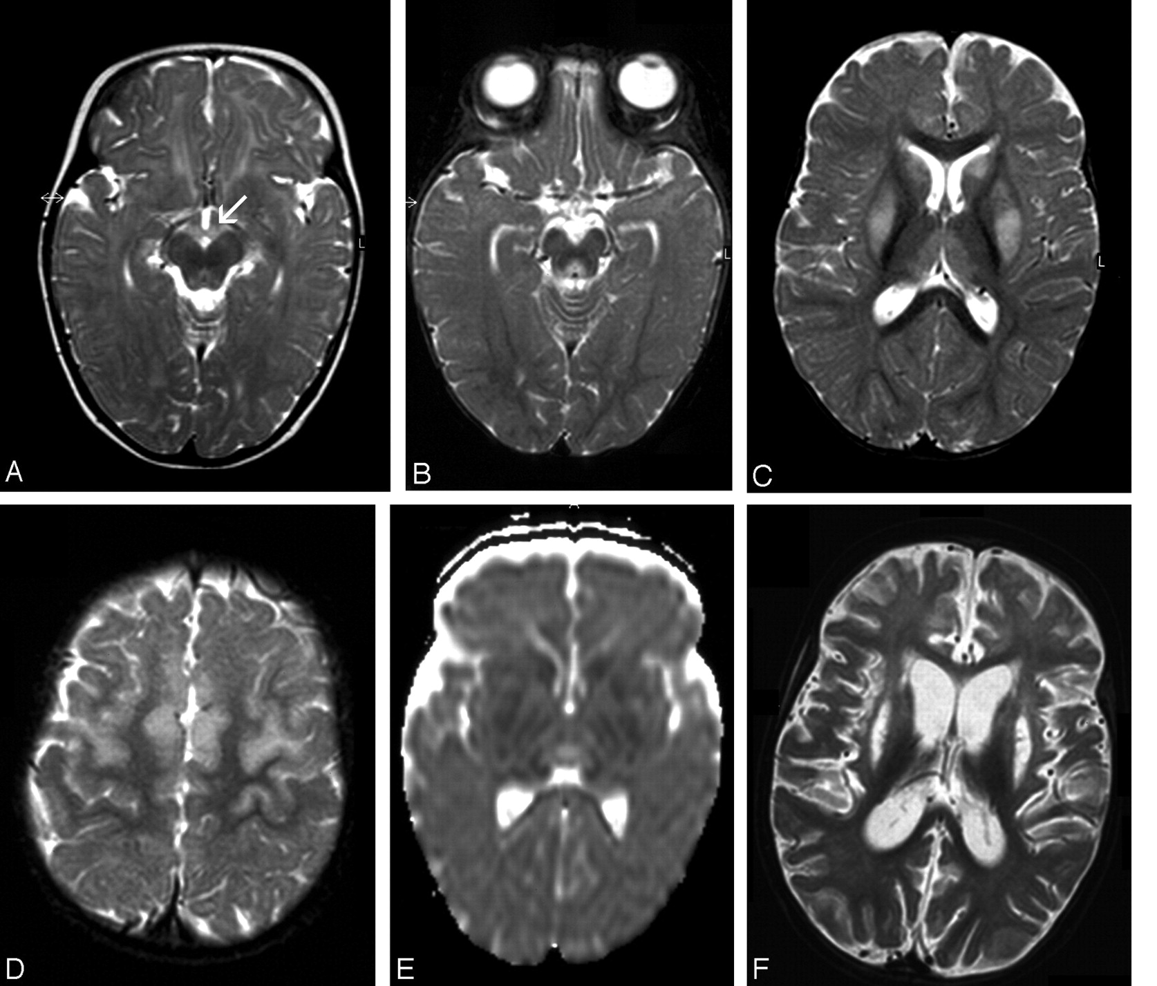
    Patient 1. Images at presentation (A) and follow-up 5 (B–E) and 45 (F) days later.

    A, Axial T2-weighted image shows abnormal hyperintensity in the mammillary bodies (arrow) and tectum.

    B–D, Axial T2-weighted images show abnormal hyperintensity in the periaqueductal region, thalami, basal ganglia, and frontal area. Lesions are bilateral and symmetric.

    E, DWI (ADC) shows restricted diffusion in the basal ganglia. Thalami have hypointensity and hyperintensity, which are presumed to represent cytotoxic and vasogenic edema, respectively.

    F, Axial T2-weighted image shows diffuse parenchymal loss, severe atrophy of the caudate nuclei, and necrosis of the putamina.

  • Fig 2.
    • Download figure
    • Open in new tab
    • Download powerpoint
    Fig 2.

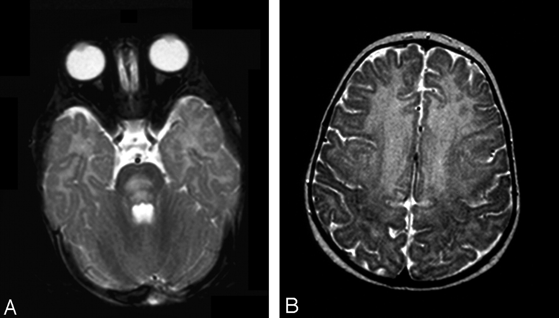
    Patient 2. Axial T2-weighted images.

    A, At presentation, large area of hyperintensity is present in the pons.

    B, Involvement of the frontal region, up to the motor cortex, is extensive.

  • Fig 3.
    • Download figure
    • Open in new tab
    • Download powerpoint
    Fig 3.

    Patient 4. Axial images at presentation.

    A, T2-weighted image.

    B, T1-weighted contrast-enhanced image shows extensive frontal injury. Note enhancement of both cortex and white matter.

  • Fig 4.
    • Download figure
    • Open in new tab
    • Download powerpoint
    Fig 4.

    Patient 5.

    A, T2-weighted image shows subtle abnormal findings, slight hyperintensity of the anterior frontal region, and localized areas of blurring of the cortical stripe.

    B, DWI (ADC) indicates restricted diffusion compatible with cytotoxic edema.

  • Fig 5.
    • Download figure
    • Open in new tab
    • Download powerpoint
    Fig 5.

    Patient 6. Proton MRS image from the periaqueductal region (TR/TE = 1500/144).

    A, At presentation. Note the negative doublet of lactate. NAA/Cr ratio is reduced 1.11.

    B, Five weeks later, the lactate doublet is no longer seen. NAA peak is higher than before.

Tables

  • Figures
    • View popup
    TABLE 1:

    Clinical findings in six infants with thiamine deficiency

    Pt.No.Age(months)GenderLactate LevelsOutcomeLength of Follow-up (months)
    BloodCSF
    18.5F↑↑Dysphagia; severe DD4
    24.5F↑↑Dysphagia; mild DD5
    310M↑↑Dysphagia; moderate DD4
    44.5FN↑Mild DD5
    52F↑↑Complete recovery2
    65F↑↑Mild DD; ataxia4
    • Note.—↑ indicates elevated; N, normal; DD, developmental delay.

    • View popup
    TABLE 2:

    Imaging findings in six children with thiamine deficiency

    PatientNo.Date ofMRPeriaqueductalDWBrain stemDWTectumMammillaryBodiesThalamiDWCaudateDWPutamenDWFrontal Cortexand WMDWVolumeLoss
    125.9.03−N−N++−N−N−N−N−
    1.10.03+C,V+N+++C,V+C+C+C−
    7.11.03+ImpN−NNecrosisAtrophy+NecrosisNHem Atrophy NecrosisNHem Atrophy NecrosisNLaminar necrosis LeukomalaciaN+
    25.11.03+N+C−++C−C−N+C−
    B1→
    10.11.03−NA−NA−++ImpNA−NA−NAImp cortexNA−
    26.11.03−N+N−Atrophy−N−N−NLaminar necrosis LeukomalaciaN+
    33.11.03+NA−NA−−+NA−NA+NA+NA+
    B1→
    17.11.03+ImpN−N−Atrophy+ImpN+WorseC+C+ImpNWorse
    425.9.03+V+N−++V−N+N+V+
    8.11.03−N−N−−ImpN+N+N+ImpNWorse
    56.11.03−N−N−NA−N−N−N−C−
    613.10.03+N−N−++N−N+N+N−
    B1→
    17.11.03−N−N−−−N−NNecrosisN+N−
    9.2.04−N−N−−−N−NAtrophyN+N+
    • Note.—+ indicates high signals on T2, FLAIR, or PD; −, normal signals; DW, diffusion-weighted images; C, cytotoxic edema; V, vasogenic edema; WM, white matter; NA, not available. N, normal or no signs of acute edema; Imp, improvement; Hem, hemorrhage; B1, start of treatment with B1.

PreviousNext
Back to top

In this issue

American Journal of Neuroradiology: 26 (7)
American Journal of Neuroradiology
Vol. 26, Issue 7
1 Aug 2005
  • Table of Contents
  • Index by author
Advertisement
Print
Download PDF
Email Article

Thank you for your interest in spreading the word on American Journal of Neuroradiology.

NOTE: We only request your email address so that the person you are recommending the page to knows that you wanted them to see it, and that it is not junk mail. We do not capture any email address.

Enter multiple addresses on separate lines or separate them with commas.
Thiamine Deficiency in Infants: MR Findings in the Brain
(Your Name) has sent you a message from American Journal of Neuroradiology
(Your Name) thought you would like to see the American Journal of Neuroradiology web site.
CAPTCHA
This question is for testing whether or not you are a human visitor and to prevent automated spam submissions.
Cite this article
Liora Kornreich, Efrat Bron-Harlev, Chen Hoffmann, Michael Schwarz, Osnat Konen, Tommy Schoenfeld, Rachel Straussberg, Elhanan Nahum, Abu-Kishk Ibrahim, Gideon Eshel, Gadi Horev
Thiamine Deficiency in Infants: MR Findings in the Brain
American Journal of Neuroradiology Aug 2005, 26 (7) 1668-1674;

Citation Manager Formats

  • BibTeX
  • Bookends
  • EasyBib
  • EndNote (tagged)
  • EndNote 8 (xml)
  • Medlars
  • Mendeley
  • Papers
  • RefWorks Tagged
  • Ref Manager
  • RIS
  • Zotero
0 Responses
Respond to this article
Share
Bookmark this article
Thiamine Deficiency in Infants: MR Findings in the Brain
Liora Kornreich, Efrat Bron-Harlev, Chen Hoffmann, Michael Schwarz, Osnat Konen, Tommy Schoenfeld, Rachel Straussberg, Elhanan Nahum, Abu-Kishk Ibrahim, Gideon Eshel, Gadi Horev
American Journal of Neuroradiology Aug 2005, 26 (7) 1668-1674;
del.icio.us logo Twitter logo Facebook logo Mendeley logo
  • Tweet Widget
  • Facebook Like
  • Google Plus One

Jump to section

  • Article
    • Abstract
    • Methods
    • Results
    • Discussion
    • Conclusion
    • References
  • Figures & Data
  • Info & Metrics
  • Responses
  • References
  • PDF

Related Articles

  • No related articles found.
  • PubMed
  • Google Scholar

Cited By...

  • Signal Change in the Mammillary Bodies after Perinatal Asphyxia
  • Cranial Ultrasonography in Infantile Encephalitic Beriberi: A Useful First-Line Imaging Tool for Screening and Diagnosis in Suspected Cases
  • Infantile Wernicke's encephalopathy
  • TEACHING NEUROIMAGES: THE FULL-BLOWN NEUROIMAGING OF WERNICKE ENCEPHALOPATHY
  • Epilepsy in children with infantile thiamine deficiency
  • Crossref
  • Google Scholar

This article has not yet been cited by articles in journals that are participating in Crossref Cited-by Linking.

More in this TOC Section

  • Comparison of Image Quality and Radiation Dose in Pediatric Temporal Bone CT Using Photon-Counting Detector CT and Energy-Integrating Detector CT
  • SyMRI & MR Fingerprinting in Brainstem Myelination
  • Gad Contrast in PEDS Seizure Evaluation
Show more Pediatrics

Similar Articles

Advertisement

Indexed Content

  • Current Issue
  • Accepted Manuscripts
  • Article Preview
  • Past Issues
  • Editorials
  • Editors Choice
  • Fellow Journal Club
  • Letters to the Editor

Cases

  • Case Collection
  • Archive - Case of the Week
  • Archive - Case of the Month
  • Archive - Classic Case

Special Collections

  • Special Collections

Resources

  • News and Updates
  • Turn around Times
  • Submit a Manuscript
  • Author Policies
  • Manuscript Submission Guidelines
  • Evidence-Based Medicine Level Guide
  • Publishing Checklists
  • Graphical Abstract Preparation
  • Imaging Protocol Submission
  • Submit a Case
  • Become a Reviewer/Academy of Reviewers
  • Get Peer Review Credit from Publons

Multimedia

  • AJNR Podcast
  • AJNR SCANtastic
  • Video Articles

About Us

  • About AJNR
  • Editorial Board
  • Not an AJNR Subscriber? Join Now
  • Alerts
  • Feedback
  • Advertise with us
  • Librarian Resources
  • Permissions
  • Terms and Conditions

American Society of Neuroradiology

  • Not an ASNR Member? Join Now

© 2025 by the American Society of Neuroradiology All rights, including for text and data mining, AI training, and similar technologies, are reserved.
Print ISSN: 0195-6108 Online ISSN: 1936-959X

Powered by HighWire