
Article Figures & Data
Figures
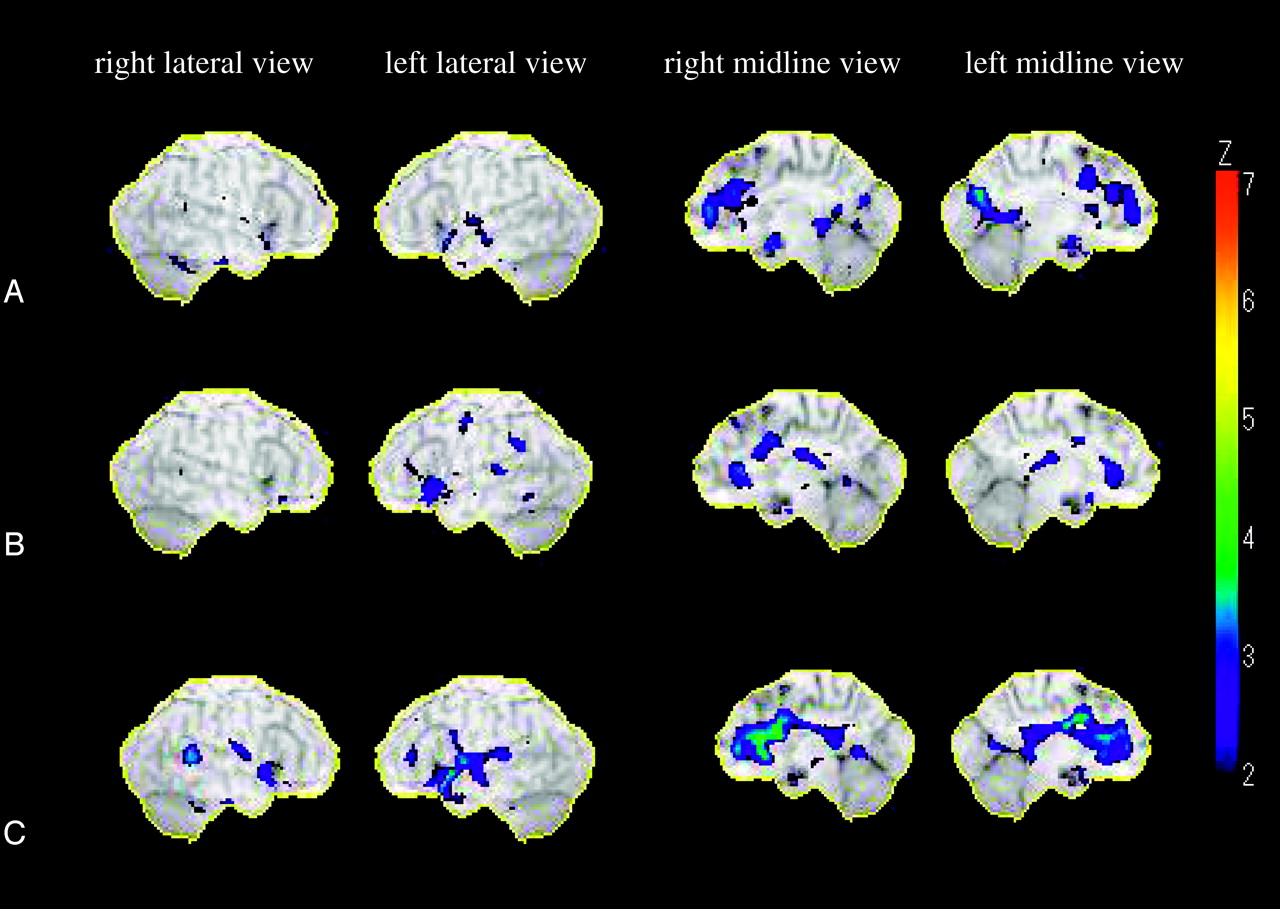
- Fig 1.
Statistical maps analyzed by 3D SSP. The color of the outer counter corresponds to a Z score of 7.
A, Relative decreases of rCBF (z score ≦2) in subjects 60–69 years of age compared with rCBF in subjects 50–59 years of age. The most extensive reduction in rCBF was observed in the right anterior cingulate gyrus. There were also decreases in rCBF in the left anterior cingulate gyrus, bilateral posterior cingulate gyri, bilateral medial frontal gyri, bilateral subcallosal gyri, left superior temporal gyrus, and left cuneus.
B, Relative decreases of rCBF (z score ≦2) in subjects 70–79 years of age compared with rCBF in subjects 60–69 years of age. The most extensive reduction in rCBF was observed in the bilateral anterior cingulate gyri. There were also decreases in rCBF in the left inferior frontal gyrus, left subcallosal gyrus, left supramarginal gyrus, and left superior temporal gyrus.
C, Relative decreases of rCBFs (z score ≦2) in subjects 70–79 years of age compared with rCBF in subjects 50–59 years of age. The most extensive reduction in rCBF was observed in the bilateral anterior cingulate gyri. There were also decreases in rCBF in the left inferior frontal gyrus, left subcallosal gyrus, left supramarginal gyrus, and left superior temporal gyrus.

Tables
Age group (y) 50–59 60–69 70–79 Number (M:F) 10 (4:6) 12 (7:5) 9 (5:4) Mean age (y) 55.2 ± 3.0 63.8 ± 2.9 73.2 ± 2.4 Okabe’s Scale, total 48.9 ± 3.0 47.0 ± 5.7 45.4 ± 6.9 Information 18.4 ± 1.6 17.5 ± 1.7 16.4 ± 1.9 Mental control 14.5 ± 1.6 12.4 ± 3.3 12.8 ± 2.9 Digit span 9.0 ± 1.6 8.9 ± 1.5 9.2 ± 0.7 Associate learning 7.0 ± 3.9 8.2 ± 3.6 7.0 ± 3.6 SDS 30.8 ± 5.9 28.9 ± 4.3 29.7 ± 5.7 Note.—Okabe Scale indicates Okabe’s Simplified Intelligence Scale (60-point scale); SDS, Zung Self-Rating Depression Scale (80-point scale).
Relative Decrease of rCBF, Age (y) 60–69 vs 50–59 70–79 vs 60–69 70–79 vs 50–59 Superior frontal gyrus L 0.80 ± 0.51 1.59 ± 0.45 0.64 ± 0.46 R 0.73 ± 0.59 1.49 ± 0.35 0.67 ± 0.41 Middle frontal gyrus L 0.60 ± 0.43 1.37 ± 0.32 0.99 ± 0.58 R 0.66 ± 0.53 1.49 ± 0.44 0.66 ± 0.48 Inferior frontal gyrus L 0.71 ± 0.53 1.97 ± 0.57 1.53 ± 0.72 R 0.53 ± 0.37 1.39 ± 0.38 1.14 ± 0.64 Medial frontal gyrus L 1.38 ± 0.84 1.62 ± 0.58 1.33 ± 0.99 R 1.37 ± 0.94 1.30 ± 0.16 1.37 ± 0.97 Subcallosal gyrus L 1.30 ± 1.04 2.08 ± 0.54 1.73 ± 0.59 R 1.47 ± 1.11 1.22 ± 0.13 0.83 ± 0.37 Superior parietal lobule L 0.24 ± 0.17 1.27 ± 0.17 0 R 0 0 0 Inferior parietal lobule L 0.12 ± 0.12 1.50 ± 0.35 0.63 ± 0.48 R 0.63 ± 0.55 1.12 ± 0.06 0.56 ± 0.40 Superior temporal gyrus L 1.19 ± 0.72 1.61 ± 0.43 1.97 ± 0.82 R 1.03 ± 0.69 1.37 ± 0.24 1.33 ± 0.96 Middle temporal gyrus L 0.95 ± 0.74 1.44 ± 0.34 1.13 ± 0.72 R 0.42 ± 0.28 1.47 ± 0.27 0.82 ± 0.69 Inferior temporal gyrus L 0.76 ± 0.42 1.56 ± 0.47 0.82 ± 0.45 R 0.92 ± 0.66 1.27 ± 0.19 1.20 ± 0.65 Cuneus L 1.29 ± 0.92 0 0.71 ± 0.63 R 0.93 ± 0.74 1.08 ± 0.04 0.36 ± 0.28 Lingual gyrus L 0.70 ± 0.58 0 0.84 ± 0.55 R 0.49 ± 0.47 1.39 ± 0.25 0.70 ± 0.38 Anterior cingulate gyrus L 1.55 ± 0.61 1.87 ± 0.59 2.51 ± 0.81 R 1.79 ± 0.73 2.02 ± 0.60 2.89 ± 0.99 Posterior cingulate gyrus L 1.31 ± 1.11 1.40 ± 0.27 1.42 ± 0.70 R 1.20 ± 0.81 1.56 ± 0.26 1.51 ± 0.48 Parahippocampal gyrus L 0.44 ± 0.24 1.43 ± 0.29 0.97 ± 0.52 R 1.26 ± 0.65 1.73 ± 0.59 0.78 ± 0.45 Superior occipital gyrus L 0.06 ± 0 0 0 R 0.76 ± 0.28 0 0.29 ± 0.15 Middle occipital gyrus L 0.47 ± 0.44 0 0.45 ± 0.19 R 0.68 ± 0.39 0 0.56 ± 0.45 Inferior occipital gyrus L 0 0 0 R 0.05 ± 0 0 0
In this issue


Jump to section
Related Articles
Cited By...
- No citing articles found.