Advertisement

Main menu

  • Home
  • Content
    • Current Issue
    • Accepted Manuscripts
    • Article Preview
    • Past Issue Archive
    • AJNR Case Collection
    • Case of the Week Archive
    • Classic Case Archive
    • Case of the Month Archive
  • Special Collections
    • Spinal CSF Leak Articles (Jan 2020-June 2024)
    • 2024 AJNR Journal Awards
    • Most Impactful AJNR Articles
  • Multimedia
    • AJNR Podcast
    • AJNR Scantastics
    • Video Articles
  • For Authors
    • Submit a Manuscript
    • Author Policies
    • Fast publishing of Accepted Manuscripts
    • Graphical Abstract Preparation
    • Manuscript Submission Guidelines
    • Imaging Protocol Submission
    • Submit a Case for the Case Collection
  • About Us
    • About AJNR
    • Editorial Board
  • More
    • Become a Reviewer/Academy of Reviewers
    • Subscribers
    • Permissions
    • Alerts
    • Feedback
    • Advertisers
    • ASNR Home
  • Other Publications
    • ajnr

User menu

  • Alerts
  • Log in

Search

  • Advanced search
American Journal of Neuroradiology

American Journal of Neuroradiology

ASHNR American Society of Functional Neuroradiology ASHNR American Society of Pediatric Neuroradiology ASSR
  • Alerts
  • Log in

Advanced Search

  • Home
  • Content
    • Current Issue
    • Accepted Manuscripts
    • Article Preview
    • Past Issue Archive
    • AJNR Case Collection
    • Case of the Week Archive
    • Classic Case Archive
    • Case of the Month Archive
  • Special Collections
    • Spinal CSF Leak Articles (Jan 2020-June 2024)
    • 2024 AJNR Journal Awards
    • Most Impactful AJNR Articles
  • Multimedia
    • AJNR Podcast
    • AJNR Scantastics
    • Video Articles
  • For Authors
    • Submit a Manuscript
    • Author Policies
    • Fast publishing of Accepted Manuscripts
    • Graphical Abstract Preparation
    • Manuscript Submission Guidelines
    • Imaging Protocol Submission
    • Submit a Case for the Case Collection
  • About Us
    • About AJNR
    • Editorial Board
  • More
    • Become a Reviewer/Academy of Reviewers
    • Subscribers
    • Permissions
    • Alerts
    • Feedback
    • Advertisers
    • ASNR Home
  • Follow AJNR on Twitter
  • Visit AJNR on Facebook
  • Follow AJNR on Instagram
  • Join AJNR on LinkedIn
  • RSS Feeds

Welcome to the new AJNR, Updated Hall of Fame, and more. Read the full announcements.


AJNR is seeking candidates for the position of Associate Section Editor, AJNR Case Collection. Read the full announcement.


Research ArticleINTERVENTIONAL

Endovascular Treatment of Intracranial Aneurysms with a Branch Arising from the Sac

B. Lubicz, F. Lefranc, M. Levivier, O. Dewitte, B. Pirotte, J. Brotchi and D. Balériaux
American Journal of Neuroradiology January 2006, 27 (1) 142-147;
B. Lubicz
  • Find this author on Google Scholar
  • Find this author on PubMed
  • Search for this author on this site
F. Lefranc
  • Find this author on Google Scholar
  • Find this author on PubMed
  • Search for this author on this site
M. Levivier
  • Find this author on Google Scholar
  • Find this author on PubMed
  • Search for this author on this site
O. Dewitte
  • Find this author on Google Scholar
  • Find this author on PubMed
  • Search for this author on this site
B. Pirotte
  • Find this author on Google Scholar
  • Find this author on PubMed
  • Search for this author on this site
J. Brotchi
  • Find this author on Google Scholar
  • Find this author on PubMed
  • Search for this author on this site
D. Balériaux
  • Find this author on Google Scholar
  • Find this author on PubMed
  • Search for this author on this site
  • Article
  • Figures & Data
  • Info & Metrics
  • Responses
  • References
  • PDF
Loading

Article Figures & Data

Figures

  • Tables
  • Fig 1.
    • Download figure
    • Open in new tab
    • Download powerpoint
    Fig 1.

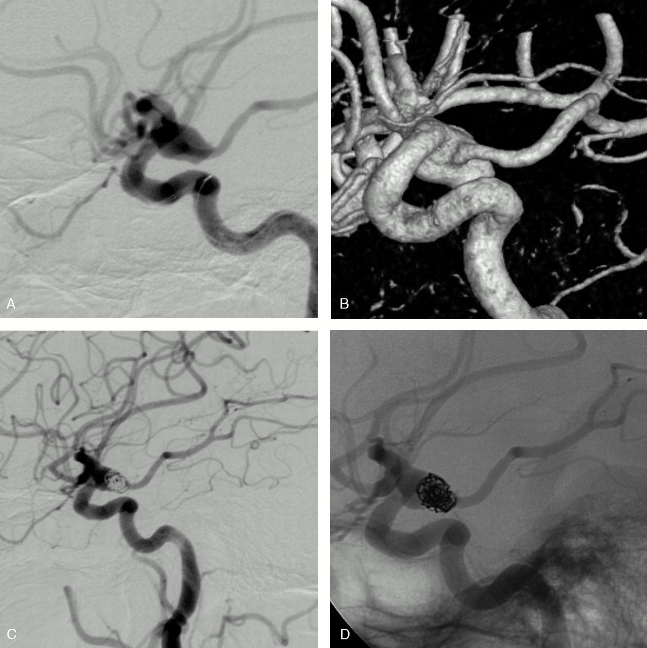
    Unruptured MCA aneurysm in a 48-year-old woman treated by selective embolization.

    A, Conventional angiogram shows a 5-mm aneurysm of the proximal MCA.

    B, 3D angiogram shows a sylvian branch arising from the sac.

    C, HyperForm remodeling balloon overinflated in the M1 segment. A part of the balloon is clearly bulging within the aneurysmal sac to protect the arterial branch.

    D–F, Final angiographic result: conventional subtracted (D), unsubtracted (E), and 3D (F) angiograms show a neck remnant and the preservation of the arterial branch.

  • Fig 2.
    • Download figure
    • Open in new tab
    • Download powerpoint
    Fig 2.

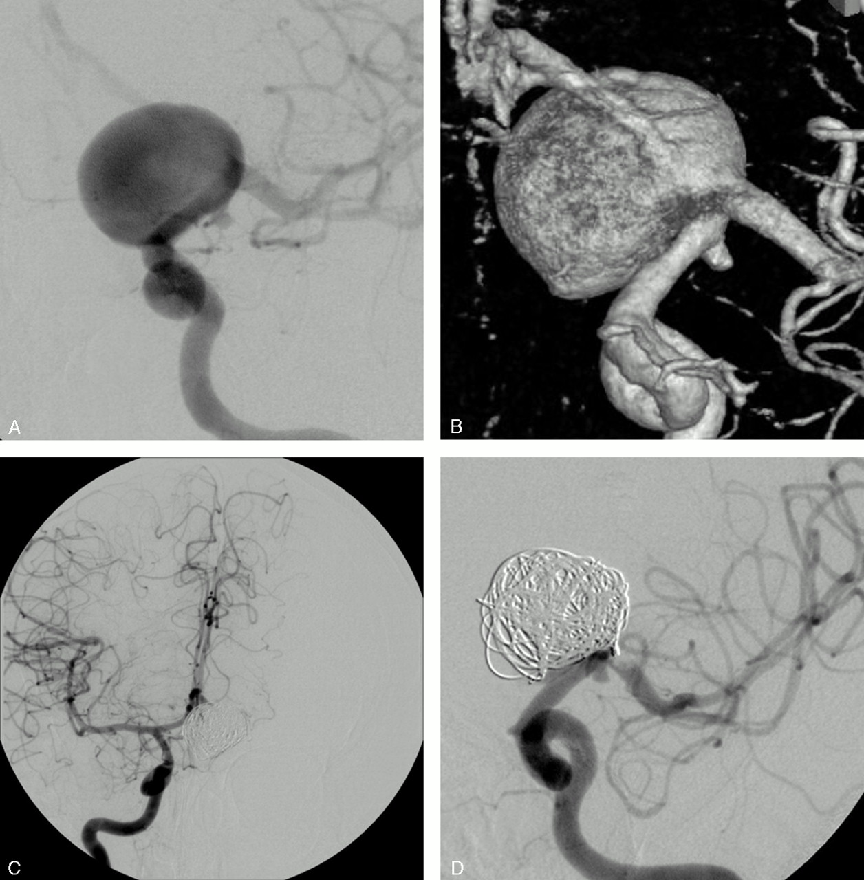
    Unruptured MCA aneurysm in a 56-year-old woman.

    Conventional (A) and 3D (B) angiograms show a wide-necked M1 aneurysm with a branch arising from the sac.

    C, Conventional angiogram at the end of the embolization shows an asymmetric reconstruction of the neck with patency of the arterial branch.

  • Fig 3.
    • Download figure
    • Open in new tab
    • Download powerpoint
    Fig 3.

    SAH in a 74-year-old woman.

    Conventional (A) and 3D (B) angiograms show a wide-necked PcomA aneurysm with the fetal-type PcomA arising from the mid-third of the sac.

    Conventional subtracted (C) and unsubtracted (D) angiograms at the end of the embolization show an incomplete aneurysm occlusion and the preservation of the PcomA.

  • Fig 4.
    • Download figure
    • Open in new tab
    • Download powerpoint
    Fig 4.

    SAH in a 64-year-old woman.

    Conventional (A) and 3D (B) angiograms show a 20-mm aneurysm of the left ICA bifurcation with the A1 segment arising from the sac.

    C, A balloon test occlusion shows the efficient collateral circulation through the AcomA.

    D, Conventional angiogram at the end of the embolization shows a complete aneurysm occlusion and the sacrifice of the origin of the left A1 segment.

  • Fig 5.
    • Download figure
    • Open in new tab
    • Download powerpoint
    Fig 5.

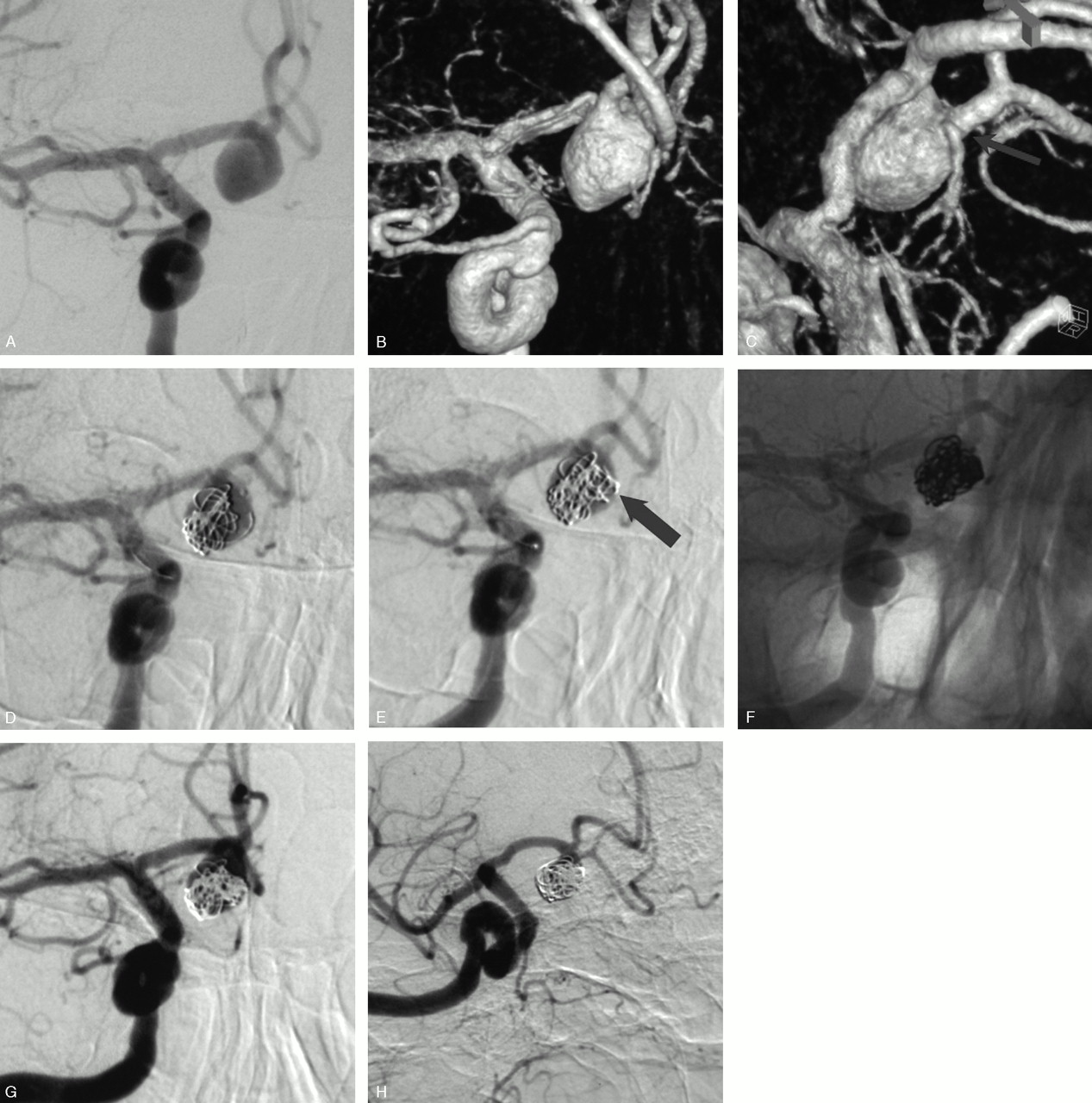
    SAH in a 73-year-old man with a ruptured AcomA aneurysm previously wrapped 4 years before.

    Conventional (A) and 3D (B and C) angiograms show a 10-mm AcomA aneurysm with the right A2 segment arising from the sac (arrow).

    D, Conventional angiogram shows a first “stabilizing” 3D coil of 10 mm placed with fewest loops in front of the arterial branch.

    E, Conventional angiogram shows a second and smaller (5-mm coil) 3D cage placed in the aneurysm inflow zone (arrow).

    Conventional unsubtracted (F) and subtracted (G) angiograms at the end of the embolization show an incomplete aneurysm occlusion and the patency of the arterial branch.

    H, Angiographic control at one week shows a further aneurysm thrombosis with preservation of the arterial branch.

Tables

  • Figures
    • View popup
    Table 1:

    Endovascular treatment of intracranial aneurysms with a branch arising from the sac: clinical and anatomic findings in 9 patients

    Patient No./Age (y)/SexClinical PresentationAneurysm Size (mm)Aneurysm LocationTreatmentClinical Outcome (GOS)
    1/73/MSAH10AcomAOcclusion of the aneurysm inflow zoneExcellent
    2/70/FIncidental9MCABalloon-assisted techniqueExcellent
    3/56/FIncidental7MCABalloon-assisted techniqueExcellent
    4/48/FIncidental5MCABalloon-assisted techniqueExcellent
    5/64/FSAH20ICA bifurcationLeft A1 sacrificeExcellent
    6/74/FSAH9PcomABalloon-assisted techniqueDeath < SAH
    7/50/FSAH3AchABalloon-assisted techniqueDeath < SAH
    8/47/FSAH4AcomABalloon-assisted techniqueExcellent
    9/43/MSAH4SCASCA sacrificeExcellent
    • Note:—AcomA indicates anterior communicating artery; PcomA, posterior communicating artery; ICA, internal carotid artery; SCA, superior cerebellar artery; AchA, anterior choroidal artery; MCA, middle cerebral artery; A1, first segment of the anterior cerebral artery; SAH, subarachnoid hemorrhage; GOS, Modified Glasgow Outcome Scale.

    • View popup
    Table 2:

    Aneurysm location and anatomic configuration

    Patient No.Aneurysm LocationArterial Branch Arising from the Sac
    1AcomAA2 segment from right ACA
    2Prebifurcation M1 segment from MCASylvian branch
    3Prebifurcation M1 segment from MCASylvian branch
    4Prebifurcation M1 segment from MCASylvian branch
    5ICA bifurcationA1 segment from left ACA
    6PcomAFetal type PcomA
    7AchAAchA
    8AcomAMedian pericallosal artery
    9BA-SCASCA
    • Note:—AcomA indicates anterior communicating artery; ACA, anterior cerebral artery; M1, first segment of the middle cerebral artery; PcomA, posterior communicating artery; ICA, internal carotid artery; BA, basilar artery; SCA, superior cerebellar artery; AchA, anterior choroidal artery; MCA, middle cerebral artery; A2, second segment of the anterior cerebral artery.

PreviousNext
Back to top

In this issue

American Journal of Neuroradiology: 27 (1)
American Journal of Neuroradiology
Vol. 27, Issue 1
January, 2006
  • Table of Contents
  • Index by author
Advertisement
Print
Download PDF
Email Article

Thank you for your interest in spreading the word on American Journal of Neuroradiology.

NOTE: We only request your email address so that the person you are recommending the page to knows that you wanted them to see it, and that it is not junk mail. We do not capture any email address.

Enter multiple addresses on separate lines or separate them with commas.
Endovascular Treatment of Intracranial Aneurysms with a Branch Arising from the Sac
(Your Name) has sent you a message from American Journal of Neuroradiology
(Your Name) thought you would like to see the American Journal of Neuroradiology web site.
CAPTCHA
This question is for testing whether or not you are a human visitor and to prevent automated spam submissions.
Cite this article
B. Lubicz, F. Lefranc, M. Levivier, O. Dewitte, B. Pirotte, J. Brotchi, D. Balériaux
Endovascular Treatment of Intracranial Aneurysms with a Branch Arising from the Sac
American Journal of Neuroradiology Jan 2006, 27 (1) 142-147;

Citation Manager Formats

  • BibTeX
  • Bookends
  • EasyBib
  • EndNote (tagged)
  • EndNote 8 (xml)
  • Medlars
  • Mendeley
  • Papers
  • RefWorks Tagged
  • Ref Manager
  • RIS
  • Zotero
0 Responses
Respond to this article
Share
Bookmark this article
Endovascular Treatment of Intracranial Aneurysms with a Branch Arising from the Sac
B. Lubicz, F. Lefranc, M. Levivier, O. Dewitte, B. Pirotte, J. Brotchi, D. Balériaux
American Journal of Neuroradiology Jan 2006, 27 (1) 142-147;
del.icio.us logo Twitter logo Facebook logo Mendeley logo
  • Tweet Widget
  • Facebook Like
  • Google Plus One

Jump to section

  • Article
    • Abstract
    • Patients and Methods
    • Results
    • Discussion
    • Conclusion
    • References
  • Figures & Data
  • Info & Metrics
  • Responses
  • References
  • PDF

Related Articles

  • No related articles found.
  • PubMed
  • Google Scholar

Cited By...

  • Combination of Multicatheter Plus Stent or Balloon for Treatment of Complex Aneurysms
  • Stent assisted coil embolization of wide-necked bilobed anterior inferior cerebellar artery aneurysm with incorporated artery arising from the dome: a technical note
  • Coil Protection Using Small Helical Coils for Wide-Neck Intracranial Aneurysms: A Novel Approach
  • Treatment of Intracranial Aneurysms Using the Pipeline Flow-Diverter Embolization Device: A Single-Center Experience with Long-Term Follow-Up Results
  • Tuberothalamic Artery Infarctions following Coil Embolization of Ruptured Posterior Communicating Artery Aneurysms with Posterior Communicating Artery Sacrifice
  • Endovascular Coil Embolization of Aneurysms with a Branch Incorporated into the Sac
  • Crossref
  • Google Scholar

This article has not yet been cited by articles in journals that are participating in Crossref Cited-by Linking.

More in this TOC Section

  • SAVE vs. Solumbra Techniques for Thrombectomy
  • CT Perfusion&Reperfusion in Acute Ischemic Stroke
  • Delayed Reperfusion Post-Thrombectomy&Thrombolysis
Show more Interventional

Similar Articles

Advertisement

Indexed Content

  • Current Issue
  • Accepted Manuscripts
  • Article Preview
  • Past Issues
  • Editorials
  • Editors Choice
  • Fellow Journal Club
  • Letters to the Editor

Cases

  • Case Collection
  • Archive - Case of the Week
  • Archive - Case of the Month
  • Archive - Classic Case

Special Collections

  • Special Collections

Resources

  • News and Updates
  • Turn around Times
  • Submit a Manuscript
  • Author Policies
  • Manuscript Submission Guidelines
  • Evidence-Based Medicine Level Guide
  • Publishing Checklists
  • Graphical Abstract Preparation
  • Imaging Protocol Submission
  • Submit a Case
  • Become a Reviewer/Academy of Reviewers
  • Get Peer Review Credit from Publons

Multimedia

  • AJNR Podcast
  • AJNR SCANtastic
  • Video Articles

About Us

  • About AJNR
  • Editorial Board
  • Not an AJNR Subscriber? Join Now
  • Alerts
  • Feedback
  • Advertise with us
  • Librarian Resources
  • Permissions
  • Terms and Conditions

American Society of Neuroradiology

  • Not an ASNR Member? Join Now

© 2025 by the American Society of Neuroradiology All rights, including for text and data mining, AI training, and similar technologies, are reserved.
Print ISSN: 0195-6108 Online ISSN: 1936-959X

Powered by HighWire