
Article Figures & Data
Figures
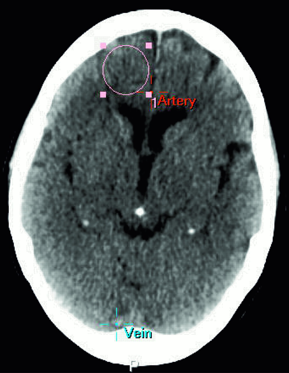
- Fig 1.
A circular region of interest with a standard size of 2.5 cm in diameter was drawn in the peripheral anterior flow territory at the level of the basal ganglia on a standard location containing both white and gray matter. The location of the measurement of the AIF (artery) and the VOF (vein) are also indicated.
- Fig 2.
Mean AUC for the VOF (A), AIF (B) and mean widths of the AIF (C) for 12-mm (○), 6-mm (▪), and 3-mm (•) section thickness in 10 patients. In all patients, a smaller section thickness results in an equal or larger AUC of the VOF (A) and AIF (B). The width of the AIF decreases slightly (C) with a smaller section thickness in most patients.
- Fig 3.
Raw attenuation curves of the optimal VOF for 12-mm (○), 6-mm (▪), and 3-mm (•) section thickness (same patient as in Fig 1).


Tables
Difference (%) P Value Difference (%) P Value Mean AUC VOF AUC of optimal VOF 6 vs 12 +17 .017 6 vs 12 +17 .014 3 vs 12 +20 .004 3 vs 12 +20 .009 3 vs 6 +4 .006 3 vs 6 +3 .056 Mean AUC AIF AUC of optimal AIF 6 vs 12 +22 .000 6 vs 12 +25 .000 3 vs 12 +34 .000 3 vs 12 +39 .000 3 vs 6 +15 .004 3 vs 6 +19 .003 Mean width AIF Width of optimal AIF 6 vs 12 −7 .063 6 vs 12 −1 .88 3 vs 12 −8 .007 3 vs 12 −2 .42 3 vs 6 −2 .328 3 vs 6 −1 .44 Note:—AUC indicates area under the curve; VOF, venous output function; AIF, arterial input function.
Section Thickness (mm) CBV (mL/100 g) 12 4.3 ± 1.53 6 3.7 ± 1.15 3 3.1 ± 0.99 CBF (mL/100 g/min) 12 61.1 ± 15.7 6 48.6 ± 12.5 3 34.4 ± 8.07 MTT (s) 12 4.2 ± 0.78 6 4.6 ± 0.79 3 5.1 ± 0.74 Note:—CBV indicates cerebral blood volume; CBF, cerebral blood flow; MTT, mean transit time. Values are means ± SD.


