Advertisement

Main menu

  • Home
  • Content
    • Current Issue
    • Accepted Manuscripts
    • Article Preview
    • Past Issue Archive
    • AJNR Case Collection
    • Case of the Week Archive
    • Classic Case Archive
    • Case of the Month Archive
  • Special Collections
    • Spinal CSF Leak Articles (Jan 2020-June 2024)
    • 2024 AJNR Journal Awards
    • Most Impactful AJNR Articles
  • Multimedia
    • AJNR Podcast
    • AJNR Scantastics
    • Video Articles
  • For Authors
    • Submit a Manuscript
    • Author Policies
    • Fast publishing of Accepted Manuscripts
    • Graphical Abstract Preparation
    • Manuscript Submission Guidelines
    • Imaging Protocol Submission
    • Submit a Case for the Case Collection
  • About Us
    • About AJNR
    • Editorial Board
  • More
    • Become a Reviewer/Academy of Reviewers
    • Subscribers
    • Permissions
    • Alerts
    • Feedback
    • Advertisers
    • ASNR Home
  • Other Publications
    • ajnr

User menu

  • Alerts
  • Log in

Search

  • Advanced search
American Journal of Neuroradiology

American Journal of Neuroradiology

ASHNR American Society of Functional Neuroradiology ASHNR American Society of Pediatric Neuroradiology ASSR
  • Alerts
  • Log in

Advanced Search

  • Home
  • Content
    • Current Issue
    • Accepted Manuscripts
    • Article Preview
    • Past Issue Archive
    • AJNR Case Collection
    • Case of the Week Archive
    • Classic Case Archive
    • Case of the Month Archive
  • Special Collections
    • Spinal CSF Leak Articles (Jan 2020-June 2024)
    • 2024 AJNR Journal Awards
    • Most Impactful AJNR Articles
  • Multimedia
    • AJNR Podcast
    • AJNR Scantastics
    • Video Articles
  • For Authors
    • Submit a Manuscript
    • Author Policies
    • Fast publishing of Accepted Manuscripts
    • Graphical Abstract Preparation
    • Manuscript Submission Guidelines
    • Imaging Protocol Submission
    • Submit a Case for the Case Collection
  • About Us
    • About AJNR
    • Editorial Board
  • More
    • Become a Reviewer/Academy of Reviewers
    • Subscribers
    • Permissions
    • Alerts
    • Feedback
    • Advertisers
    • ASNR Home
  • Follow AJNR on Twitter
  • Visit AJNR on Facebook
  • Follow AJNR on Instagram
  • Join AJNR on LinkedIn
  • RSS Feeds

Welcome to the new AJNR, Updated Hall of Fame, and more. Read the full announcements.


AJNR is seeking candidates for the position of Associate Section Editor, AJNR Case Collection. Read the full announcement.


OtherBRAIN

Acute and Chronic Brain Infarcts on MR Imaging in a 20-Year-Old Woman with Acute Posterior Multifocal Placoid Pigment Epitheliopathy

A.N. Bugnone, F. Hartker, M. Shapiro, H.S. Pineless and G. Velez
American Journal of Neuroradiology January 2006, 27 (1) 67-69;
A.N. Bugnone
  • Find this author on Google Scholar
  • Find this author on PubMed
  • Search for this author on this site
F. Hartker
  • Find this author on Google Scholar
  • Find this author on PubMed
  • Search for this author on this site
M. Shapiro
  • Find this author on Google Scholar
  • Find this author on PubMed
  • Search for this author on this site
H.S. Pineless
  • Find this author on Google Scholar
  • Find this author on PubMed
  • Search for this author on this site
G. Velez
  • Find this author on Google Scholar
  • Find this author on PubMed
  • Search for this author on this site
  • Article
  • Figures & Data
  • Info & Metrics
  • Responses
  • References
  • PDF
Loading

Abstract

SUMMARY: A 20-year-old woman recently diagnosed with acute posterior multifocal placoid pigment epitheliopathy developed headaches, weakness, and paresthesias. MR imaging of the brain revealed an acute infarct (demonstrated by diffusion-weighted images) in the head of the right caudate nucleus, a chronic infarct with encephalomalacia in the body of the corpus callosum, and multiple foci of abnormal signal intensity in the white matter of the centrum semiovale.

Acute posterior multifocal placoid pigment epitheliopathy (APMPPE) is a chorioretinal disease that has been associated with multiple neurologic complications, including headaches, aseptic meningitis, transient ischemic attacks, and stroke.1 The case presented in this article is unique not only because APMPPE is a rare entity, but also because it is unusual for these patients to develop ischemic brain infarcts and even more unusual to depict them on the acute stage on MR imaging of the brain, especially when the infarcts are clinically silent, as in our case. Moreover, to the best of our knowledge, the neuroimaging findings of APMPPE have not been reported in the radiologic literature.

Case Report

A 20-year-old woman presented for neurology consultation complaining of multiple acute episodes of right-sided paresthesia and right-sided weakness affecting the face, arm, and leg, as well as language disturbance, photophobia, phonophobia, and intense headaches. She had been diagnosed with APMPPE 1.5 months earlier and was treated by her ophthalmologist with a short course of steroids. The patient had been having headaches for several months. On presentation, physical examination revealed visual acuity of 20/25 of the left eye and 20/70 of the right eye. Sensory and motor examination was unremarkable at that time. No CSF or laboratory studies were performed.

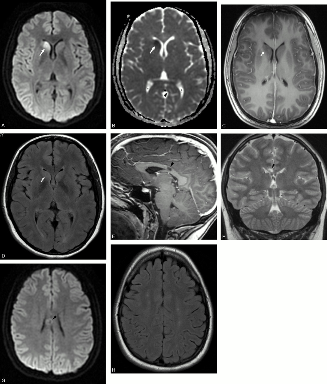
MR imaging of the brain was performed 40 days after the initial episode of neurologic deficit and 3 months after diagnosis of APMPPE, on a 3T magnet (Signa Excite; GE Medical Systems, Milwaukee, Wisc). The head of the right caudate nucleus demonstrated restricted diffusion, normal T1-weighted signal intensity, and slightly increased T2-weighted and fluid-attenuated inversion recovery (FLAIR) signal intensity (Fig 1A–D). No mass or mass effect was seen. There was no abnormal enhancement after the intravenous administration of gadolinium-based contrast material. These findings were consistent with an acute stroke of the head of the right caudate nucleus. There was a focal lesion in the posterior aspect of the body of the corpus callosum, slightly to the right of the midline, which was isointense to CSF in the center and slight hyperintense to gray matter on the T2-weighted and FLAIR sequences in the periphery, without restricted diffusion or abnormal enhancement (Figs 1E–G). This lesion was consistent with a chronic ischemic stroke with central cystic encephalomalacia. In addition, there were multiple foci of increased signal intensity on the T2-weighted and FLAIR sequences in the periventricular white matter and subcortical white matter of the centrum semiovale bilaterally and symmetrically, without abnormal enhancement or restricted diffusion (Fig 1H). These lesions most likely represented microvascular ischemic demyelination, because of central nervous system (CNS) vasculitis, though other demyelinating processes, such as multiple sclerosis, could also demonstrate similar findings in a young patient. There was no evidence of abnormal meningeal enhancement.

Fig 1.
  • Download figure
  • Open in new tab
  • Download powerpoint
Fig 1.

A, Axial diffusion-weighted image (b = 1000) demonstrates increased signal intensity in the head of the right caudate nucleus (arrow).

B, Axial apparent diffusion coefficient map demonstrates decreased apparent diffusion in the head of the right caudate nucleus (arrow).

C, Axial contrast-enhanced 3D fast-spoiled gradient (FSPGR) T1-weighted image (flip angle/TR/TE, 13/5/1.2) after intravenous gadolinium demonstrates normal signal intensity and no enhancement in the head of the right caudate nucleus (arrow).

D, Axial FLAIR image (TR/TE/TI, 8200/123/2000) demonstrates mild increased signal intensity in the head of the right caudate nucleus (arrow).

E, Sagittal contrast-enhanced 3D-FSPGR T1-weighted image (FL/TR/TE, 13/5/1.2) after intravenous gadolinium demonstrates a nonenhancing lesion isointense to CSF in the body of the corpus callosum (arrowhead).

F, Coronal fast spin-echo T2-weighted (TR/TE, 4000/103) image demonstrates the corpus callosum lesion with signal intensity isointense to CSF (arrowhead).

G, Axial diffusion-weighted (b = 1000) image at the level of the lesion in the corpus callosum demonstrates no restricted diffusion (arrowhead).

H, Axial FLAIR image (TR/TE/TI, 8200/123/2000) at the level of the centrum semiovale demonstrates multiple small foci of high signal intensity in the white matter.

Follow-up neurology visits 3 months after the onset of symptoms disclosed that her headaches had resolved spontaneously. Her paresthesias also resolved 2.5 months after her symptoms appeared. She still had subjective eye complaints regarding her visual field deficits, and was scheduled for follow-up with her neuro-ophthalmologist.

Discussion

APMPPE typically affects young adult patients without sex predilection and presents with bilateral acute loss of visual acuity, usually after a flulike syndrome.2 On fundoscopic examination, multifocal yellow-white placoid lesions at the level of the pigment epithelium and choroid of the retina are seen.2 It is a vasculitis of the choroid that secondarily affects the retina and pigment epithelium3 and has been associated with other systemic manifestations of vasculitis.4 The etiology of the vasculitis causing APMPPE is unknown, though various conjectures have associated it with viral infections, Lyme disease, postvaccination autoimmune response, collagen vascular diseases, such as lupus, juvenile rheumatoid arthritis, and other systemic processes, such as sarcoidosis.1

Cerebral ischemia in patients with APMPPE is unusual and manifests clinically as strokes, or less frequently as transient ischemic attacks, with cerebral vasculitis the presumed mechanism of injury.5–9 These patients usually have evidence of inflammation of the CSF or aseptic meningitis.1 Infarcts usually affect the basal ganglia or the posterior circulation.1,7 Large ischemic strokes can occur and have been reported to result in patient death.10,11 The onset of stroke usually occurs within 4–5 months of the onset of ocular disease, though is typically concomitant.1 Strokes can also occur or recur during the tapering of immunosuppressive therapy.1

Caudate nucleus infarcts are uncommon, because of the rich blood supply from the anterior and lateral lenticulostriate arteries and the recurrent artery of Heubner. Caudate nucleus infarctions are more frequently caused by small vessel disease than emboli11 and have been reported in patients with APMPPE.1 The patient we are presenting had an ischemic infarction of the head of the right caudate nucleus, which is presumed to be due to small vessel vasculitis, on the basis of the underlying diagnosis of APMPPE.

Corpus callosum infarctions are also very rare, because of the rich blood supply from the paired pericallosal and posterior pericallosal arteries, and are also most frequently related to small vessel disease.12 We assume the lesion in the corpus callosum in this case also represents an old infarct with central cystic encephalomalacia due to vasculitis secondary to the underlying diagnosis of APMPPE.

In light of the clinical and imaging context, the periventricular and subcortical white matter lesions seen in our case are most likely due to microvascular ischemic demyelination due to CNS vasculitis. This pattern can also be seen in patients with demyelination related to other vasculitides, multiple sclerosis, or chronic small vessel disease (usually in older patients).

The differential diagnosis in a young patient with multiple ischemic infarcts in different stages, small-vessel supratentorial white matter disease, and loss of visual acuity includes recurrent emboli, vasculitis (systemic lupus erythematosus, Takayasu arteritis, Susac syndrome, Behcet syndrome, neurosyphilis, and APMPPE) and thrombophilias (antiphospholipid antibodies, sickle cell disease). Multiple or recurrent emboli in young patients are usually related to congenital heart disease. Vasculitis from systemic lupus erythematosus can result in multiple brain infracts and in optic neuropathy, which may account for visual symptoms.13 Takayasu arteritis is a large vessel vasculitis that occurs most frequently in adolescents and young adults; patients usually have visual symptoms and can complicate with strokes believed to be secondary to microemboli.14 Patients typically present with extremity claudication, asymmetric or diminished pulses, and renovascular hypertension.14 Susac syndrome (encephalopathy, branch retinal artery occlusions, and hearing loss) has constant multifocal supratentorial white matter lesions and corpus callosum involvement (usually central), and frequent deep gray nuclei involvement. Sensorineural hearing loss (due to choclear involvement) is constant.15 Behcet syndrome is a disease of young adults that may involve the brain (neuro-Behcet), frequently affecting the basal ganglia and/or brain stem.16 Recurrent aphtous oral ulcers are constant and necessary to establish the diagnosis; genital ulcers and eye lesions (uveitis or retinal vasculitis) are frequent.16 Syphilis may result in retinal vasculitis17 as well as brain vasculitis leading to stroke.18 Antiphospholipid antibodies and sickle cell disease are recognized risk factors for recurrent brain infarcts that have also been related to eye infarcts and visual symptoms.19,20 Giant cell arteritis is another vasculitis that can also produce visual loss and ischemic CNS infarcts, but it typically occurs in patients older than 50 years of age.21

In conclusion, APMPPE-related CNS vasculitis should be considered in young patients with stroke (especially when multiple, in the basal ganglia, posterior circulation, or when they do not follow a particular vascular distribution), particularly when there is associated acute loss of visual acuity or history of APMPPE.

References

  1. ↵
    Comu S, Verstraeten T, Rinkoff JS, et al. Neurological manifestations of acute posterior multifocal placoid pigment epitheliopathy. Stroke 1996;27:996–1001
    Abstract/FREE Full Text
  2. ↵
    Gass JDM. Acute posterior multifocal placoid pigment epitheliopathy. Arch Ophthalmol 1968;80:177–85
    CrossRefPubMed
  3. ↵
    Holt WS, Regan CD, Trempe C. Acute posterior multifocal placoid pigment epitheliopathy. Am J Ophthalmol 1976;81:403–12
    PubMed
  4. ↵
    Hsu CT, Harlan JB, Goldberg MF, et al. Acute posterior multifocal placoid pigment epitheliopathy associated with a systemic necrotizing vasculitis. Retina 2003;23:64–68
    CrossRefPubMed
  5. ↵
    Smith CH, Savino PJ, Beck RW, et al. Acute posterior multifocal placoid pigment epitheliopathy and cerebral vasculitis. Arch Neurol 1983;40:48–50
    CrossRefPubMed
  6. Weinstein JM, Bresnick GH, Bell CL, et al. Acute posterior multifocal placoid pigment epitheliopathy with cerebral vasculitis. J Clin Neuroophthalmol 1988;8:195–201
    PubMed
  7. ↵
    Bewermeyer H, Nelles G, Huber M, et al. Pontine infarction in acute posterior multifocal placoid epitheliopathy. J Neurol 1993;241:22–26
    CrossRefPubMed
  8. Al Kawi A, Wang DZ, Kishore K, et al. A case of ischemic cerebral infarction associated with acute posterior multifocal placoid pigment epitheliopathy, CNS vasculitis, vitamin B(12) deficiency and homocysteinemia. Cerebrovasc Dis 2004 21;18:338–39
    PubMed
  9. ↵
    Wilson CA, Choromokos EA, Sheppard R. Acute posterior multifocal placoid pigment epitheliopathy and cerebral vasculitis. Arch Ophthalmol 1988;106:796–800
    CrossRefPubMed
  10. ↵
    Hammer ME, Grizzard WS, Travies D. Death associated with acute, multifocal, placoid pigment epitheliopathy. Arch Ophthalmol 1989;107:170–71
    CrossRefPubMed
  11. ↵
    Kumral E, Evyapan D, Balkir K. Acute caudate vascular lesions. Stroke 1999;30:100–108
    Abstract/FREE Full Text
  12. ↵
    Kasow DL, Destian S, Braun C, et al. Corpus callosum infarcts with atypical clinical and radiologic presentations. AJNR Am J Neuroradiol 2000;21:1876–80
    Abstract/FREE Full Text
  13. ↵
    Sklar EM, Schatz NJ, Glaser JS, et al. MR of vasculitis-induced optic neuropathy. AJNR Am J Neuroradiol 1996;17:121–28
    Abstract
  14. ↵
    Kumral E, Evyapan D, Aksu K, et al. Microembolus in patients with Takayasu’s arteritis. Stroke 2002;33:712–16
    Abstract/FREE Full Text
  15. ↵
    Susac JO, Murtagh FR, Egan RA, et al. MRI findings in Susac’s syndrome. Neurology 2003;61:1783–87
    Abstract/FREE Full Text
  16. ↵
    Ackman-Demor G, Serdaroglu P, Tasci B. Clinical patterns of neurological involvement in Behcet’s disease: evaluation of 200 patients: the Neuro-Behcet Study Group. Brain 1999;122:2171–82
    Abstract/FREE Full Text
  17. ↵
    Yokoi M, Kase M. Retinal vasculitis due to secondary syphilis. Jpn J Ophthalmol 2004;48:65–67
    CrossRefPubMed
  18. ↵
    Timmermans M, Carr J. Neurosyphilis in the modern era. J Neurol Neurosurg Psychiatry 2004;75:1727–30
    Abstract/FREE Full Text
  19. ↵
    Levine SR, Deegan MJ, Futrell N, et al. Cerebrovascular and neurologic disease associated with antiphospholipid antibodies: 48 cases. Neurology 1990;40:1181–89
    Abstract/FREE Full Text
  20. ↵
    Fine LC, Petrovic V, Irvine AR, et al. Spontaneous central retinal artery occlusion in hemoglobin sickle cell disease. Am J Ophthalmol 2000;129:680–81
    CrossRefPubMed
  21. ↵
    Mohan N, Kerr G. Spectrum of giant cell vasculitis. Curr Rheumatol Rep 2000;2:390–95
    PubMed
  • Received January 2, 2005.
  • Accepted after revision January 26, 2005.
  • Copyright © American Society of Neuroradiology
View Abstract
PreviousNext
Back to top

In this issue

American Journal of Neuroradiology: 27 (1)
American Journal of Neuroradiology
Vol. 27, Issue 1
January, 2006
  • Table of Contents
  • Index by author
Advertisement
Print
Download PDF
Email Article

Thank you for your interest in spreading the word on American Journal of Neuroradiology.

NOTE: We only request your email address so that the person you are recommending the page to knows that you wanted them to see it, and that it is not junk mail. We do not capture any email address.

Enter multiple addresses on separate lines or separate them with commas.
Acute and Chronic Brain Infarcts on MR Imaging in a 20-Year-Old Woman with Acute Posterior Multifocal Placoid Pigment Epitheliopathy
(Your Name) has sent you a message from American Journal of Neuroradiology
(Your Name) thought you would like to see the American Journal of Neuroradiology web site.
CAPTCHA
This question is for testing whether or not you are a human visitor and to prevent automated spam submissions.
Cite this article
A.N. Bugnone, F. Hartker, M. Shapiro, H.S. Pineless, G. Velez
Acute and Chronic Brain Infarcts on MR Imaging in a 20-Year-Old Woman with Acute Posterior Multifocal Placoid Pigment Epitheliopathy
American Journal of Neuroradiology Jan 2006, 27 (1) 67-69;

Citation Manager Formats

  • BibTeX
  • Bookends
  • EasyBib
  • EndNote (tagged)
  • EndNote 8 (xml)
  • Medlars
  • Mendeley
  • Papers
  • RefWorks Tagged
  • Ref Manager
  • RIS
  • Zotero
0 Responses
Respond to this article
Share
Bookmark this article
Acute and Chronic Brain Infarcts on MR Imaging in a 20-Year-Old Woman with Acute Posterior Multifocal Placoid Pigment Epitheliopathy
A.N. Bugnone, F. Hartker, M. Shapiro, H.S. Pineless, G. Velez
American Journal of Neuroradiology Jan 2006, 27 (1) 67-69;
del.icio.us logo Twitter logo Facebook logo Mendeley logo
  • Tweet Widget
  • Facebook Like
  • Google Plus One

Jump to section

  • Article
    • Abstract
    • Case Report
    • Discussion
    • References
  • Figures & Data
  • Info & Metrics
  • Responses
  • References
  • PDF

Related Articles

  • No related articles found.
  • PubMed
  • Google Scholar

Cited By...

  • Recurring meningoencephalitis in sinusitis-associated acute posterior multifocal placoid pigment epitheliopathy under prednisone tapering
  • Crossref
  • Google Scholar

This article has not yet been cited by articles in journals that are participating in Crossref Cited-by Linking.

More in this TOC Section

  • Qualitative and Quantitative Analysis of MR Imaging Findings in Patients with Middle Cerebral Artery Stroke Implanted with Mesenchymal Stem Cells
  • Multimodal CT Provides Improved Performance for Lacunar Infarct Detection
  • Statin Therapy Does Not Affect the Radiographic and Clinical Profile of Patients with TIA and Minor Stroke
Show more Brain

Similar Articles

Advertisement

Indexed Content

  • Current Issue
  • Accepted Manuscripts
  • Article Preview
  • Past Issues
  • Editorials
  • Editors Choice
  • Fellow Journal Club
  • Letters to the Editor

Cases

  • Case Collection
  • Archive - Case of the Week
  • Archive - Case of the Month
  • Archive - Classic Case

Special Collections

  • Special Collections

Resources

  • News and Updates
  • Turn around Times
  • Submit a Manuscript
  • Author Policies
  • Manuscript Submission Guidelines
  • Evidence-Based Medicine Level Guide
  • Publishing Checklists
  • Graphical Abstract Preparation
  • Imaging Protocol Submission
  • Submit a Case
  • Become a Reviewer/Academy of Reviewers
  • Get Peer Review Credit from Publons

Multimedia

  • AJNR Podcast
  • AJNR SCANtastic
  • Video Articles

About Us

  • About AJNR
  • Editorial Board
  • Not an AJNR Subscriber? Join Now
  • Alerts
  • Feedback
  • Advertise with us
  • Librarian Resources
  • Permissions
  • Terms and Conditions

American Society of Neuroradiology

  • Not an ASNR Member? Join Now

© 2025 by the American Society of Neuroradiology All rights, including for text and data mining, AI training, and similar technologies, are reserved.
Print ISSN: 0195-6108 Online ISSN: 1936-959X

Powered by HighWire