Advertisement

Main menu

  • Home
  • Content
    • Current Issue
    • Accepted Manuscripts
    • Article Preview
    • Past Issue Archive
    • AJNR Case Collection
    • Case of the Week Archive
    • Classic Case Archive
    • Case of the Month Archive
  • Special Collections
    • Spinal CSF Leak Articles (Jan 2020-June 2024)
    • 2024 AJNR Journal Awards
    • Most Impactful AJNR Articles
  • Multimedia
    • AJNR Podcast
    • AJNR Scantastics
    • Video Articles
  • For Authors
    • Submit a Manuscript
    • Author Policies
    • Fast publishing of Accepted Manuscripts
    • Graphical Abstract Preparation
    • Manuscript Submission Guidelines
    • Imaging Protocol Submission
    • Submit a Case for the Case Collection
  • About Us
    • About AJNR
    • Editorial Board
  • More
    • Become a Reviewer/Academy of Reviewers
    • Subscribers
    • Permissions
    • Alerts
    • Feedback
    • Advertisers
    • ASNR Home
  • Other Publications
    • ajnr

User menu

  • Alerts
  • Log in

Search

  • Advanced search
American Journal of Neuroradiology

American Journal of Neuroradiology

ASHNR American Society of Functional Neuroradiology ASHNR American Society of Pediatric Neuroradiology ASSR
  • Alerts
  • Log in

Advanced Search

  • Home
  • Content
    • Current Issue
    • Accepted Manuscripts
    • Article Preview
    • Past Issue Archive
    • AJNR Case Collection
    • Case of the Week Archive
    • Classic Case Archive
    • Case of the Month Archive
  • Special Collections
    • Spinal CSF Leak Articles (Jan 2020-June 2024)
    • 2024 AJNR Journal Awards
    • Most Impactful AJNR Articles
  • Multimedia
    • AJNR Podcast
    • AJNR Scantastics
    • Video Articles
  • For Authors
    • Submit a Manuscript
    • Author Policies
    • Fast publishing of Accepted Manuscripts
    • Graphical Abstract Preparation
    • Manuscript Submission Guidelines
    • Imaging Protocol Submission
    • Submit a Case for the Case Collection
  • About Us
    • About AJNR
    • Editorial Board
  • More
    • Become a Reviewer/Academy of Reviewers
    • Subscribers
    • Permissions
    • Alerts
    • Feedback
    • Advertisers
    • ASNR Home
  • Follow AJNR on Twitter
  • Visit AJNR on Facebook
  • Follow AJNR on Instagram
  • Join AJNR on LinkedIn
  • RSS Feeds

Welcome to the new AJNR, Updated Hall of Fame, and more. Read the full announcements.


AJNR is seeking candidates for the position of Associate Section Editor, AJNR Case Collection. Read the full announcement.


OtherBRAIN

Perfusion MR Imaging of an Intracranial Collision Tumor Confirmed by Image-Guided Biopsy

P. Jun, J. Garcia, T. Tihan, M.W. McDermott and S. Cha
American Journal of Neuroradiology January 2006, 27 (1) 94-97;
P. Jun
  • Find this author on Google Scholar
  • Find this author on PubMed
  • Search for this author on this site
J. Garcia
  • Find this author on Google Scholar
  • Find this author on PubMed
  • Search for this author on this site
T. Tihan
  • Find this author on Google Scholar
  • Find this author on PubMed
  • Search for this author on this site
M.W. McDermott
  • Find this author on Google Scholar
  • Find this author on PubMed
  • Search for this author on this site
S. Cha
  • Find this author on Google Scholar
  • Find this author on PubMed
  • Search for this author on this site
  • Article
  • Figures & Data
  • Info & Metrics
  • Responses
  • References
  • PDF
Loading

Article Figures & Data

Figures

  • Fig 1.
    • Download figure
    • Open in new tab
    • Download powerpoint
    Fig 1.
    • Download figure
    • Open in new tab
    • Download powerpoint
    Fig 1.

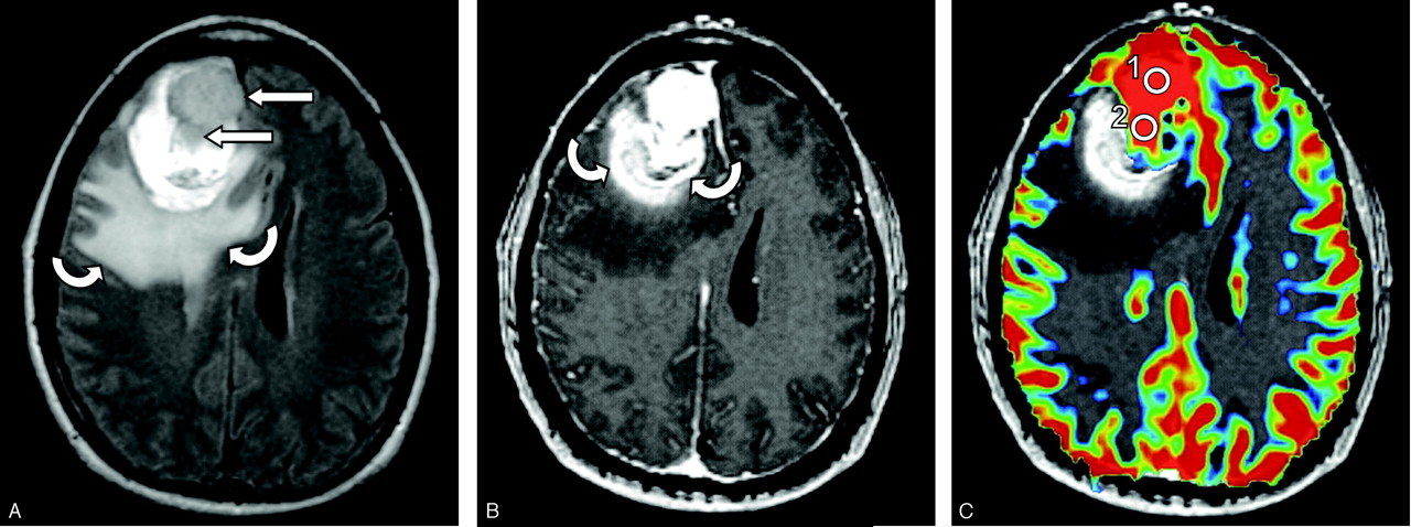
    Images of a 56-year-old woman with a history of breast carcinoma presenting with 2 weeks of headache, nausea, and vomiting. Image-guided open biopsy demonstrated a collision tumor between a typical meningioma and metastatic breast carcinoma.

    A, Axial fluid-attenuated inversion recovery (10,000/148/2,200 milliseconds [TR/TE/TI]) image. A heterogeneously intense, right anterior frontal bilobed mass (straight arrows) is seen with surrounding edema and hemorrhage (curved arrows).

    B, Axial postcontrast spoiled gradient-recalled (SPGR; 34/8) T1-weighted image. The mass demonstrates uniform contrast enhancement with a posterior rim of hemorrhage (curved arrows).

    C, Perfusion MR color map overlaid onto the corresponding axial postcontrast section. Relative cerebral blood volume is increased within the bilobed enhancing lesion. Two regions of interest, labeled 1 and 2, are defined on the anterior and posterior lobes of the tumor.

    D, T2* susceptibility signal intensity-time curves. The anterior (1) and posterior (2) lobes demonstrate differences in the degree of signal intensity drop and pattern of signal intensity recovery.

  • Fig 2.
    • Download figure
    • Open in new tab
    • Download powerpoint
    Fig 2.

    Intraoperative screen captures from the StealthStation neuronavigation system. The anterior lobe, corresponding to region of interest 1, is targeted for image-guided open biopsy.

  • Fig 3.
    • Download figure
    • Open in new tab
    • Download powerpoint
    Fig 3.

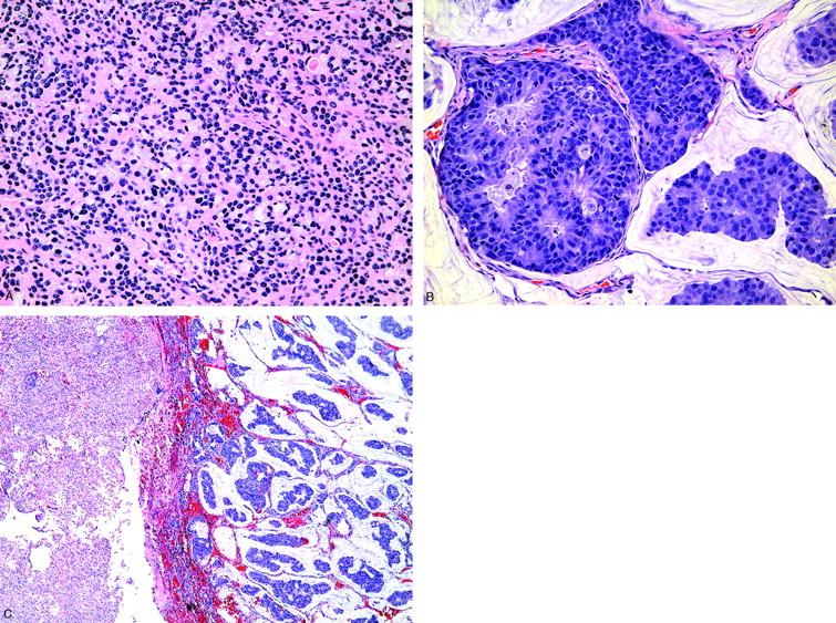
    Histopathologic sections from the anterior and posterior lobes corresponding to regions of interest 1 and 2, respectively.

    A, Anterior biopsy shows hyperchromatic tumor cells arranged in sheets consistent for a typical meningioma (hematoxylin-eosin, magnification ×200).

    B, Posterior biopsy demonstrates pleomorphic tumor cells arranged in attenuated clusters with pseudoglandular formation. This is histologically identical to the patient’s prior breast carcinoma (hematoxylin-eosin, magnification ×200).

    C, Section through the resected tumor reveals the interface between the typical meningioma and metastatic breast carcinoma (hematoxylin-eosin, magnification ×40).

PreviousNext
Back to top

In this issue

American Journal of Neuroradiology: 27 (1)
American Journal of Neuroradiology
Vol. 27, Issue 1
January, 2006
  • Table of Contents
  • Index by author
Advertisement
Print
Download PDF
Email Article

Thank you for your interest in spreading the word on American Journal of Neuroradiology.

NOTE: We only request your email address so that the person you are recommending the page to knows that you wanted them to see it, and that it is not junk mail. We do not capture any email address.

Enter multiple addresses on separate lines or separate them with commas.
Perfusion MR Imaging of an Intracranial Collision Tumor Confirmed by Image-Guided Biopsy
(Your Name) has sent you a message from American Journal of Neuroradiology
(Your Name) thought you would like to see the American Journal of Neuroradiology web site.
CAPTCHA
This question is for testing whether or not you are a human visitor and to prevent automated spam submissions.
Cite this article
P. Jun, J. Garcia, T. Tihan, M.W. McDermott, S. Cha
Perfusion MR Imaging of an Intracranial Collision Tumor Confirmed by Image-Guided Biopsy
American Journal of Neuroradiology Jan 2006, 27 (1) 94-97;

Citation Manager Formats

  • BibTeX
  • Bookends
  • EasyBib
  • EndNote (tagged)
  • EndNote 8 (xml)
  • Medlars
  • Mendeley
  • Papers
  • RefWorks Tagged
  • Ref Manager
  • RIS
  • Zotero
0 Responses
Respond to this article
Share
Bookmark this article
Perfusion MR Imaging of an Intracranial Collision Tumor Confirmed by Image-Guided Biopsy
P. Jun, J. Garcia, T. Tihan, M.W. McDermott, S. Cha
American Journal of Neuroradiology Jan 2006, 27 (1) 94-97;
del.icio.us logo Twitter logo Facebook logo Mendeley logo
  • Tweet Widget
  • Facebook Like
  • Google Plus One

Jump to section

  • Article
    • Abstract
    • Case Report
    • Discussion
    • References
  • Figures & Data
  • Info & Metrics
  • Responses
  • References
  • PDF

Related Articles

  • No related articles found.
  • PubMed
  • Google Scholar

Cited By...

  • No citing articles found.
  • Crossref
  • Google Scholar

This article has not yet been cited by articles in journals that are participating in Crossref Cited-by Linking.

More in this TOC Section

  • Evaluating the Effects of White Matter Multiple Sclerosis Lesions on the Volume Estimation of 6 Brain Tissue Segmentation Methods
  • White Matter Alterations in the Brains of Patients with Active, Remitted, and Cured Cushing Syndrome: A DTI Study
  • Enhanced Axonal Metabolism during Early Natalizumab Treatment in Relapsing-Remitting Multiple Sclerosis
Show more Brain

Similar Articles

Advertisement

Indexed Content

  • Current Issue
  • Accepted Manuscripts
  • Article Preview
  • Past Issues
  • Editorials
  • Editors Choice
  • Fellow Journal Club
  • Letters to the Editor

Cases

  • Case Collection
  • Archive - Case of the Week
  • Archive - Case of the Month
  • Archive - Classic Case

Special Collections

  • Special Collections

Resources

  • News and Updates
  • Turn around Times
  • Submit a Manuscript
  • Author Policies
  • Manuscript Submission Guidelines
  • Evidence-Based Medicine Level Guide
  • Publishing Checklists
  • Graphical Abstract Preparation
  • Imaging Protocol Submission
  • Submit a Case
  • Become a Reviewer/Academy of Reviewers
  • Get Peer Review Credit from Publons

Multimedia

  • AJNR Podcast
  • AJNR SCANtastic
  • Video Articles

About Us

  • About AJNR
  • Editorial Board
  • Not an AJNR Subscriber? Join Now
  • Alerts
  • Feedback
  • Advertise with us
  • Librarian Resources
  • Permissions
  • Terms and Conditions

American Society of Neuroradiology

  • Not an ASNR Member? Join Now

© 2025 by the American Society of Neuroradiology All rights, including for text and data mining, AI training, and similar technologies, are reserved.
Print ISSN: 0195-6108 Online ISSN: 1936-959X

Powered by HighWire