
Article Figures & Data
Figures
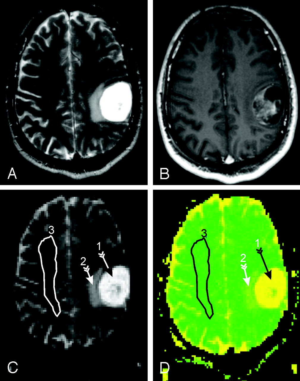
- Fig 1.
A, Top left, axial T2-weighted MR image of a section containing the lesion (mixed glioneurocytoma grade II) in patient 4 in the Table.
B, Top right, postcontrast T1-weighted image of the same section.
C, Bottom left, DTI (b = 0) image from the same level as A and B and the contralateral NAWM ROIs used for the MD and FA analyses 3.
D, Bottom right, color-coded MD map of the same section showing the marked elevated MD in the tumor and surrounding tissue, analogous to regions 1 and 2 in C. Note that the ROI selected does not contain any apparent tissue abnormalities.
- Fig 2.
Scatterplot of WBNAA versus MD for the 10 patients (black circles) and 9 controls (white circles). The WBNAA deficit strongly inversely correlates with MD increase (r = −0.879, P = .01) in patients (WBNAA = −18.69.MD + 27.65, solid line) but not in controls (r = 0.012, P = .975, dashed line).


Tables
Patient information
Patient Age (Years)/Sex Pathology Tumor Resection Volume (mL) WBNAA mmol/L MD (mm2s−1) Clinical Follow-up: Status, Time Elapsed Since Surgery 1 55/M Anaplastic Mixed GGNC (III) 45 8.4 1.068 Deceased, 6 months 2 49/F GBM (IV) 59 9.5 .940 Deceased, 12 months 3 35/M GBM (IV) 99 10.7 .923 Deceased, 12 months 4 37/M Mixed GN (II) N/A 8.6 .953 Stable, 17 months 5 30/M Mixed GN (II) 9 9.2 .982 Stable, 15 months 6 23/F GGNC-DNT (I) 32 12.6 .894 Stable, 10 months 7 39/F Mixed GN (II) 37 12.2 .917 Stable, 12 months 8 74/M GBM (IV) 75 7.8 1.065 Deceased, 9 months 9 62/M Anaplastic GGNC (III) 41 7.9 1.009 Stable, 10 months 10 32/F Mixed GGNC (II) N/A 9.8 .872 Stable, 12 months Note:—WBNAA indicates whole-brain N-acetylaspartate; MD, mean diffusivity; GGNC, ganglioglioneurocytoma; DNT, dysembryoplastic neuroepithelial tumor; GN, glioneurocytoma; DFA, diffuse fibrillary astrocytoma; GBM, glioblastoma multiforme; N/A, below our measurement sensitivity of 9 cm3.


