Advertisement

Main menu

  • Home
  • Content
    • Current Issue
    • Accepted Manuscripts
    • Article Preview
    • Past Issue Archive
    • AJNR Case Collection
    • Case of the Week Archive
    • Classic Case Archive
    • Case of the Month Archive
  • Special Collections
    • Spinal CSF Leak Articles (Jan 2020-June 2024)
    • 2024 AJNR Journal Awards
    • Most Impactful AJNR Articles
  • Multimedia
    • AJNR Podcast
    • AJNR Scantastics
    • Video Articles
  • For Authors
    • Submit a Manuscript
    • Author Policies
    • Fast publishing of Accepted Manuscripts
    • Graphical Abstract Preparation
    • Manuscript Submission Guidelines
    • Imaging Protocol Submission
    • Submit a Case for the Case Collection
  • About Us
    • About AJNR
    • Editorial Board
  • More
    • Become a Reviewer/Academy of Reviewers
    • Subscribers
    • Permissions
    • Alerts
    • Feedback
    • Advertisers
    • ASNR Home
  • Other Publications
    • ajnr

User menu

  • Alerts
  • Log in

Search

  • Advanced search
American Journal of Neuroradiology

American Journal of Neuroradiology

ASHNR American Society of Functional Neuroradiology ASHNR American Society of Pediatric Neuroradiology ASSR
  • Alerts
  • Log in

Advanced Search

  • Home
  • Content
    • Current Issue
    • Accepted Manuscripts
    • Article Preview
    • Past Issue Archive
    • AJNR Case Collection
    • Case of the Week Archive
    • Classic Case Archive
    • Case of the Month Archive
  • Special Collections
    • Spinal CSF Leak Articles (Jan 2020-June 2024)
    • 2024 AJNR Journal Awards
    • Most Impactful AJNR Articles
  • Multimedia
    • AJNR Podcast
    • AJNR Scantastics
    • Video Articles
  • For Authors
    • Submit a Manuscript
    • Author Policies
    • Fast publishing of Accepted Manuscripts
    • Graphical Abstract Preparation
    • Manuscript Submission Guidelines
    • Imaging Protocol Submission
    • Submit a Case for the Case Collection
  • About Us
    • About AJNR
    • Editorial Board
  • More
    • Become a Reviewer/Academy of Reviewers
    • Subscribers
    • Permissions
    • Alerts
    • Feedback
    • Advertisers
    • ASNR Home
  • Follow AJNR on Twitter
  • Visit AJNR on Facebook
  • Follow AJNR on Instagram
  • Join AJNR on LinkedIn
  • RSS Feeds

Welcome to the new AJNR, Updated Hall of Fame, and more. Read the full announcements.


AJNR is seeking candidates for the position of Associate Section Editor, AJNR Case Collection. Read the full announcement.


OtherBRAIN

Cerebral Alveolar Echinoccosis Mimicking Primary Brain Tumor

S. Senturk, K.K. Oguz, F. Soylemezoglu and S. Inci
American Journal of Neuroradiology February 2006, 27 (2) 420-422;
S. Senturk
  • Find this author on Google Scholar
  • Find this author on PubMed
  • Search for this author on this site
K.K. Oguz
  • Find this author on Google Scholar
  • Find this author on PubMed
  • Search for this author on this site
F. Soylemezoglu
  • Find this author on Google Scholar
  • Find this author on PubMed
  • Search for this author on this site
S. Inci
  • Find this author on Google Scholar
  • Find this author on PubMed
  • Search for this author on this site
  • Article
  • Figures & Data
  • Info & Metrics
  • Responses
  • References
  • PDF
Loading

Abstract

We present a case of cerebral infestation by Echinococcosis multilocularis mimicking an infiltrative primary brain tumor. A heavily calcified mass invading the midbrain enhanced in a cauliflower-like fashion with small peripheral nodules present on MR imaging. Perfusion-weighted MR imaging revealed low relative cerebral blood volume within the calcified lesion and peripheral hyperemia. Single-voxel proton MR spectroscopy with an echo time of 135 milliseconds was normal.

Two species of the genus Echinococcus are the most clinically relevant forms: the cystic echinococcosis (hydatid cyst) caused by Echinococcosis granulosus (EG) and alveolar echinococcosis (AE) caused by E. multilocularis (EM). Human AE is an uncommon zoonotic infestation caused by intrahepatic growth of the EM parasitic larvae. The metacestode spreads from the inevitably involved liver to other organs such as the lungs and brain and bone by infiltration and metastasis. Cerebral metastasis is very rare, being reported in only 1% of patients.1–3 Cerebral infestation by EM and EG differ: AE occurs in adults who live in rural areas, whereas hydatid disease affects mostly children. EG infestation is usually self-limited, whereas EM infestation may appear as an infiltrative neoplasm in both the liver and brain, which makes total surgical removal difficult.4–9 This report presents findings of brain CT and MR examinations, in addition to perfusion-weighted MR (pMRI) and proton MR spectroscopy imaging findings of cerebral AE.

Case Report

A 26-year-old woman was referred to our hospital with a complaint of headache lasting for 2 months. Neurologic examination revealed confusion, anisocoria, and right hemiparesis. The Babinsky sign was present on the right side. Routine blood tests revealed mild leukocytosis with a white blood cell count of 14,600.

She had a long history of a liver cystic disease, for which she had been surgically treated in 1992 and 1998 at an outside institution; however, neither written information about her medical history nor a report of pathologic examination about her liver disease was available. Abdominal CT performed in our hospital, however, revealed irregular cysts at the capsular surface near the right hepatic lobectomy margin (not shown).

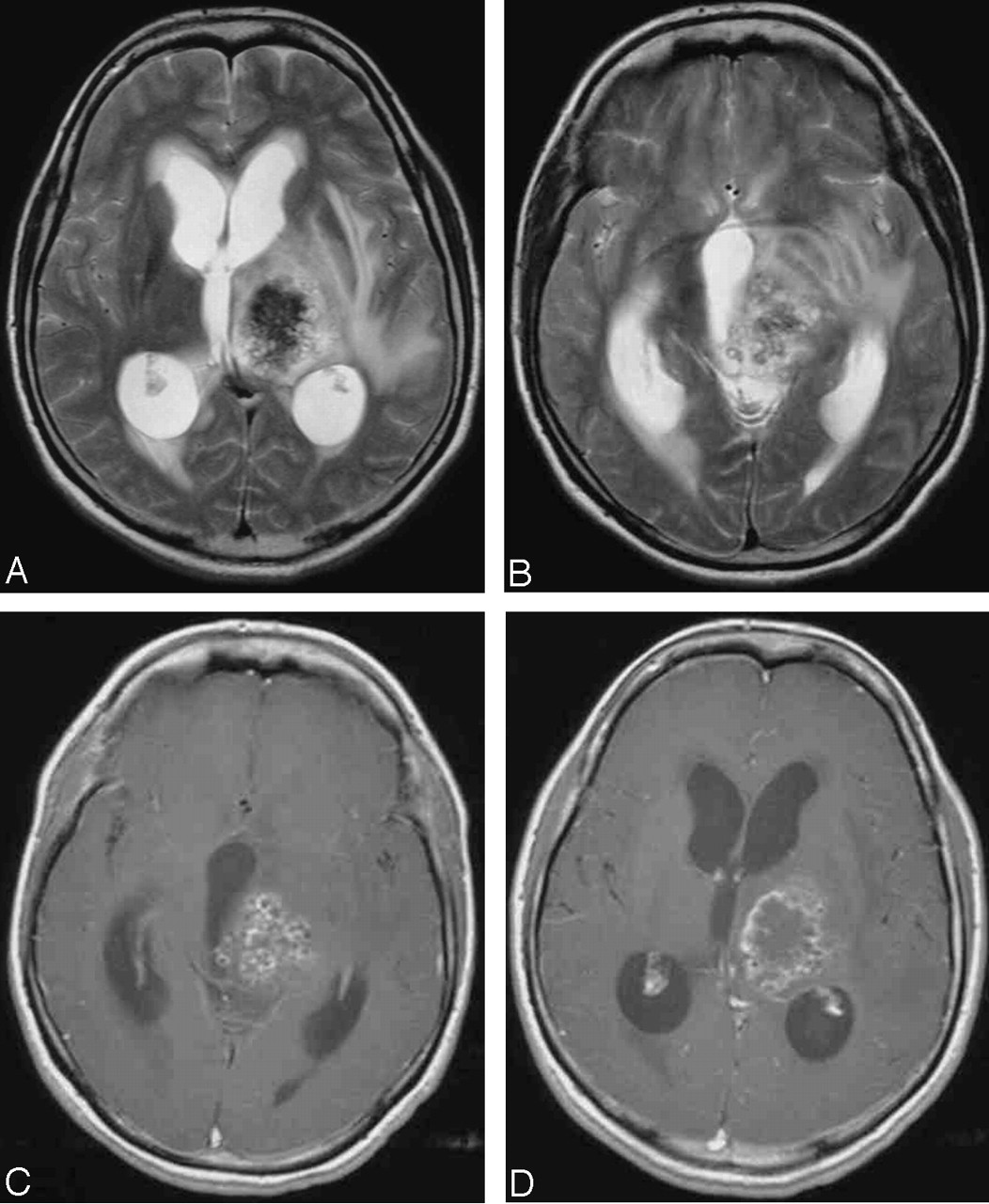
Initial cranial CT showed a calcified mass in the left thalamus with profound edema (Fig 1). Conventional MR imaging demonstrated left thalamic mass infiltrating the surrounding tissue and down to the midbrain. The lesion was isointense to cerebral white matter with nodular low-signal-intensity areas on T1-weighted images. T2- weighed images revealed a heterogeneous low-intensity mass with peripheral high signal intensity consistent with edema (Fig 2A, -B). Calcification on CT corresponded to nodular hypointense areas in the lesion on both T1- and T2 -weighed images. Irregular cauliflower-like enhancement of the nodules and an enhancing rim all around the mass were observed (Fig 2C, -D).

Fig 1.
  • Download figure
  • Open in new tab
  • Download powerpoint
Fig 1.

Cranial CT shows left thalamic calcified mass (arrow) with profound edema.

Fig 2.
  • Download figure
  • Open in new tab
  • Download powerpoint
Fig 2.

Axial T2-weighted imaging (TR/TE, 3080/93 milliseconds) shows calcified mass with microcysts extending between the left thalamus (A) and the midbrain (B). Axial T1-weighted postcontrast imaging (TR/TE, 650/9 milliseconds) reveals irregular enhancement of the nodules in a cauliflower-like pattern (C) and an enhancing rim around the mass (D).

Perfusion MR imaging revealed low relative cerebral blood volume (rCBV) within the lesion, whereas higher values were obtained from peripheral areas of the lesion, most probably representing inflammation, later demonstrated on pathologic specimen (Fig 3). Baseline artifacts were present in MR spectroscopy (TR/TE, 3000/135; single voxel; voxel size, 15 × 15 × 15 mm) because of heterogenity of the lesion with massive calcification. A voxel obtained at the periphery, which consisted of both enhancing and nonenhancing portions of the mass, however, showed normal N-acetylaspartate/creatine (1.10) and choline/creatine (0.71) ratios, which suggested that the lesion was of non-neoplastic origin.

Fig 3.
  • Download figure
  • Open in new tab
  • Download powerpoint
Fig 3.

rCBV map of perfusion-weighted MR imaging (EPI [TR/TE, 1430/46 milliseconds]; NEX, 1; section thickness, 5 mm; intersection gap,10 mm; mtx, 128 ×128) demonstrates low rCBV within the lesion (arrow) located at left thalamus with higher values peripherally (thin arrows).

The lesion was solid, multilocular, and white-colored macroscopically and was grossly and totally removed during surgery. The patient was discharged as neurologically intact except for slight right homonymus hemianopsia.

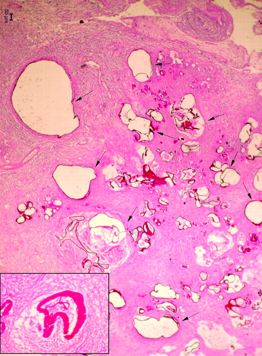
Histologic examination revealed a multilobulated lesion composed of multiple cysts characterized by a thick laminated-noncellular outer layer exhibiting focal discontinuities. Cuticle was periodic acid–Schiff (PAS) positive, and a granular layer was present. Amorphous necrotic debris was surrounded by attenuated gliosis with reactive astroglial proliferation. Epitheloid and giant cells containing multiple granulomas of different size were detected. Dense lymphoplasmocytic cell infiltrate rich in eosinophilic leukocytes was observed in the surrounding brain. Morphologic findings revealed degenerating AE cysts with reabsorption (Fig 4).

Fig 4.
  • Download figure
  • Open in new tab
  • Download powerpoint
Fig 4.

Multiple cysts (arrows) in the brain parenchyma surrounded by necrosis and giant cell granulomatous reaction (PAS, ×4). Inset, PAS-positive cuticular membranes (PAS, ×40).

Discussion

The cyst of AE grows by external budding of the germinal membrane with progressive infiltration of the surrounding tissue, and this feature differs from EG, which is usually self-limited.2 Difficulty in diagnosis stems from resemblance of AE infestation to malignancy as a result invasion of adjacent structures, destructive tissue growth, and metastasis in distant organs. Lesions of AE have alveolar structures composed of numerous irregular cysts with a diameter between 1 and 20 mm and not sharply demarcated from surrounding tissue. Central cycstic cavities can result from necrosis and liquefaction of the inner part. Irregularly thickened and partially calcified wall is present in most cases.1 Clinical features of cerebral AE—including headache, seizure, increased intracranial pressure, and focal neurologic deficits, depending on the location of the lesion—are those of any space-occupying lesion.

There are just a few published reports of separate cases on CT and MR imaging features. Yet, to the best of our knowledge, there has been no report of pMRI or MR spectroscopy findings. On CT and MR imaging the lesions mainly appear as solid and semisolid or sometimes as a multilocular cystic mass.7–13 Disseminated intracerebral AE lesions mostly appear at the terminal phase of the disease.7,9,12 Calcification and surrounding edema are common findings of the lesions as observed in the present case. Although not always seen, contrast enhancement of the lesion has been proposed because of blood-brain barrier distruption caused by the inflammatory reaction.11 Peripheral ringlike, heterogenous, nodular, and cauliflower-like enhancement patterns have all been reported in different presentations of the infestation.8–10,12 In our case, T1-weighted postcontrast imaging showed that the mass was enhancing irregularly in a cauliflower-like pattern with small enhancing nodules. In addition, there was a rimlike enhancement surrounding the mass. With these findings the mass was indistinguishable from an ependymoma especially in the absence of knowledge about the patient’s history of liver disease. pMRI revealed low rCBV within the lesion with higher values in the periphery. The low rCBV might have resulted from attenuated gliosis and granulomas shown on histologic examination, which also give the concrete nature of the lesion observed in the operation. More peripheral areas with increased blood volume, on the other hand, might be related to surrounding parenchymal inflammation observed on histologic specimen (Fig 4).

A few reports on proton MR spectroscopy in differential diagnosis of cystic cerebral lesions displayed resonances from lactate, succinate, and acetate, as well as pyruvate, in cestodal cysts.14–17 The mass we report, however, was solid, and MR spectroscopy of a voxel at the periphery of the lesion, which contained a partly enhancing component and accompanying reactive inflammatory edema, was sufficient to show its nontumoral nature (a glial tumor or a metastasis), though it did not give an additional finding to suggest its infectious origin.

Our case differs from previously reported cases in that the epicenter of the mass was the left thalamus and it infiltrated the brain stem. Involvement of the deep gray matter, a feature of this case, has not been present in any previously reported cases. The locations of reported AE lesions are lobar and posterior fossae.7–9,11,12

It is known that human parasitic zoonoses sometimes cause diagnostic and therapeutic problems. Despite newly developed neuroradiologic and serologic methods, these entities are rarely diagnosed preoperatively.16,18 Differential diagnosis includes tumors and infectious lesions such as tuberculosis and bacterial abscess. Because AE infestation simulates malignancy by its invasive nature in the brain, gliomas and metastases are also included in the differential diagnosis.

Geographic prevalence, clinical history of hepatic involvement, and serologic tests are required for diagnosis. Intra-axial lesions imaging as grapelike multilocular cystic masses, cauliflower-like enhancing, or infiltrative heavily calcified masses with small nodules in patients with known liver disease should suggest the cerebral involvement of AE infestation. Moreover, the recently developed neuroradiologic tools such as pMRI and MR spectroscopy help to differentiate these cerebral space-occupying lesions from malignant tumors, which remain the main differential in our patient.

References

  1. ↵
    Ammann RW, Eckert J. Echinococosus. Gastroenterol Clin North Am 1996;25:655–89
    CrossRefPubMed
  2. ↵
    Kammerer WS. Echinococcosis affecting the central nervous system. Semin Neurol 1993;13:144–47
    PubMed
  3. ↵
    Bresson-Hadni S, Vuitton DA, Bartholomot B, et al. Twenty-year history of alveolar echinococcosis: analysis of a series of 117 patients from eastern France. Eur J Gastoenterol Hepatol 2000;12:327–36
    PubMed
  4. ↵
    Gomori JH, Cohen D, Eyd A, et al. Water lily sign in CT of cerebral hydatid disease: a case report. Neuroradiology 1988;30:358
    CrossRefPubMed
  5. Haliloglu M, Saatci I, Akhan O, et al. Spectrum of imaging findings in pediatric hydatid disease. AJR Am J Roentgenol 1997;169:1627–31
    PubMed
  6. Aydin Y, Barlas O, Yolas C, et al. Alveolar hydatid disease of the brain: report of four cases. J Neurosurg 1986;65:115–19
    PubMed
  7. ↵
    Algros MP, Majo F, Bresson-Hadni S, et al. Intracerebral alveolar echinococcosis. Infection 2003;31:63–65
    CrossRefPubMed
  8. ↵
    Bensaid AH, Dietemann JL, Filippe de la Palavesa MM, et al. Intracerebral alveolar echinococcosis: CT and MRI. Neuroradiology 1994;36:289–91
    PubMed
  9. ↵
    Piotin M, Cattin F, Miralbes S, et al. Disseminated intracerebral alveolar echinococcosis. Neuroradiology 1997;39:431–33
    PubMed
  10. ↵
    Bükte Y, KemanoFlu S, NazaroFlu H, et al. Cerebral hydatid disease: CT and MRI findings. Swiss Med Wkly 2004;134:459–67
    PubMed
  11. ↵
    Reitner P, Szolar DH, Schmid M. Systemic manifestations of echinococcus alveolaris infection. J Comput Assist Tomogr 1996;6:1030–32
  12. ↵
    TunacH M, TunacH A, Engin G, et al. MRI of cerebral alveolar echinococcosis. Neuroradiology 1999;41:844–46
    CrossRefPubMed
  13. ↵
    Pamir MN, Özer AF, Keles GE, et al. Cerebral echinococcosis multilocularis. J Neurosurg Sci 1991;35:161–64
    PubMed
  14. ↵
    Shukla-Dave A, Gupta RK, Roy R, et al. Prospective evaluation of in vivo proton MR spectroscopy in differentiation of similar appearing intracranial cystic lesions. Magn Reson Imaging 2001;19:103–10
    CrossRefPubMed
  15. Chang KH, Song IC, Kim SH, et al. In vivo single-voxel proton MR spectroscopy in intracranial cystic masses. AJNR Am J Neuroradiol 1998;19:401–405
    Abstract
  16. ↵
    Talan-Hranilovic J, Sajko T, Negovetic L, et al. Cerebral cysticercosis and echinococcosis: a preoperative diagnostic dilemma. Arch Med Res 2002;33:590–94
    PubMed
  17. ↵
    Jayakumar PN, Srikanth SG, Chandrashekar HS, et al. Pvruvate: An in vivo marker of cestodal infestation of the human brain on proton MR spectroscopy. J Magn Reson Imaging 2003;18:675–80
    PubMed
  18. ↵
    Ho A, Ma L, Schantz PM, et al. Differential serodiagnosis for cystic and alveolar echinococcosis using fractions of E. granulosus cyst fluid (Antigen B) and E. multilocularis protoscolex (EM18). Am J Trop Med Hyg 1996;60:188–92
  • Received February 22, 2005.
  • Accepted after revision March 30, 2005.
  • Copyright © American Society of Neuroradiology
View Abstract
PreviousNext
Back to top

In this issue

American Journal of Neuroradiology: 27 (2)
American Journal of Neuroradiology
Vol. 27, Issue 2
February, 2006
  • Table of Contents
  • Index by author
Advertisement
Print
Download PDF
Email Article

Thank you for your interest in spreading the word on American Journal of Neuroradiology.

NOTE: We only request your email address so that the person you are recommending the page to knows that you wanted them to see it, and that it is not junk mail. We do not capture any email address.

Enter multiple addresses on separate lines or separate them with commas.
Cerebral Alveolar Echinoccosis Mimicking Primary Brain Tumor
(Your Name) has sent you a message from American Journal of Neuroradiology
(Your Name) thought you would like to see the American Journal of Neuroradiology web site.
CAPTCHA
This question is for testing whether or not you are a human visitor and to prevent automated spam submissions.
Cite this article
S. Senturk, K.K. Oguz, F. Soylemezoglu, S. Inci
Cerebral Alveolar Echinoccosis Mimicking Primary Brain Tumor
American Journal of Neuroradiology Feb 2006, 27 (2) 420-422;

Citation Manager Formats

  • BibTeX
  • Bookends
  • EasyBib
  • EndNote (tagged)
  • EndNote 8 (xml)
  • Medlars
  • Mendeley
  • Papers
  • RefWorks Tagged
  • Ref Manager
  • RIS
  • Zotero
0 Responses
Respond to this article
Share
Bookmark this article
Cerebral Alveolar Echinoccosis Mimicking Primary Brain Tumor
S. Senturk, K.K. Oguz, F. Soylemezoglu, S. Inci
American Journal of Neuroradiology Feb 2006, 27 (2) 420-422;
del.icio.us logo Twitter logo Facebook logo Mendeley logo
  • Tweet Widget
  • Facebook Like
  • Google Plus One

Jump to section

  • Article
    • Abstract
    • Case Report
    • Discussion
    • References
  • Figures & Data
  • Info & Metrics
  • Responses
  • References
  • PDF

Related Articles

  • No related articles found.
  • PubMed
  • Google Scholar

Cited By...

  • Diagnosis and treatment of cardiac echinococcosis
  • Crossref
  • Google Scholar

This article has not yet been cited by articles in journals that are participating in Crossref Cited-by Linking.

More in this TOC Section

  • Fast Contrast-Enhanced 4D MRA and 4D Flow MRI Using Constrained Reconstruction (HYPRFlow): Potential Applications for Brain Arteriovenous Malformations
  • Quiet PROPELLER MRI Techniques Match the Quality of Conventional PROPELLER Brain Imaging Techniques
  • Predictors of Reperfusion in Patients with Acute Ischemic Stroke
Show more Brain

Similar Articles

Advertisement

Indexed Content

  • Current Issue
  • Accepted Manuscripts
  • Article Preview
  • Past Issues
  • Editorials
  • Editors Choice
  • Fellow Journal Club
  • Letters to the Editor

Cases

  • Case Collection
  • Archive - Case of the Week
  • Archive - Case of the Month
  • Archive - Classic Case

Special Collections

  • Special Collections

Resources

  • News and Updates
  • Turn around Times
  • Submit a Manuscript
  • Author Policies
  • Manuscript Submission Guidelines
  • Evidence-Based Medicine Level Guide
  • Publishing Checklists
  • Graphical Abstract Preparation
  • Imaging Protocol Submission
  • Submit a Case
  • Become a Reviewer/Academy of Reviewers
  • Get Peer Review Credit from Publons

Multimedia

  • AJNR Podcast
  • AJNR SCANtastic
  • Video Articles

About Us

  • About AJNR
  • Editorial Board
  • Not an AJNR Subscriber? Join Now
  • Alerts
  • Feedback
  • Advertise with us
  • Librarian Resources
  • Permissions
  • Terms and Conditions

American Society of Neuroradiology

  • Not an ASNR Member? Join Now

© 2025 by the American Society of Neuroradiology All rights, including for text and data mining, AI training, and similar technologies, are reserved.
Print ISSN: 0195-6108 Online ISSN: 1936-959X

Powered by HighWire