
Article Figures & Data
Figures
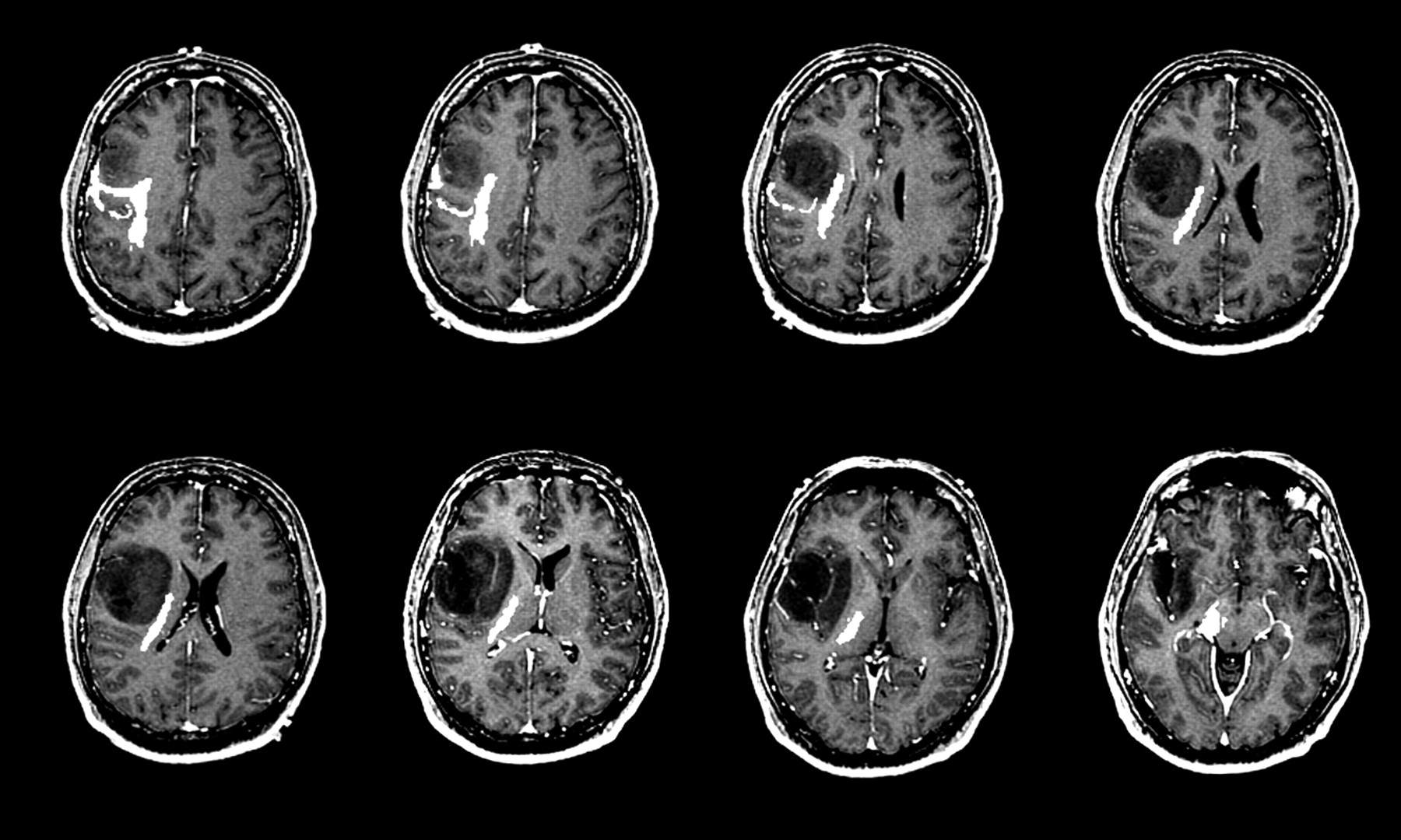
- Fig 1.
ADC and astrocytoma grade. WHO grade II (top row), grade III (middle row), and grade IV (bottom row) astrocytomas. Axial postcontrast T1-weighted images (left column), FLAIR images (middle column), and ADC maps (right column) demonstrate typical examples of 3 different grades of astrocytoma. With increasing tumor grade, the tumor ADC value of grade III astrocytoma is lower (black arrows) than that of grade II and the grade IV astrocytoma has the lowest (white arrow).
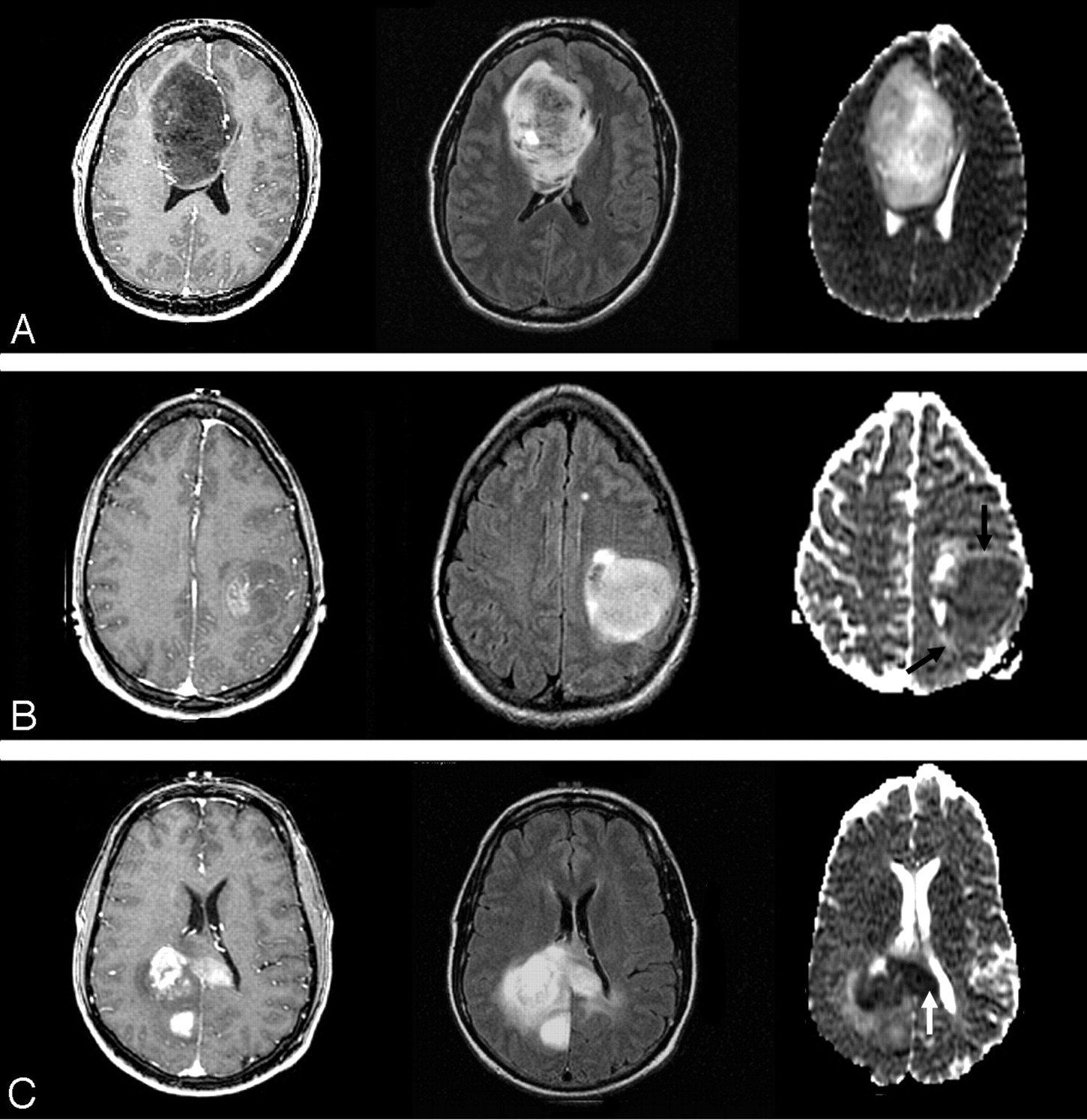
- Fig 2.
A 51-year-old man with right frontal glioblastoma multiforme.
A, Preoperative axial postcontrast T1-weighted image shows a centrally necrotic, peripherally enhancing right frontal lobe mass compressing the adjacent lateral ventricle.
B, Immediately postoperative axial postcontrast T1-weighted image confirms gross total resection of the enhancing portion of the mass.
C, Immediately postoperative axial diffusion-weighted image demonstrates a wedge-shaped area of abnormal reduced diffusion (white arrow) within the surgical bed.
D, One-month postoperative axial postcontrast T1-weighted image reveals irregular area (black arrow) of abnormal contrast enhancement within the margins of the surgical bed simulating the appearance of a recurrent tumor.
E, Two-month postoperative axial postcontrast T1-weighted image shows complete resolution of the abnormal contrast enhancement and a gliotic cavity (arrowhead).
- Fig 3.
A 37-year-old man with right frontal low-grade astrocytoma. A series of axial postcontrast T1-weighted images of the brain demonstrate a nonenhancing right frontal insular mass. The diffusion tensor tractogram, which has been coregistered and overlaid onto the contrast-enhanced T1-weighted images, demonstrates corticospinal tract (white marks) that is displaced but not invaded by the tumor.
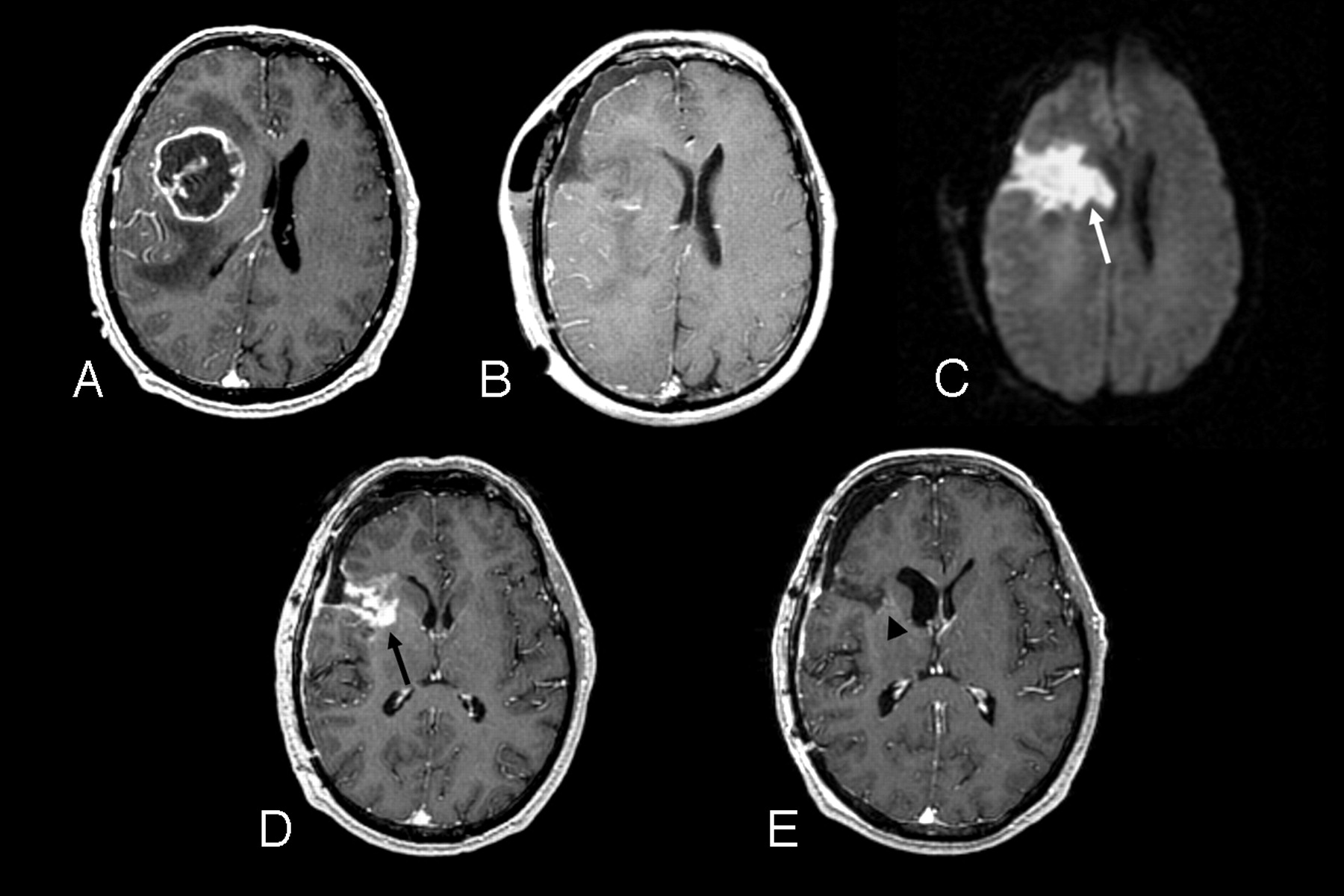
- Fig 4.
A 59-year-old woman with right superior frontal mass initially diagnosed as low-grade astrocytoma.
A, Axial postcontrast T1-weighted image shows an ill-defined nonenhancing mass within the right superior frontal lobe.
B, Axial FLAIR image demonstrates homogeneous T2 prolongation within the mass and a single-voxel proton-spectroscopic imaging within the center of the mass reveals a marked increase in lactate (Lac) and choine (Cho) metabolites and a decrease in N-acetylaspartate (NAA) metabolite.
C, 3D, lactate-edited, proton spectroscopic image of the same tumor location confirms the presence of lactate metabolite within the tumor. Re-evaluation of the tissue specimen revealed a few mitotic figures and vascular hyperplasia, and the tumor was upgraded to anaplastic astrocytoma.
D, Serial axial postcontrast T1-weighted images during 1-year period show emergence of subtle enhancement at the posterior surgical margin at 9-month follow-up (white arrow), which rapidly progresses into an aggressive grade IV astrocytoma (black arrow).
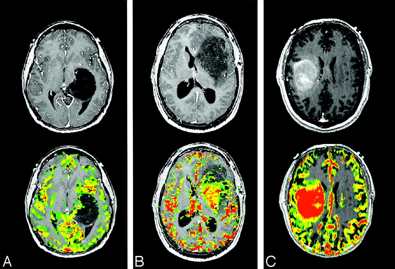
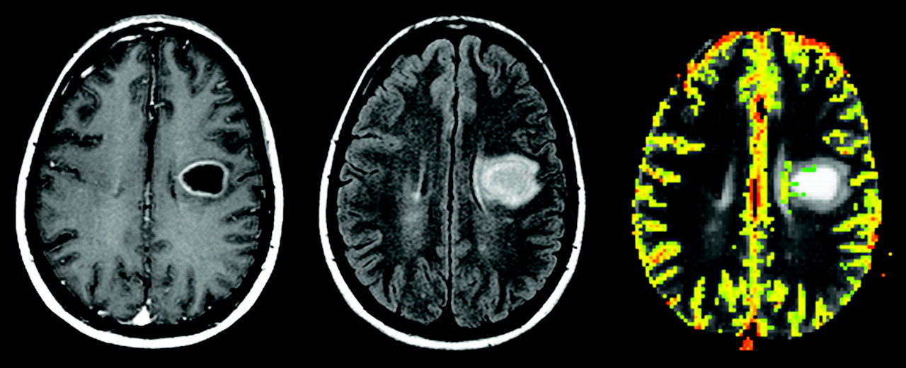
- Fig 5.
Relative cerebral blood volume (rCBV) maps of astrocytoma grade II, grade III, and grade IV (left to right).
Top row, Axial postcontrast T1-weighted images of grade II (left), grade III (middle), and grade IV (right) astrocytoma demonstrate definite contrast enhancement associated with only the grade IV tumor.
Bottom row, Axial rCBV color maps coregistered with axial postcontrast T1-weighted image show increasing tumor blood volume abnormality with increasing tumor grade.
- Fig 6.
A 34-year-old man with left frontal grade II oligodendroglioma. Axial postcontrast T1-weighted image (left) shows a heterogeneous, mildly enhancing (black arrow) left frontal mass. Axial rCBV map at the same anatomic location demonstrates marked increased in tumor blood volume (white arrow).
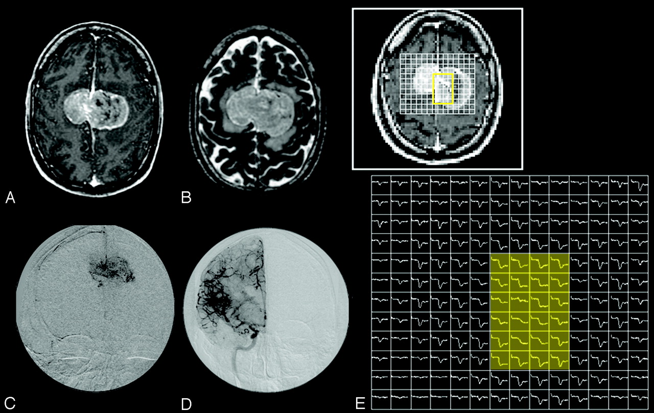
- Fig 7.
A 5-year-old girl with right lateral ventricle choroids plexus papilloma.
A, Axial postcontrast T1-weighted image shows an avidly enhancing, intraventricular mass.
B, Dynamic susceptibility-weighted contrast-enhanced image during the maximum bolus demonstrates marked signal intensity drop within the tumor because of increased tumor vascularity.
C, Susceptibility-weighted signal intensity time curves from regions of interest placed over the tumor (green) and the contralateral normal brain (pink) show marked difference in signal intensity recovery between the 2 regions. The tumor vasculature lacks blood-brain barrier and there is immediate and marked leakage of contrast agent during the bolus phase (arrows) and minimal return to the baseline.
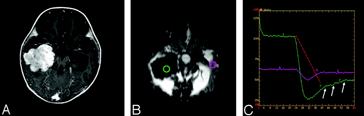
- Fig 8.
A 42-year-old man with bifalcine meningioma.
A and B, Axial postcontrast T1-weighted (A) and T2-weighted (B) images show a large enhancing, bifalcine, extra-axial mass.
C and D, Catheter angiographic images from selective external carotid artery (C) and internal carotid artery (D) demonstrate abnormal tumor vascularity. The central aspect of the tumor appears to be supplied predominantly by the external carotid artery.
E, 2D array of susceptibility-weighted signal intensity time curve over the tumor shows different patterns of signal intensity recovery. The central region of the tumor (yellow shaded area), which is supplied by the external carotid artery, has markedly lower degree of signal intensity recovery than the tumor periphery supplied by the internal carotid artery.
- Fig 9.
A 31-year-old woman with left frontal lobe tumefactive demyelinating lesion. Axial postcontrast T1-weighted (left) and FLAIR (middle) images show a rim enhancing left frontal mass. Relative cerebral blood volume map demonstrates lack of increased in blood volume within the lesion.


In this issue


Jump to section
- Article
- Unique Features of Brain Tumors
- Classification of Brain Tumors
- Physiology-Based MR Imaging
- DWI and Postoperative Injury
- ADC and Peritumoral Edema: Vasogenic versus Infiltrative
- Diffusion Tensor Imaging and White Matter Tractography
- Proton MR Spectroscopic Imaging
- Lactate-Edited 1H-MR Spectroscopy
- MR Spectroscopy Summary
- Perfusion MR Imaging
- DSC MR Imaging
- DCE T1-Weighted Permeability Imaging
- Conclusion
- Footnotes
- References
- Figures & Data
- Info & Metrics
- Responses
- References
Related Articles
Cited By...
- Advanced Distance-Resolved Evaluation of the Perienhancing Tumor Areas with FLAIR Hyperintensity Indicates Different ADC Profiles by MGMT Promoter Methylation Status in Glioblastoma
- Acoustic tumor paint for real-time imaging, surgical guidance and recurrence monitoring of brain tumors with ultrasound
- Grading of Pediatric Intracranial Tumors: Are Intravoxel Incoherent Motion and Diffusional Kurtosis Imaging Superior to Conventional DWI?
- Sequential Apparent Diffusion Coefficient for Assessment of Tumor Progression in Patients with Low-Grade Glioma
- Diagnostic Accuracy of T1-Weighted Dynamic Contrast-Enhanced-MRI and DWI-ADC for Differentiation of Glioblastoma and Primary CNS Lymphoma
- The Combined Performance of ADC, CSF CXC Chemokine Ligand 13, and CSF Interleukin 10 in the Diagnosis of Central Nervous System Lymphoma
- Direct Cranial Nerve Involvement by Gliomas: Case Series and Review of the Literature
- Evaluation of Microvascular Permeability with Dynamic Contrast-Enhanced MRI for the Differentiation of Primary CNS Lymphoma and Glioblastoma: Radiologic-Pathologic Correlation
- Comparison of Perfusion, Diffusion, and MR Spectroscopy between Low-Grade Enhancing Pilocytic Astrocytomas and High-Grade Astrocytomas
- Variability of Proliferation and Diffusion in Different Lung Cancer Models as Measured by 3'-Deoxy-3'-18F-Fluorothymidine PET and Diffusion-Weighted MR Imaging
- A Prognostic Model Based on Preoperative MRI Predicts Overall Survival in Patients with Diffuse Gliomas
- Value of 18F-3,4-Dihydroxyphenylalanine PET/MR Image Fusion in Pediatric Supratentorial Infiltrative Astrocytomas: A Prospective Pilot Study
- Differentiation of Primary Central Nervous System Lymphomas and Glioblastomas: Comparisons of Diagnostic Performance of Dynamic Susceptibility Contrast-Enhanced Perfusion MR Imaging without and with Contrast-Leakage Correction
- Correlation of MRI-Derived Apparent Diffusion Coefficients in Newly Diagnosed Gliomas with [18F]-Fluoro-L-Dopa PET: What Are We Really Measuring with Minimum ADC?
- Combined Diffusion and Perfusion MR Imaging as Biomarkers of Prognosis in Immunocompetent Patients with Primary Central Nervous System Lymphoma
- MR Imaging of Neoplastic Central Nervous System Lesions: Review and Recommendations for Current Practice
- The Added Value of Apparent Diffusion Coefficient to Cerebral Blood Volume in the Preoperative Grading of Diffuse Gliomas
- Does MR Perfusion Imaging Impact Management Decisions for Patients with Brain Tumors? A Prospective Study
- Imaging biomarkers of angiogenesis and the microvascular environment in cerebral tumours
- Prediction of Response to Chemoradiation Therapy in Squamous Cell Carcinomas of the Head and Neck Using Dynamic Contrast-Enhanced MR Imaging