
Article Figures & Data
Figures
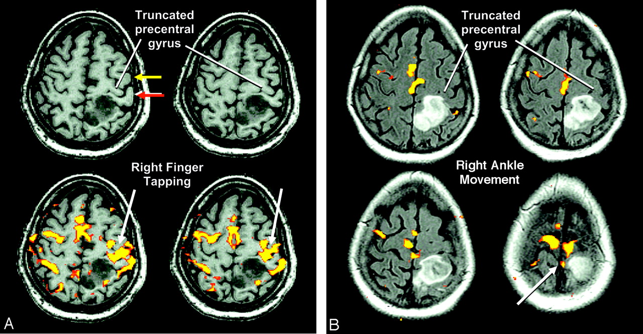
- Fig 1.
A, Axial SPGR images showing truncation of the left precentral gyrus (upper row) and activation to sequential finger tapping (white arrows). The left postcentral gyrus (red arrow) extends anteriorly around a truncated precentral gyrus, and comes near the posterior aspect of the left middle frontal gyrus (yellow arrow). Nearly continuous activity spanning from truncated precentral gyrus to overlapping postcentral gyrus is notable. Demarcating the boundary between M1 and S1 cortex is not possible at preoperative functional MRI (fMRI). B, Activation (white arrow) to right ankle movement is shown superimposed onto fluid-attenuated inversion recovery (FLAIR) images, yielding only slight activity. Slighter than typically observed activity to the lower-extremity task may be due to a combination of tumor-induced neurovascular uncoupling and the limitations of the task, and probably underestimates the actual distal lower-extremity M1 cortical field. In A and B, the proximity of eloquent lower-extremity and upper-extremity M1 cortex to the anterior-medial and anterior-lateral borders of the mass, respectively, are notable.
- Fig 2.
Axial FLAIR (top row) and color-coded FA maps (bottom row) demonstrating the proximity of the tumor to corticospinal fibers. The color-coded FA maps hinder visualization of the tumor, which implies preserved FA and function of involved white matter tracts. Thus, comparing FLAIR images to the color-coded FA maps is critical in fully appreciating the proximity of tumor borders to motor fibers. Based on anatomic relationships and DTI data, the white matter arising from lower-extremity M1 cortex wraps around the anterior-medial edge of the mass, whereas white matter descending (blue) from upper- and lower-extremity M1 cortex (arrows) is in proximity to the anterior border of the mass. White matter arising from corresponding postcentral gyrus wraps around the anterior-lateral border of the mass. In light of the results of the intraoperative mapping, it is possible that postcentral gyrus white matter contains motor fibers. T, tumor. White matter orientation color encoding: blue, ascending-descending fiber orientation; red, right-left fiber orientation; green, anteroposterior fiber orientation.
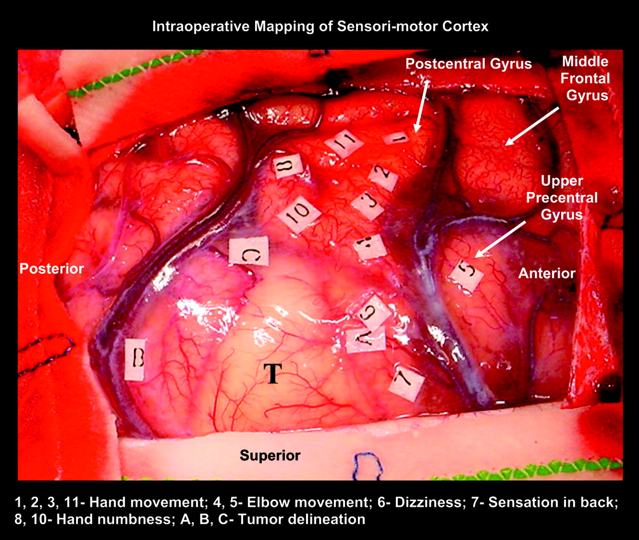
- Fig 3.
Intraoperative images of the brain tumor outlined by sonography and demarcated on the surface by letters A, B, and C. The postcentral gyrus extending anteriorly to approximate the posterior border of the middle frontal gyrus, hiding the superior-lateral portion of the precentral gyrus from view, is notable. Electrocortical stimulation of the upper precentral gyrus (position 5) resulted in proximal upper-extremity movement. Electrocortical stimulation at positions 1, 2, 3, and 11 within the anterior aspect of the postcentral gyrus resulted in hand movements. Electrocortical stimulation at positions 8 and 10 in the posterior aspect of the postcentral gyrus resulted in right-hand numbness. Stimulation at position 7 within the postcentral gyrus involved by the tumor resulted in a reproducible abnormal sensation of the upper back. T, tumor beneath visible cortex.


In this issue


Jump to section
Related Articles
Cited By...
- No citing articles found.