
Article Figures & Data
Figures
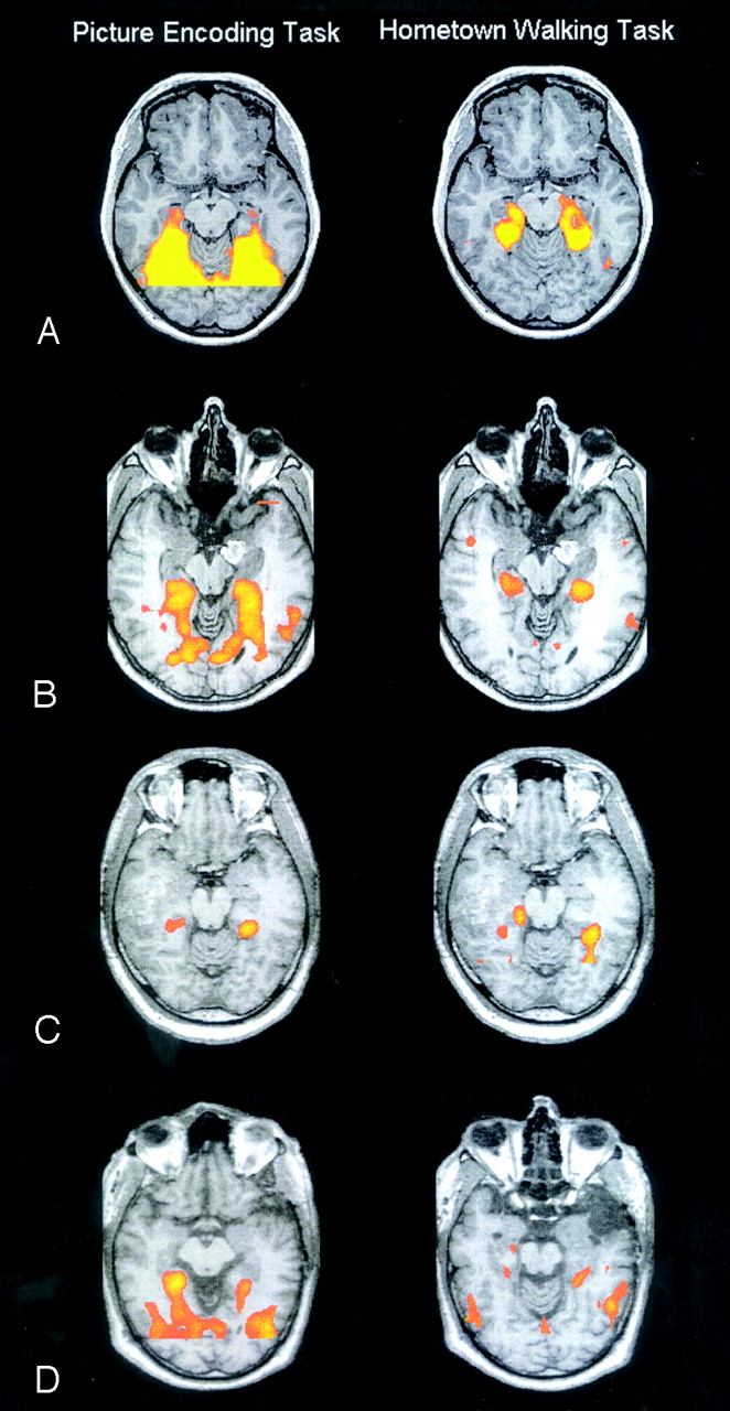
- Fig 1.
Functional activation results for the picture-encoding task (left) and the hometown-walking task (right). All images are in radiologic orientation. Activation thresholds were at 0.00001 for groups and 0.001 for individual patients.
A, Results for the control group.
B, Results for patient 2 showing symmetric bilateral activation of the temporal lobe for both tasks.
C, Results for patient 6 showing asymmetric activation of the temporal lobe for both tasks.
D, Results for patient 19 showing a clear discordance between the results of both tasks, where the picture-encoding tasks showed a contralesional representation of memory, the hometown-walking task showed an ipsilesional representation of memory.
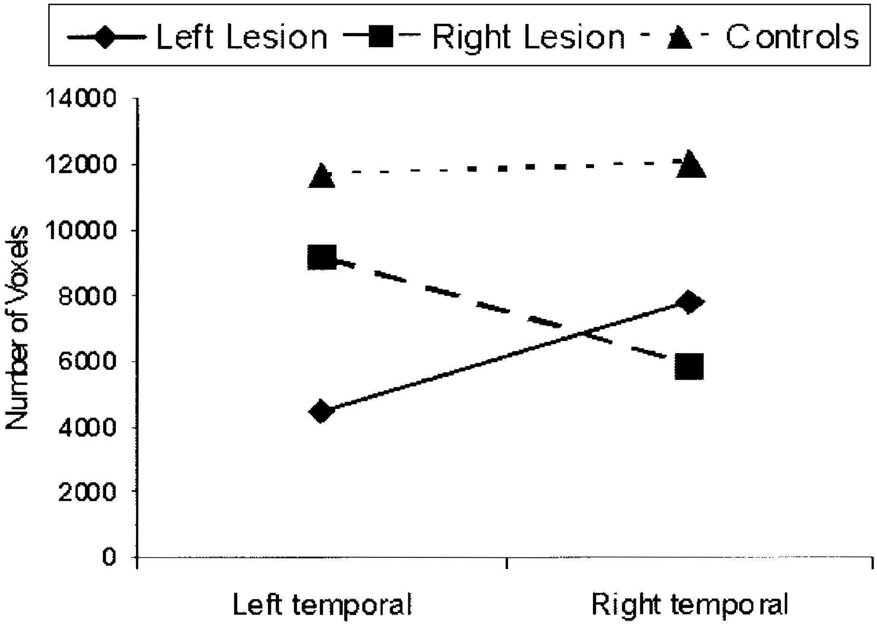
- Fig 2.
Mean number of voxels activated for both tasks in each group.


Tables
Patient No./Ae (y)/Sex Temporal Lobe Etiology Hippocampal Formation Affected TLE L1 Hometown Walking Task L1 Picture Encoding Task 1/30/M L Glioblastoma multiforme grade IV Yes No −93.35 −40.90 2/28/M L Cavernous angioma Yes No 9.30 8.20 3/44/F L Glioblastoma multiforme grade IV Yes Yes −83.57 −35.13 4/61/M R Glioblastoma multiforme grade IV Yes No 61.77 98.37 5/61/M R Cavernous angioma Yes Yes 18.31 30.69 6/39/M R Glioblastoma multiforme grade IV Yes No 29.17 31.99 7/51/F L Cavernous angioma No Yes −36.20 −29.57 8/36/M R Low-grade astrocytoma Yes No 16.83 9/70/M L Glioblastoma multiforme grade IV Yes Yes −43.71 −75.25 10/64/M L AVM Yes No 11.82 11/30/F L Oligoastrocytoma grade II Yes Yes −100 12/39/F R Hippocampal sclerosis Yes Yes 17.05 −2.14 13/72/M L High-grade glioma No No −36.35 −85.52 14/43/M L AVM Yes Yes −23.66 15/55/F R Oligodendroglioma grade II Yes Yes 57.54 48.55 16/34/M R Low-grade astrocytoma Yes Yes 19.56 6.18 17/27/F L Meningothelial meningioma No No −9.21 −99.00 18/33/F R Cavernoma No Yes 8.85 −29.61 19/47/M L Glioblastoma multiforme grade IV Yes Yes 53.27 −35.66 20/42/F L Cavernous angioma Yes Yes −64.09 1.26 21/26/F L Medial temporal lobe atrophy Yes Yes −8.67 −8.68 22/37/F L AVM Yes No −100 −100 23/50/M L Tumoral lesion* Yes No −40.29 −25.44 24/46/F R Hippocampal sclerosis Yes Yes 54.92 25/23/M R Low-grade glioma Yes Yes 40.09 8.79 Controls 1/26/F 17.86 9.01 2/60/F −7.28 3.23 3/62/F −4.01 11.66 4/26/F 6.51 −14.00 5/28/F 5.60 −7.07 6/45/M −5.70 −13.23 7/40/F −5.71 0.47 8/47/F −10.02 18.37 9/39/F 0.24 3.02 10/59/M 7.94 −7.87 11/43/F −14.96 −6.35 12/45/F −12.93 17.99 Note:—TLE indicates temporal lobe epilepsy; LI, lateralization index; AVM, arteriovenous malformation.
* No histological diagnosis.
Brain area Picture-Encoding Task Hometown-Walking Task BA Talairach Coordinate Mean t Value BA Talairach Coordinate Mean t Value x y z x y z Supplementary motor area 6 6 9 50 4.80 6 9 12 48 5.28 Lateral frontal gyrus (R) 6 36 10 30 5.02 6 24 −1 55 4.95 Lateral frontal gyrus (L) 6 −26 −4 45 4.85 6 −27 −10 54 5.32 Parahippocampal gyrus (R) 19, 27, 28, 30, 35, 36 25 −37 −5 5.28 19, 27, 28, 30, 35, 36, 37 22 −29 −8 5.45 Parahippocampal gyrus (L) 19, 28, 30, 36, 37 −22 −42 −5 5.75 19, 27, 28, 30, 35, 36, 37 −20 −34 −7 5.41 Lingual gyrus (R) 18, 19 14 −57 0 6.02 Lingual gyrus (L) 18,19 −11 −56 1 5.56 Fusiform gyrus (R) 19, 20, 37 33 −50 −10 6.06 Fusiform gyrus (L) 19, 20, 37 −31 −49 −11 6.10 Posterior cingulate (R) 23, 29, 30, 31 5 −52 8 5.62 23, 29, 30, 31 9 −49 11 6.09 Posterior cingulate (L) 23, 29, 30, 31 −5 −52 8 5.13 23, 29, 30, 31 −6 −49 9 6.29 Hippocampus (R) 27 −16 −13 4.41 27 −24 −9 4.52 Hippocampus (L) −27 −16 −13 5.44 −26 −24 −6 4.60 Thalamus (R) 5 −23 3 8.90 11 −24 14 5.18 Thalamus (L) −17 −28 2 4.98 −10 −22 10 5.48 Note:—Areas are detailed as a function of gyri and Broadmann’s maps. BA indicates Broadmann areas; L, left hemisphere; R, right hemisphere.
- Table 3:
Correlations between recognition scores on the picture- encoding task and number of left and right voxels activated in both tasks
Mean (±SD) Picture-Encoding Task Hometown-Walking Task Left Right Left Right Recognition store 4.13 (2.46) .21 56* .44 .45 RAVLT First trial 4.71 (1.90) .65** .36 .63** .14 Five trials 12.07 (3.64) .62** .40 .67** .28 Recall (30 min) 9.36 (4.58) .57* .40 .48 .20 Rey figure Copy 64.08 (31.16) −.02 .00 .14 .30 Recall 15.77 (13.14) .17 .58* .37 .38 Digit span 8.07 (2.3) .54* .58* .22 .30 Note:—
* P < .05,
** P < .01.
RAVLT indicates Rey Auditory Verbal Learning Task.


