
Article Figures & Data
Figures
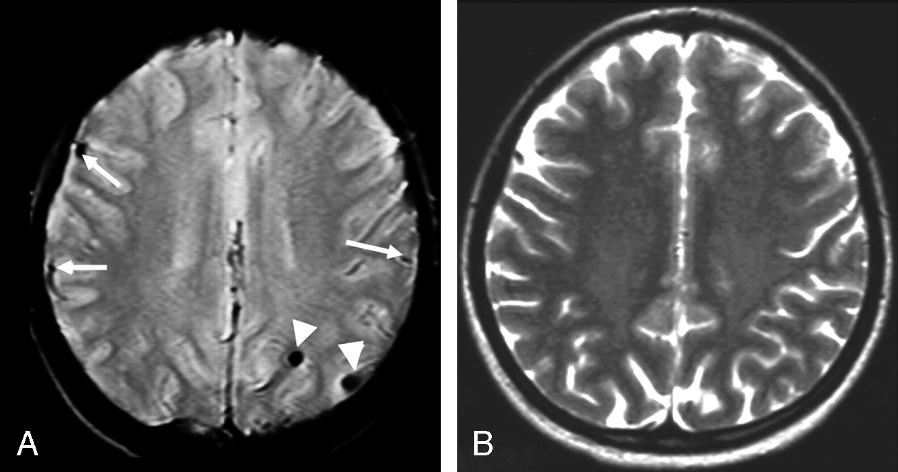
- Fig 1.
Twenty-year-old woman diagnosed 14 years previously and treated with intrathecal MTX (total dose = 72 mg) and cranial radiation therapy (18 Gy). A, Transverse gradient echo image shows 2 lesions compatible with old hemorrhages (arrowheads). The structures indicated by the arrows are blood vessels. B, Transverse T2-weighted fast spin-echo image at the same level appears normal.
- Fig 2.
Twenty-seven-year-old man diagnosed 18 years previously and treated with intrathecal MTX (total dose = 300 mg) and cranial radiation therapy (24 Gy). On coronal FLAIR image, there is grade 1 leukoencephalopathy (arrows), defined as patchy, mildly increased signal intensity in the periventricular white matter, without involvement of the subcortical U-fibers.


Tables
ALL Patients Control Patients Student t test Number 42 22 Number (%) male 27 (64) 15 (68) Age at examination (y) Range 6.9–27.6 7.2–31.8 P = .16 Mean ± SD 17.4 ± 4.6 15.4 ± 5.5 Age at diagnosis (y) Range 1.2–13.7 0.5–13.0 P = .46 Mean ± SD 5.2 ± 2.9 5.9 ± 4.0 Interval from diagnosis (y) Range 5.0–18.8 5.6–20.4 P = .015 Mean ± SD 12.2 ± 3.6 9.5 ± 4.2 Note:—ALL indicates acute lymphoblastic leukemia.
- Table 2:
Location of hemorrhages detected at gradient echo imaging in 42 long-term survivors of acute lymphoblastic leukemia
Region Number (%) Subregion Number Cerebral hemispheres 51 (82%) Frontal lobe 23 Parietal lobe 10 Temporal lobe 10 Occipital lobe 6 Insula 2 Deep brain structures 5 (8%) Corpus callosum 2 Pineal 1 Thalamus 1 Caudate nucleus 1 Posterior fossa 6 (10%) Cerebellum 5 Brain stem 1 Total 62 (100%) Sequence No. of hemorrhagic foci detected Gradient echo 62 T2-weighted spin echo 9 Fluid-attenuated inversion recovery 7 T1-weighted spin echo 3


