
Article Figures & Data
Figures
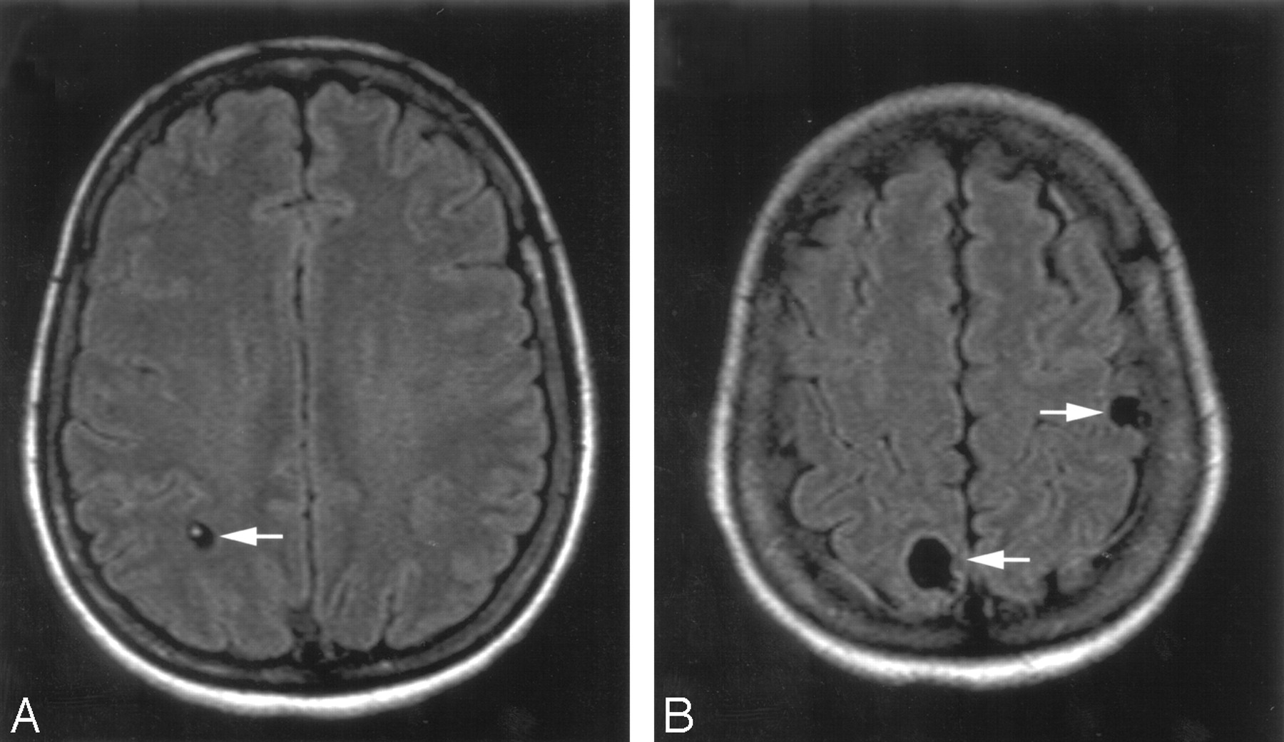
- Fig 1.
Fluid-attenuated inversion recovery (FLAIR) imaging studies before therapy. Three of 11 cystic lesions are shown: living parasite in a right parietal cystic lesion (white arrow, A); another right parietal and a left parietal cortical cystic lesion (white arrows, B).
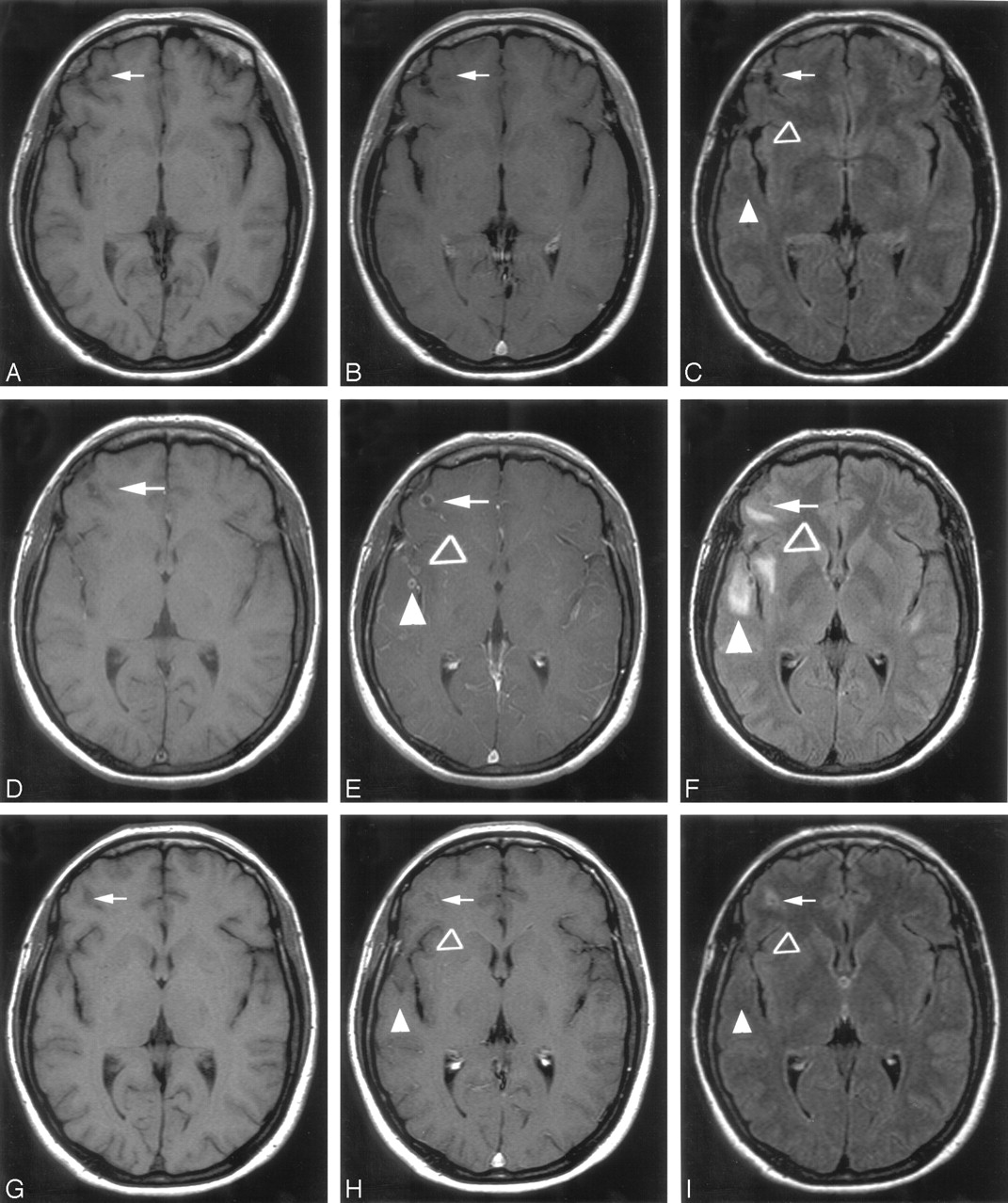
- Fig 2.
Treatment response of the patient shown by MR imaging. Imaging was performed before treatment (A–C) and 10 days (D–F) and 3 months (G–I) after therapy. A–C, A small cystic right frontal lesion without contrast enhancement (A, B) or edema (white arrow, C) is seen; in the right perisylvian area, hypointense lesions medially (open triangle) and laterally to the right sylvian fissure are seen on fluid-attenuated inversion recovery (FLAIR) but not on T1-weighted images. D–F, Soon after therapy, ring enhancement (D, E) and edema (F) are detected in the right frontal lesion (white arrow); ring enhancement with severe edema is seen in the perisylvian lesions (open and white triangles; E, F). G–I, Three months after treatment, a slight enhancement (G, H) and edema (I) are still present frontally (white arrow); no lesion is seen in the perisylvian area (open and white triangles; H, I). A, D, G are T1-weighted images without contrast; B, E, H, T1-weighted images with contrast; and C, F, I, FLAIR images.
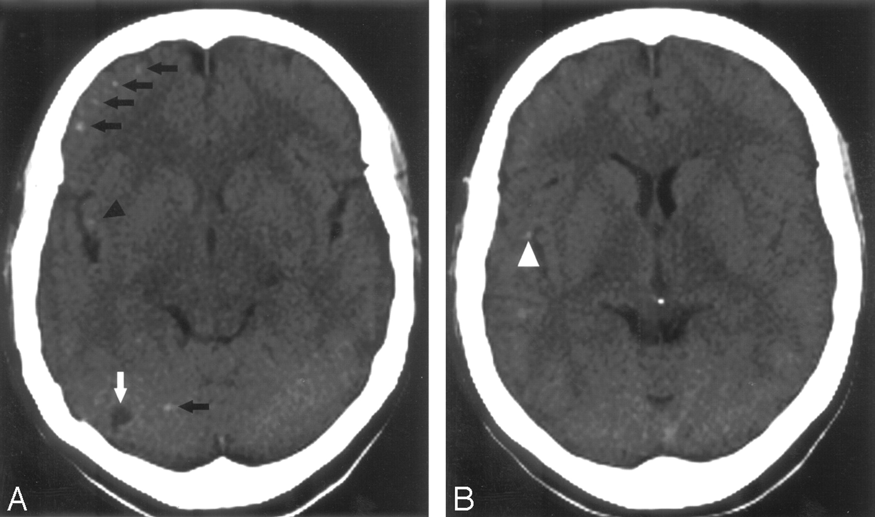
- Fig 3.
CT scans obtained 3 months before therapy. The calcifications medially (triangle, A) and laterally (white triangle, B) to the right perisylvian fissure correspond to the perisylvian lesions shown in Fig 2 D–F. Note several calcifications not showing an inflammatory reaction after therapy (black arrows, A) and an additional right cerebellar lesion (white arrow, A).


In this issue


Jump to section
Related Articles
Cited By...
- Intraventricular Taenia solium Cysts Presenting with Bruns Syndrome and Indications for Emergent Neurosurgery
- Calcified Neurocysticercus, Perilesional Edema, and Histologic Inflammation
- Corticosteroid Withdrawal Precipitates Perilesional Edema around Calcified Taenia solium Cysts
- A Calcified Taenia solium Granuloma Associated with Recurrent Perilesional Edema Causing Refractory Seizures: Histopathological Features