Advertisement

Main menu

  • Home
  • Content
    • Current Issue
    • Accepted Manuscripts
    • Article Preview
    • Past Issue Archive
    • AJNR Case Collection
    • Case of the Week Archive
    • Classic Case Archive
    • Case of the Month Archive
  • Special Collections
    • Spinal CSF Leak Articles (Jan 2020-June 2024)
    • 2024 AJNR Journal Awards
    • Most Impactful AJNR Articles
  • Multimedia
    • AJNR Podcast
    • AJNR Scantastics
    • Video Articles
  • For Authors
    • Submit a Manuscript
    • Author Policies
    • Fast publishing of Accepted Manuscripts
    • Graphical Abstract Preparation
    • Manuscript Submission Guidelines
    • Imaging Protocol Submission
    • Submit a Case for the Case Collection
  • About Us
    • About AJNR
    • Editorial Board
  • More
    • Become a Reviewer/Academy of Reviewers
    • Subscribers
    • Permissions
    • Alerts
    • Feedback
    • Advertisers
    • ASNR Home
  • Other Publications
    • ajnr

User menu

  • Alerts
  • Log in

Search

  • Advanced search
American Journal of Neuroradiology

American Journal of Neuroradiology

ASHNR American Society of Functional Neuroradiology ASHNR American Society of Pediatric Neuroradiology ASSR
  • Alerts
  • Log in

Advanced Search

  • Home
  • Content
    • Current Issue
    • Accepted Manuscripts
    • Article Preview
    • Past Issue Archive
    • AJNR Case Collection
    • Case of the Week Archive
    • Classic Case Archive
    • Case of the Month Archive
  • Special Collections
    • Spinal CSF Leak Articles (Jan 2020-June 2024)
    • 2024 AJNR Journal Awards
    • Most Impactful AJNR Articles
  • Multimedia
    • AJNR Podcast
    • AJNR Scantastics
    • Video Articles
  • For Authors
    • Submit a Manuscript
    • Author Policies
    • Fast publishing of Accepted Manuscripts
    • Graphical Abstract Preparation
    • Manuscript Submission Guidelines
    • Imaging Protocol Submission
    • Submit a Case for the Case Collection
  • About Us
    • About AJNR
    • Editorial Board
  • More
    • Become a Reviewer/Academy of Reviewers
    • Subscribers
    • Permissions
    • Alerts
    • Feedback
    • Advertisers
    • ASNR Home
  • Follow AJNR on Twitter
  • Visit AJNR on Facebook
  • Follow AJNR on Instagram
  • Join AJNR on LinkedIn
  • RSS Feeds

Welcome to the new AJNR, Updated Hall of Fame, and more. Read the full announcements.


AJNR is seeking candidates for the position of Associate Section Editor, AJNR Case Collection. Read the full announcement.


Research ArticleINTERVENTIONAL

Microcatheter Navigation and Thrombolysis in Acute Symptomatic Cervical Internal Carotid Occlusion

A. Srinivasan, M. Goyal, P. Stys, M. Sharma and C. Lum
American Journal of Neuroradiology April 2006, 27 (4) 774-779;
A. Srinivasan
  • Find this author on Google Scholar
  • Find this author on PubMed
  • Search for this author on this site
M. Goyal
  • Find this author on Google Scholar
  • Find this author on PubMed
  • Search for this author on this site
P. Stys
  • Find this author on Google Scholar
  • Find this author on PubMed
  • Search for this author on this site
M. Sharma
  • Find this author on Google Scholar
  • Find this author on PubMed
  • Search for this author on this site
C. Lum
  • Find this author on Google Scholar
  • Find this author on PubMed
  • Search for this author on this site
  • Article
  • Figures & Data
  • Info & Metrics
  • Responses
  • References
  • PDF
Loading

Article Figures & Data

Figures

  • Tables
  • Fig 1.
    • Download figure
    • Open in new tab
    • Download powerpoint
    Fig 1.

    A, Diagnostic angiogram of the left internal carotid artery (ICA) reveals good flow across the anterior communicating artery (AcomA) but poor filling of the right MCA territory because of a thrombus demonstrated on an earlier CT angiogram.

    B, Successful recanalization of right MCA is seen after administration of 9 mg of tissue plasminogen activator (tPA) into the middle cerebral artery (MCA).

    C, Diagnostic angiogram of the left ICA reveals cross-filling through the AcomA into the right MCA. There is probably dilution of contrast in the MCA by nonopacified blood from the ipsilateral posterior communicating artery because good patency of the MCA was demonstrated on the earlier microcatheter injection.
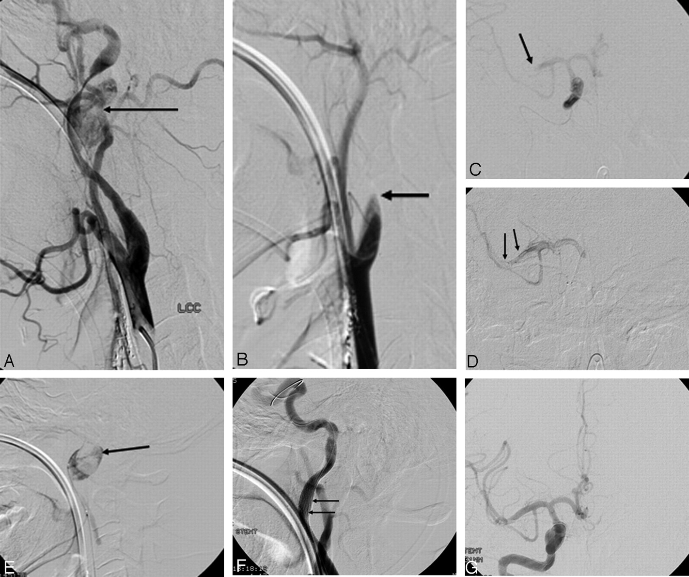
  • Fig 2.
    • Download figure
    • Open in new tab
    • Download powerpoint
    Fig 2.

    A, Lateral angiogram of the left common carotid artery (CCA) in a 24-year-old man with acute left hemiplegia reveals a pseudoaneurysm (arrow) in the distal cervical internal carotid artery (ICA) with narrowing of the parent artery.

    B, The right CCA angiogram shows complete occlusion of the right ICA (arrow) just beyond the bulb.

    C, Microcatheter injection demonstrates a thrombus in the distal M1 segment (arrow) of the middle cerebral artery (MCA).

    D, Administration of 7.5 mg of intra-arterial tissue plasminogen activator (IA tPA) through the microcatheter, which was advanced into the face of the thrombus, resulted in lysis of thrombus and restoration of antegrade flow.

    E, Microcatheter injections demonstrate a pseudoaneurysm in the distal cervical right ICA (arrow).

    F and G, Repeat angiogram through the guiding catheter after successful treatment by using 2 overlapping covered stents reveals no filling of the pseudoaneurysm and normal flow in the MCA. The patient recovered complete power on the left side within minutes of the procedure. The left ICA dissection and pseudoaneurysm were managed conservatively and, at 2 months after the procedure, the patient had no neurologic deficits.

Tables

  • Figures
    • View popup
    Table 1:

    Clinical and imaging features of 7 patients with occluded internal carotid artery and acute stroke

    Patient No./Age (y)/SexSite of Occlusion (CTA)Presence of Penumbra (CTP)NIHSS at PresentationNIHSS at Follow-up
    1/54/MLICA and LMCA: M1Yes2215 (1 mo)
    2/51/MLICA and LMCA: M1 and M2Yes151 (14 mo)
    3/62/FRICA and RMCA: M1Yes210 (6 mo)
    4/78/FLICA and LMCA: M1Yes110 (5 mo)
    5/70/FRICA*Yes†171 (14 mo)
    6/28/FLICA and LMCA: M1No perfusion study‡170 (40 mo)
    7/24/MRICA and RMCA: M1No perfusion study120 (1 mo)
    • Note:—CTA indicates computed tomographic angiography; CTP, computed tomographic perfusion; NIHSS, National Institutes of Health Stroke Scale; RICA, right internal carotid artery; LICA, left internal carotid artery; RMCA, right middle cerebral artery; LMCA, left middle cerebral artery.

    • * Seen on MR angiogram.

    • † Seen on MR perfusion study.

    • ‡ No perfusion imaging performed since the intervention “pre-dated routine perfusion imaging.”

    • View popup
    Table 2:

    Intervention and outcomes in the 7 patients with acute stroke due to occluded ICA

    Patient No.IA tPA (mg)Stenting and angioplasty of ICALysis of MCA Clot through occluded ICATIMI Flow after ThromboysisPost Rx CTOutcome (mRS) at 30 days
    120YesYes2Basal ganglia infarct4
    210YesYes3Patchy parietal infarcts3
    39NoYes3Basal ganglia infarct0
    40*YesNo3Basal ganglia infarct0
    518NoYes3Basal ganglia infarct0
    610NoNo3Evolving frontal bleed1
    715Yes†Yes3Small frontal lobe infarct0
    • Note:—ICA indicates internal carotid artery; IA tPA, intra-arterial tissue plasminogen activator; MCA, middle cerebral artery; CT, computed tomography; mRS, modified Rankin Scale.

    • * This patient received 38 mg intravenous tPA prior to angiography.

    • † ICA pseudoaneurysm was treated with 2 overlapping covered stents.

PreviousNext
Back to top

In this issue

American Journal of Neuroradiology: 27 (4)
American Journal of Neuroradiology
Vol. 27, Issue 4
April 2006
  • Table of Contents
  • Index by author
Advertisement
Print
Download PDF
Email Article

Thank you for your interest in spreading the word on American Journal of Neuroradiology.

NOTE: We only request your email address so that the person you are recommending the page to knows that you wanted them to see it, and that it is not junk mail. We do not capture any email address.

Enter multiple addresses on separate lines or separate them with commas.
Microcatheter Navigation and Thrombolysis in Acute Symptomatic Cervical Internal Carotid Occlusion
(Your Name) has sent you a message from American Journal of Neuroradiology
(Your Name) thought you would like to see the American Journal of Neuroradiology web site.
CAPTCHA
This question is for testing whether or not you are a human visitor and to prevent automated spam submissions.
Cite this article
A. Srinivasan, M. Goyal, P. Stys, M. Sharma, C. Lum
Microcatheter Navigation and Thrombolysis in Acute Symptomatic Cervical Internal Carotid Occlusion
American Journal of Neuroradiology Apr 2006, 27 (4) 774-779;

Citation Manager Formats

  • BibTeX
  • Bookends
  • EasyBib
  • EndNote (tagged)
  • EndNote 8 (xml)
  • Medlars
  • Mendeley
  • Papers
  • RefWorks Tagged
  • Ref Manager
  • RIS
  • Zotero
0 Responses
Respond to this article
Share
Bookmark this article
Microcatheter Navigation and Thrombolysis in Acute Symptomatic Cervical Internal Carotid Occlusion
A. Srinivasan, M. Goyal, P. Stys, M. Sharma, C. Lum
American Journal of Neuroradiology Apr 2006, 27 (4) 774-779;
del.icio.us logo Twitter logo Facebook logo Mendeley logo
  • Tweet Widget
  • Facebook Like
  • Google Plus One

Jump to section

  • Article
    • Abstract
    • Patients and Methods
    • Results
    • Discussion
    • References
  • Figures & Data
  • Info & Metrics
  • Responses
  • References
  • PDF

Related Articles

  • No related articles found.
  • PubMed
  • Google Scholar

Cited By...

  • Acute carotid stenting in patients undergoing thrombectomy: a systematic review and meta-analysis
  • Relay-balloon technique for recanalization of acute symptomatic proximal internal carotid artery occlusion with short balloon-tipped guiding catheter landing zone
  • Response to Letter by Nedeltchev et al Regarding Article, "Short-Term Outcomes After Symptomatic Internal Carotid Artery Occlusion"
  • Crossref
  • Google Scholar

This article has not yet been cited by articles in journals that are participating in Crossref Cited-by Linking.

More in this TOC Section

  • SAVE vs. Solumbra Techniques for Thrombectomy
  • CT Perfusion&Reperfusion in Acute Ischemic Stroke
  • Delayed Reperfusion Post-Thrombectomy&Thrombolysis
Show more Interventional

Similar Articles

Advertisement

Indexed Content

  • Current Issue
  • Accepted Manuscripts
  • Article Preview
  • Past Issues
  • Editorials
  • Editors Choice
  • Fellow Journal Club
  • Letters to the Editor

Cases

  • Case Collection
  • Archive - Case of the Week
  • Archive - Case of the Month
  • Archive - Classic Case

Special Collections

  • Special Collections

Resources

  • News and Updates
  • Turn around Times
  • Submit a Manuscript
  • Author Policies
  • Manuscript Submission Guidelines
  • Evidence-Based Medicine Level Guide
  • Publishing Checklists
  • Graphical Abstract Preparation
  • Imaging Protocol Submission
  • Submit a Case
  • Become a Reviewer/Academy of Reviewers
  • Get Peer Review Credit from Publons

Multimedia

  • AJNR Podcast
  • AJNR SCANtastic
  • Video Articles

About Us

  • About AJNR
  • Editorial Board
  • Not an AJNR Subscriber? Join Now
  • Alerts
  • Feedback
  • Advertise with us
  • Librarian Resources
  • Permissions
  • Terms and Conditions

American Society of Neuroradiology

  • Not an ASNR Member? Join Now

© 2025 by the American Society of Neuroradiology All rights, including for text and data mining, AI training, and similar technologies, are reserved.
Print ISSN: 0195-6108 Online ISSN: 1936-959X

Powered by HighWire