Advertisement

Main menu

  • Home
  • Content
    • Current Issue
    • Accepted Manuscripts
    • Article Preview
    • Past Issue Archive
    • AJNR Case Collection
    • Case of the Week Archive
    • Classic Case Archive
    • Case of the Month Archive
  • Special Collections
    • Spinal CSF Leak Articles (Jan 2020-June 2024)
    • 2024 AJNR Journal Awards
    • Most Impactful AJNR Articles
  • Multimedia
    • AJNR Podcast
    • AJNR Scantastics
    • Video Articles
  • For Authors
    • Submit a Manuscript
    • Author Policies
    • Fast publishing of Accepted Manuscripts
    • Graphical Abstract Preparation
    • Manuscript Submission Guidelines
    • Imaging Protocol Submission
    • Submit a Case for the Case Collection
  • About Us
    • About AJNR
    • Editorial Board
  • More
    • Become a Reviewer/Academy of Reviewers
    • Subscribers
    • Permissions
    • Alerts
    • Feedback
    • Advertisers
    • ASNR Home
  • Other Publications
    • ajnr

User menu

  • Alerts
  • Log in

Search

  • Advanced search
American Journal of Neuroradiology

American Journal of Neuroradiology

ASHNR American Society of Functional Neuroradiology ASHNR American Society of Pediatric Neuroradiology ASSR
  • Alerts
  • Log in

Advanced Search

  • Home
  • Content
    • Current Issue
    • Accepted Manuscripts
    • Article Preview
    • Past Issue Archive
    • AJNR Case Collection
    • Case of the Week Archive
    • Classic Case Archive
    • Case of the Month Archive
  • Special Collections
    • Spinal CSF Leak Articles (Jan 2020-June 2024)
    • 2024 AJNR Journal Awards
    • Most Impactful AJNR Articles
  • Multimedia
    • AJNR Podcast
    • AJNR Scantastics
    • Video Articles
  • For Authors
    • Submit a Manuscript
    • Author Policies
    • Fast publishing of Accepted Manuscripts
    • Graphical Abstract Preparation
    • Manuscript Submission Guidelines
    • Imaging Protocol Submission
    • Submit a Case for the Case Collection
  • About Us
    • About AJNR
    • Editorial Board
  • More
    • Become a Reviewer/Academy of Reviewers
    • Subscribers
    • Permissions
    • Alerts
    • Feedback
    • Advertisers
    • ASNR Home
  • Follow AJNR on Twitter
  • Visit AJNR on Facebook
  • Follow AJNR on Instagram
  • Join AJNR on LinkedIn
  • RSS Feeds

Welcome to the new AJNR, Updated Hall of Fame, and more. Read the full announcements.


AJNR is seeking candidates for the position of Associate Section Editor, AJNR Case Collection. Read the full announcement.


Research ArticlePEDIATRICS

Diffusion Tensor Imaging of Tract Involvement in Children with Pontine Tumors

K.J. Helton, N.S. Phillips, R.B. Khan, F.A. Boop, R.A. Sanford, P. Zou, C.S. Li, J.W. Langston and R.J. Ogg
American Journal of Neuroradiology April 2006, 27 (4) 786-793;
K.J. Helton
  • Find this author on Google Scholar
  • Find this author on PubMed
  • Search for this author on this site
N.S. Phillips
  • Find this author on Google Scholar
  • Find this author on PubMed
  • Search for this author on this site
R.B. Khan
  • Find this author on Google Scholar
  • Find this author on PubMed
  • Search for this author on this site
F.A. Boop
  • Find this author on Google Scholar
  • Find this author on PubMed
  • Search for this author on this site
R.A. Sanford
  • Find this author on Google Scholar
  • Find this author on PubMed
  • Search for this author on this site
P. Zou
  • Find this author on Google Scholar
  • Find this author on PubMed
  • Search for this author on this site
C.S. Li
  • Find this author on Google Scholar
  • Find this author on PubMed
  • Search for this author on this site
J.W. Langston
  • Find this author on Google Scholar
  • Find this author on PubMed
  • Search for this author on this site
R.J. Ogg
  • Find this author on Google Scholar
  • Find this author on PubMed
  • Search for this author on this site
  • Article
  • Figures & Data
  • Info & Metrics
  • Responses
  • References
  • PDF
Loading

Article Figures & Data

Figures

  • Tables
  • Fig 1.
    • Download figure
    • Open in new tab
    • Download powerpoint
    Fig 1.

    MR imaging and DTI of a 15-year-old patient with diffuse pontine tumor.

    A, Sagittal T1-weighted image. The pontine tumor is diffusely infiltrative. B, Axial T2-weighted image. The pons appears expanded and hyperintense with an area of focal necrosis. C, Axial ADC map. The tumor demonstrates elevated diffusion, and the necrosis is extremely hyperintense. D, Axial FA map. The tumor demonstrates diminished fractional anisotropy, and necrosis is hypointense.

  • Fig 2.
    • Download figure
    • Open in new tab
    • Download powerpoint
    Fig 2.

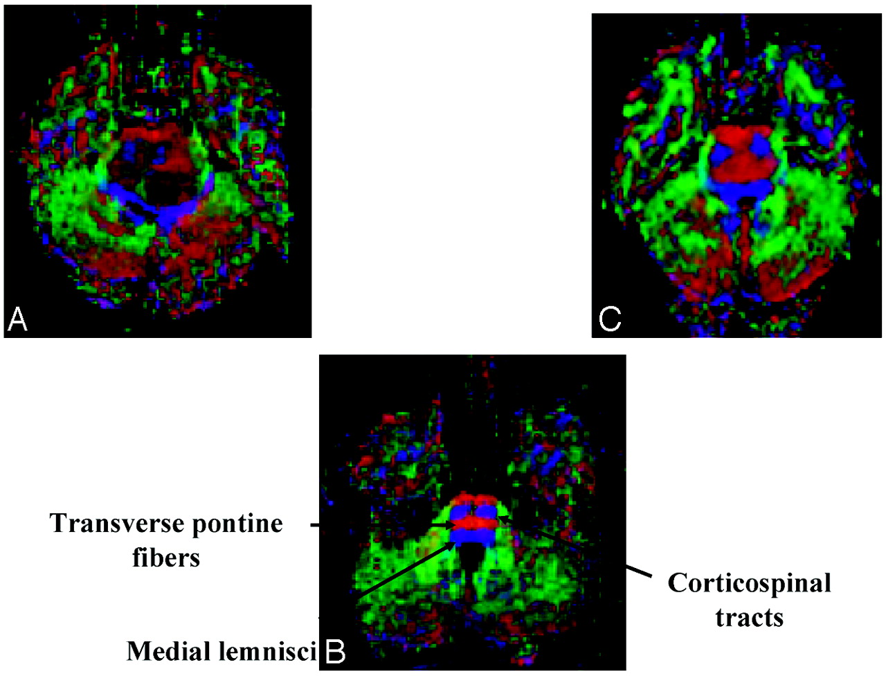
    Axial diffusion tensor color maps of the brain stem at the level of the middle cerebellar peduncles.

    A, Image of patient with a pontine tumor showing destruction of the normal anisotropy of the corticospinal tracts and posterior displacement of the medial lemnisci.

    B, Control image showing normal corticospinal tracts, transverse pontine fibers, and medial lemnisci.

    C, Image of patient with a pontine tumor showing a diffusely infiltrating pattern.

  • Fig 3.
    • Download figure
    • Open in new tab
    • Download powerpoint
    Fig 3.

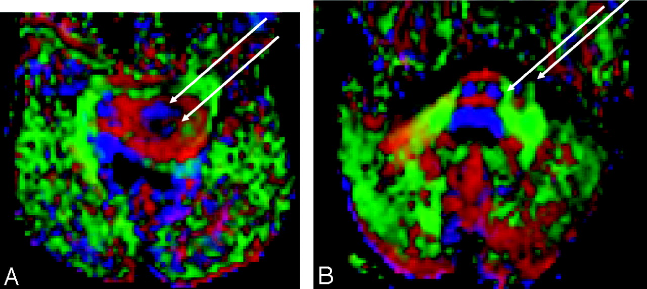
    Axial diffusion tensor color maps demonstrating tract invasion.

    A, Image of a 6-year-old patient with a diffusely infiltrating pontine tumor. The left corticospinal tract is enlarged, compared with the right, and tumor infiltration separates the corticospinal (anterior arrow) and corticobulbar (posterior arrow) components.

    B, Image of an 8-year-old patient with a focally exophytic pontine tumor. Mild lateral expansion of the left corticospinal tract (medial arrow), with focally exophytic tumor (lateral arrow).

  • Fig 4.
    • Download figure
    • Open in new tab
    • Download powerpoint
    Fig 4.

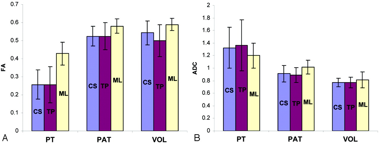
    Graphs show diffusion tensor imaging parameters for major white matter tracts in the brain stem.

    A, Fractional anisotropy (FA). B, Apparent diffusion coefficient (ADC). Data are shown for the corticospinal (CS, blue bars), transverse pontine (TP, red bars), and medial lemnisci (ML, yellow bars). Subject groups included patients with pontine tumors, patient control (PAT), and healthy volunteer (VOL) groups.

Tables

  • Figures
    • View popup
    Table 1:

    Comparison of conventional MR imaging and diffusion tensor imaging measures in 7 pediatric patients with pontine tumors

    Patient No.Tumor CharacteristicsConventional MR ImagingDiffusion Tensor Imaging
    LocationSize (cm)T1WT2WEnhancementFocalityNecrosisMass EffectHydrocephalusCorticospinal TractsTransverse Pontine TractsSensory Tracts
    1Midbrain/pons4.6 × 3.6HypoHyperNoNoNoModerateShunt in placeBilateral sev exp, distinct CBSev exp of left tractPosterior displ, noninfiltrated
    2Pons3.0 × 3.4HypoHyperNoNoNoMinimalNoMild exp, lateral displ of right tractSev bilateral expPosterior displ, noninfiltrated
    Mild reduction, medial deviation, and slightly isotropic left tract
    3Pons2.8 × 3.1HypoHyperNoNoNoMinimalNoBilateral sev exp, minimal posterior displ of right tractMild bilateral expNormal
    4Pons3.2 × 4.3Sev hypoSev hyperNoNoNoMinimalNoBilateral sev expSev bilateral expBilateral grossly posterior displ (left > right)
    5Pons3.8 × 4.5Sev hypoSev hyperSurrounding necrosisNoYes*ModerateMildBilateral sev exp, distinct CBSev bilateral exp, necrosisBilateral grossly posterior displ (left > right)
    6Pons3.9 × 4.7Sev hypoSev hyperNoNoNoMildNoBilateral sev exp, posterior displ, distinct right CBSev prominently anterior expBilateral grossly posterior displ (left > right)
    7†Pons1.6 × 1.0HypoHyperMinimal focalYesNoMinimalNoMild exp of left tractNormalNormal
    1.0 × 0.8
    • Note:—hypo indicates hypointense; hyper, hyperintense; sev, severe; exp, expansion; CB, corticobulbar tracts; displ, displacement.

    • * The necrotic region measured 2.2 × 2.5 cm.

    • † This patient’s tumor had an exophytic component that measured 1.0 × 0.8 cm.

    • View popup
    Table 2:

    Color map assessment of diffusion tensor images of brain stem white matter tracts in 7 pediatric patients with pontine tumors*

    Patient No.Corticospinal TractsTransvers Pontine TractsMedial Lemnisci
    LeftRightLeftRightLeftRight
    1D/IIIIDD
    2IX/IIIDD
    3IIIID/ID/I
    4IIX/IX/IDD
    5X/IX/IX/IX/III
    6IX/IIINI
    7INNNNN
    • * The condition of the tracts was classified as follows: D, displaced; E, edematous; I, infiltrated; N, normal; X, destroyed.

    • View popup
    Table 3:

    Neurologic deficits in 7 pediatric patients with pontine tumors

    Patient No.Neurologic Deficit (Grade* and Location)
    CorticospinalCranial NervesSensoryAtaxia
    1Mild (left)Mild (left) VIN/AAbsent
    2Mild (right)M/S (left) VI, VII, IXN/AAbsent
    3AbsentAbsentN/AAbsent
    4AbsentM/S (bilateral) VI (left), VII, IXAbsentM/S (bilateral), worse on left
    5AbsentM/S left gaze, VI, VII, and partial IXAbsentSevere gait, moderate bilateral limb dysmetria
    6Mild (bilateral), worse on leftM/S (bilateral) VI, IX, worse on rightN/AMild gait, left limb dysmetria
    7AbsentAbsentAbsentMild limb dysmetria
    • Note:—N/A indicates not available.

    • * Deficits were classified into 2 grades: absent/mild, and M/S, moderate/severe.

PreviousNext
Back to top

In this issue

American Journal of Neuroradiology: 27 (4)
American Journal of Neuroradiology
Vol. 27, Issue 4
April 2006
  • Table of Contents
  • Index by author
Advertisement
Print
Download PDF
Email Article

Thank you for your interest in spreading the word on American Journal of Neuroradiology.

NOTE: We only request your email address so that the person you are recommending the page to knows that you wanted them to see it, and that it is not junk mail. We do not capture any email address.

Enter multiple addresses on separate lines or separate them with commas.
Diffusion Tensor Imaging of Tract Involvement in Children with Pontine Tumors
(Your Name) has sent you a message from American Journal of Neuroradiology
(Your Name) thought you would like to see the American Journal of Neuroradiology web site.
CAPTCHA
This question is for testing whether or not you are a human visitor and to prevent automated spam submissions.
Cite this article
K.J. Helton, N.S. Phillips, R.B. Khan, F.A. Boop, R.A. Sanford, P. Zou, C.S. Li, J.W. Langston, R.J. Ogg
Diffusion Tensor Imaging of Tract Involvement in Children with Pontine Tumors
American Journal of Neuroradiology Apr 2006, 27 (4) 786-793;

Citation Manager Formats

  • BibTeX
  • Bookends
  • EasyBib
  • EndNote (tagged)
  • EndNote 8 (xml)
  • Medlars
  • Mendeley
  • Papers
  • RefWorks Tagged
  • Ref Manager
  • RIS
  • Zotero
0 Responses
Respond to this article
Share
Bookmark this article
Diffusion Tensor Imaging of Tract Involvement in Children with Pontine Tumors
K.J. Helton, N.S. Phillips, R.B. Khan, F.A. Boop, R.A. Sanford, P. Zou, C.S. Li, J.W. Langston, R.J. Ogg
American Journal of Neuroradiology Apr 2006, 27 (4) 786-793;
del.icio.us logo Twitter logo Facebook logo Mendeley logo
  • Tweet Widget
  • Facebook Like
  • Google Plus One

Jump to section

  • Article
    • Abstract
    • Methods
    • Results
    • Discussion
    • Conclusion
    • Footnotes
    • References
  • Figures & Data
  • Info & Metrics
  • Responses
  • References
  • PDF

Related Articles

  • No related articles found.
  • PubMed
  • Google Scholar

Cited By...

  • A Novel Methodology for Applying Multivoxel MR Spectroscopy to Evaluate Convection-Enhanced Drug Delivery in Diffuse Intrinsic Pontine Gliomas
  • Quantitative Diffusion-Weighted and Dynamic Susceptibility-Weighted Contrast-Enhanced Perfusion MR Imaging Analysis of T2 Hypointense Lesion Components in Pediatric Diffuse Intrinsic Pontine Glioma
  • Apparent Diffusion and Fractional Anisotropy of Diffuse Intrinsic Brain Stem Gliomas
  • Crossref
  • Google Scholar

This article has not yet been cited by articles in journals that are participating in Crossref Cited-by Linking.

More in this TOC Section

  • Comparison of Image Quality and Radiation Dose in Pediatric Temporal Bone CT Using Photon-Counting Detector CT and Energy-Integrating Detector CT
  • SyMRI & MR Fingerprinting in Brainstem Myelination
  • Dandy-Walker Phenotype with Brainstem Involvement
Show more Pediatrics

Similar Articles

Advertisement

Indexed Content

  • Current Issue
  • Accepted Manuscripts
  • Article Preview
  • Past Issues
  • Editorials
  • Editors Choice
  • Fellow Journal Club
  • Letters to the Editor

Cases

  • Case Collection
  • Archive - Case of the Week
  • Archive - Case of the Month
  • Archive - Classic Case

Special Collections

  • Special Collections

Resources

  • News and Updates
  • Turn around Times
  • Submit a Manuscript
  • Author Policies
  • Manuscript Submission Guidelines
  • Evidence-Based Medicine Level Guide
  • Publishing Checklists
  • Graphical Abstract Preparation
  • Imaging Protocol Submission
  • Submit a Case
  • Become a Reviewer/Academy of Reviewers
  • Get Peer Review Credit from Publons

Multimedia

  • AJNR Podcast
  • AJNR SCANtastic
  • Video Articles

About Us

  • About AJNR
  • Editorial Board
  • Not an AJNR Subscriber? Join Now
  • Alerts
  • Feedback
  • Advertise with us
  • Librarian Resources
  • Permissions
  • Terms and Conditions

American Society of Neuroradiology

  • Not an ASNR Member? Join Now

© 2025 by the American Society of Neuroradiology All rights, including for text and data mining, AI training, and similar technologies, are reserved.
Print ISSN: 0195-6108 Online ISSN: 1936-959X

Powered by HighWire