
Abstract
BACKGROUND AND PURPOSE: The purpose of this study was to compare the volumetric results of intracranial aneurysms obtained by calculation of the volume of an ellipsoid with those obtained with 3D rotational angiography (3D-RA).
METHODS: First, the precision of 3D-RA in the assessment of volumetric measurement of intracranial aneurysm had to be established. The 3D-RA gave an overestimation of 4% to 5.5% of the actual volume of a spherical object. Then, 484 consecutive human intracranial aneurysms were studied with 3D-RA, allowing the determination of their volume. In the meantime, aneurysm dimensions (height and width) were measured on the 3D pictures generated by the 3D-RA. The aneurysm volumes were calculated (Vcalc) by considering the aneurysm shape to be ellipsoidal.
RESULTS: The calculated aneurysm volume (Vcalc) overestimated by 15 ± 38% the volume given by 3D-RA. Taking into account a 10% margin of error, 227 (47%) aneurysms were overestimated by 44 ± 34%, whereas 113 (23%) aneurysms were underestimated by 25 ± 12%. Only 144 (30%) aneurysms had calculated and 3D-RA results within the limits of 10% of discrepancy. Concordance was good for pericallosal and basilar tip aneurysms (mean overestimation of 6 ± 22% and 8 ± 27%, respectively). Conversely, there was a high discrepancy between calculated and 3D-RA results for posterior communicating artery aneurysms (mean overestimation of 22 ± 44%).
CONCLUSION: The calculation of the volume based on aneurysm dimensions is relatively accurate for pericallosal and basilar tip aneurysms, probably owing to their spherical or elliptic shape. Conversely, this formula is not adequate for irregularly shaped lesions, such as posterior communicating aneurysms.
The last decade has seen the endovascular treatment of aneurysms gain increasing acceptance within the neurosurgical community, and, in many centers, coiling has become the first-line treatment option for both ruptured and unruptured aneurysms. To assess the degree of aneurysm packing with coils, it has become imperative to determine preoperatively the volume of the aneurysm to be embolized. In many centers, digital subtraction angiography with 3D reconstruction rotational angiography (3D-RA) is now available to serve as a tool to plan the strategy for either surgical or endovascular treatment. This technology gives the capability, by using 3D-RA volumetric measurement, to obtain precise volumetric measurements.1,2 Conversely, this capability is not always available, and aneurysm volume calculation is performed under the assumption of the aneurysm as an ellipsoidal geometric form. Previous studies referred to aneurysm volume to assess aneurysm coil packing based on the ellipsoid approximation.3–5 Many aneurysms are irregularly shaped; so far, there is no definitive evidence of the accuracy of this method of aneurysm volume calculation. To determine whether the aneurysmal volume calculation by using ellipsoid approximation is a valid method, we compared the results obtained with 3D-RA and those obtained with ellipsoid approximation.
Methods
To begin, we had to establish whether our 3D-RA system was precise enough to be able to provide us with accurate volume measurements. As a start, our system was tested with 2 iron balls of 5 and 50 mm in diameter (Fig 1), which were imaged by using the same acquisition parameters routinely used in the clinical setting (Integris 3D-RA; Philips, Best, the Netherlands). The 3D-RA technique overestimated the volume of the 50-mm ball by 4% and that of the 5-mm ball by 5.5% (Fig 2). This small discrepancy between the actual volumes and those obtained with the 3D-RA system software therefore raised concerns and called into question the validity of the latter method. Nevertheless, we accepted the 4% to 5.5% discrepancy as acceptable and decided to use the volume values calculated with the latter methods as the correct values. Then, based upon 498 aneurysms that were consecutively treated selectively with coils in our department from January 2002 to March 2004, 484 had a 3D-RA, which enabled the aneurysm volume measurement before endovascular treatment. This means that, in 14 (3%) aneurysms, the quality of the 3D pictures was insufficient (poor aneurysm filling with contrast material, image blurring) to allow volume determination. Injection protocol was as follows: injection rate of 4 mL/s for a total of iodinated contrast material of 21 mL through a 5F catheter inserted in the proximal internal carotid artery or in the proximal dominant vertebral artery, with a 1-s delay between the beginning of the injection and the beginning of the rotational scan. The aneurysm volume was measured as follows: before the coiling procedure, a rotational angiography with a 17-cm image intensifier was performed. Physical limitations due to blood flow and injection characteristics led to a collection of images that exhibited variable contrast across a single rotational dataset. Each resulting projection image, however, was then automatically postprocessed with global histogram so that the equalization of each projection image appeared similar in every image of the rotational dataset. A magnification of 33% of the initial 3D dataset was reconstructed in a matrix of 2563. In this magnified 3D reconstruction, the aneurysm was manually segmented from the parent artery, and volume was calculated by using the 3D-RA volumetric measurement of the system software (Fig 3). The manual outlining and segmentation of the aneurysm from the parent artery was performed by the same neuroradiologist (M.P.) for all aneurysms on a prospective basis. The volume of the aneurysm model was automatically given by the built-in software by multiplying the total number of voxels belonging to the segmented aneurysm by the corresponding elementary voxel of the aneurysm volume. In the meantime, the volume (Vcalc) of each aneurysm was calculated according to the following equation corresponding to the calculation of the volume of an ellipsoid:  where a denoted the largest horizontal diameter and b the largest vertical diameter as measured on the 3D-RA images, with a and b being oriented perpendicularly. The locations of the 484 aneurysms were middle cerebral artery (n = 125), anterior communicating artery (n = 116), posterior communicating artery (n = 55), ophthalmic artery (n = 42), basilar tip (n = 35), supraclinoid (other than posterior communicating and ophthalmic arteries) internal carotid artery (n = 33), cavernous sinus (n = 25), carotid bifurcation (n = 23), pericallosal artery (n = 11), laterobasal (n = 5), posterior inferior cerebellar artery (n = 2), superior cerebellar artery (n = 5), vertebral junction (n = 4), and posterior cerebral artery (n = 3). Of these 484 aneurysms, 385 were <10 mm in maximum size, whereas 99 had a maximum diameter ≥10 mm. Mean volume misestimations according to aneurysm location were calculated and compared. Differences in misestimations due to aneurysm location were statistically analyzed by using the Kruskal-Wallis test (nonparametric test of analysis of variance). Differences in misestimation according to aneurysm sizes were statistically analyzed by using the Mann-Whitney test (nonparametric test for continuous variables, which are not normally distributed). P values <.05 were considered significant.

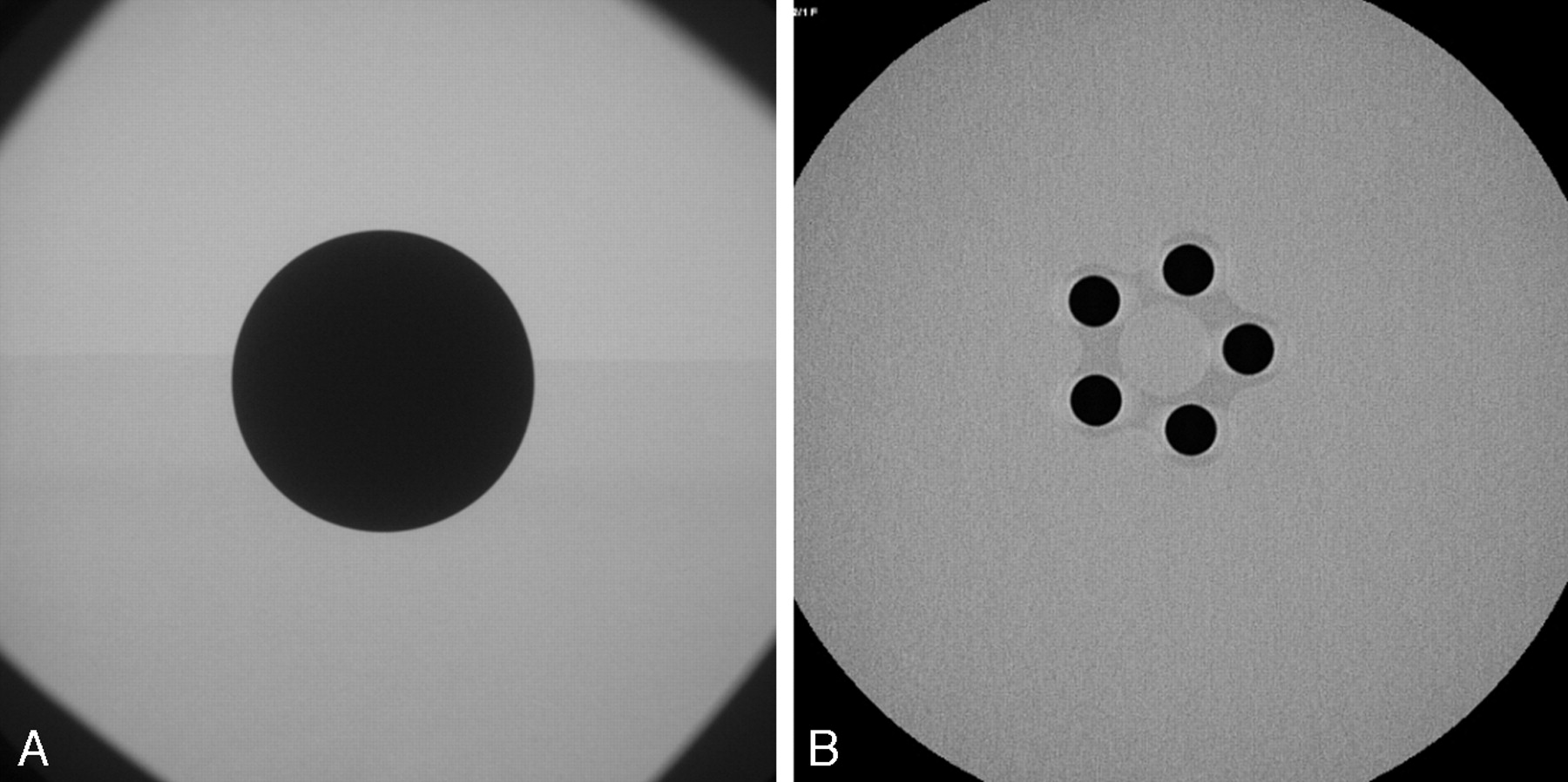
A, Radiograph of the 50-mm-diameter iron ball that served for the calibration of the 3D rotational angiography (3D-RA) system.
B, Radiograph of the 5-mm-diameter iron ball that also served for the calibration of the 3D-RA system.

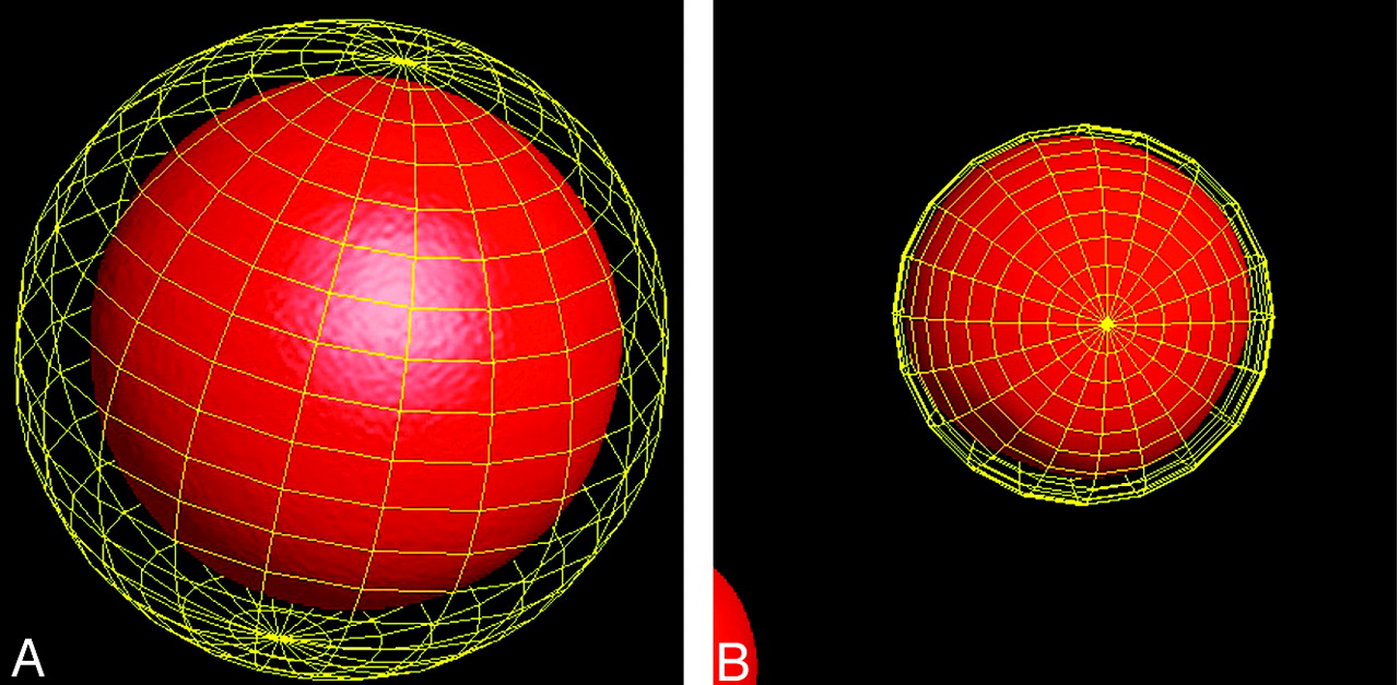
A, Volume determination with the 3D-RA software of the 50-mm-diameter iron ball displayed with surface-shaded display. The software is giving a volume of 67.875 mm3, whereas the ellipsoid calculation is giving 65.45 mm3.
B, Volume determination with the 3D-RA software of the 5-mm-diameter iron ball displayed with surface shaded display. The software is giving a volume of 69 mm3, whereas the ellipsoid calculation is giving 65.5 mm3.

Manual segmentation of a 12-mm large carotid ophthalmic artery aneurysm with the 3D rotational angiography (3D-RA) volumetric measurement software.
A, Volume-rendering display of a 3D-RA representation of the aneurysm obtained after rotational angiography of the right ICA. The gross contour of the aneurysm is first extracted with the cutting graphical tool of the software (yellow line).
B, Cutting of the aneurysm is refined to extract only the contour of the aneurysm (yellow line).
C, Finally, the volume is given by placing the segmented aneurysm inside the sphere giving a volume of 591 mm3, which corresponds to a 34% overestimation provided by the ellipsoid approximation.
Results
The 484 aneurysms had maximum diameters ranging from 2 to 29 mm (mean, 8 ± 4 mm) with a volume as measured on 3D-RA varying from 4 to 4264 mm3 (mean, 223 ± 436 mm3). Taking into account a maximum discrepancy of 10% between the calculated aneurysm volume (Vcalc) and the 3D-RA volume, 227 (47%) aneurysms were overestimated by 44 ± 34%, whereas 113 (23%) aneurysms were underestimated by 25 ± 12%. Only 144 (30%) aneurysms had calculation and 3D-RA volumetric measurement results within the limits of 10% of discrepancy (with 0.25% ± 6% of accuracy). Concordance was good for basilar tip aneurysms (mean overestimation of 8 ± 27%). Conversely, there was a high discrepancy between mathematical and 3D-RA results for posterior communicating artery aneurysms (mean overestimation of 22 ± 44%). The results according to various aneurysm locations are summarized in the Table. Overall, the Vcalc overestimated by 15 ± 38% the volume given by 3D-RA. Although there was no statistically significant difference in aneurysm volume misestimating between the various anatomical locations, the sole variable that statistically correlated with an increased misestimating was an aneurysm size ≥10 mm.
Volume misestimating of ellipsoid calculation regarding volume provided by 3D measurements
Discussion
Coil packing attenuation has been shown to be a critical factor for the long-term circulatory exclusion of aneurysms.3–7 Therefore, such endovascular techniques in which implants are used to treat the aneurysmal sac might benefit from the exact quantification of the aneurysm volume to guide the intervention. Moreover, the emergent use of liquid embolic polymers as a potential alternative to coiling in some circumstances might also substantially benefit from the volumetric assessment of the aneurysm cavity.8,9 3D-RA, which consists of the 3D reconstruction of a rotational angiographic dataset, combines the anatomic resolution of RA with 3D-rendering techniques previously available with only CT or MR angiography. It consists of the computation of 3D voxels from pixels derived from projection images,10 and, consequently, the precision and accuracy of the 3D reconstruction relies on the realistic and constant representation of the object of interest on the projection images throughout the entire rotational cycle.11 In recent years, 3D-RA has shown to be very effective in the treatment planning, either endovascular or surgical, of aneurysms.12–18 It is also superior in the performance of sophisticated tasks, such as measuring the volume of in vitro aneurysms.1,2
Potential Pitfalls of 3D-RA
First, 2 iron balls with diameters of 5 and 50 mm were used to calibrate the system. For this shape, the approximation by an ellipsoid should give the correct volume. The small discrepancy between the real volumes and those given by the 3D-RA software were 4% and 5.5%, respectively. Nevertheless, these small discrepancies should therefore raise concerns and actually question the validity of the latter method. Moreover, the differences between iron and the contrast agent used in interventional procedures might have some influence on the calibration method. We decided, however, to use the volume values calculated with the latter methods as the correct values. The precision of 3D-RA gave a slight overestimation of 4% and 5.5%, but this assertion was based on a single measurement of the 2 iron balls. Thus, the accuracy of the 3D-RA was perhaps overrated. It is also important to notice that 3D-RA volume measurement might have some variability based on display choices made by the operator; however, this variability was probably small, because the same operator performed the volume measurements. To standardize the display of the 3D pictures of the aneurysms, window settings were adjusted to suppress background noise and give sharp delineation of the aneurysm margins. Although the aneurysmal volume calculation was not an accurate method by which to approach the actual volume of the aneurysm, it is important to mention that some clinically relevant issues might reduce the quality of aneurysm filling and subsequently hamper the aneurysm volume determination obtained in vivo with the 3D-RA.19 3D-RA reconstructions may suffer from technical and physiologic limitations that lead to the collection of images with variable contrast across a single rotational dataset.11 Possible distortions of the acquisition system due to equipment vibration, along with the dilution of contrast material during blood flow and the degradation of data due to pulsation and inhomogeneity of the surrounding tissues may hinder image quality. Such degradation generally produces a blurring of the data, which results in more difficult segmentation of the structure of interest, such as an aneurysm. This happened in nearly 3% of aneurysms of our series, where 3D-RA image quality did not allow aneurysm volume determination.
To maximize the filling of the aneurysmal sac with contrast material during the entire rotational cycle, it has been demonstrated that the quality of the bolus contrast material injection (quantity, flow rate) improved visualization of the aneurysms on the 3D reconstructions compared with continuous-flow technique. The quantity and flow rate we used were in accordance with these recommendations.19 The visualization of saccular aneurysms with 3D-RA relies on the intra-aneurysmal hemodynamics, which depend on the geometric relationship between the aneurysm and the parent artery.20–25 In bifurcation and terminal aneurysms, Strother et al21 reported a rapid intra-aneurysmal circulation without vortex formation or stasis, whereas in lateral aneurysms, they described a regular flow pattern with inflow at the distal extent of the ostium, slow vortical flow in the center, and outflow along the aneurysm walls. Whereas aneurysms with a bifurcation geometry (such as those found in most lesions in the middle cerebral artery, anterior communicating artery, pericallosal, internal carotid artery bifurcation, basilar tip locations) can be depicted with high accuracy, the visualization of lateral aneurysms (internal carotid artery in the cavernous sinus, ophthalmic, and other supraclinoid aneurysms) with a small neck may suffer sometimes from incomplete contrast filling. This possible restriction may be the source of error in volume measurement, which depends on the homogeneity of contrast in the voxels of the reconstruction volume.
Conclusion
The standard equation of an ellipsoid that is used for the volume calculation of intracranial aneurysm is not valid in most aneurysms that are irregularly shaped. The spread of 3D-RA facilities will help in the exact determination of aneurysm dimensions and volume and will consequently provide more precise results concerning the rate of aneurysm packing whether with coils or other implants.
References
- Received April 25, 2005.
- Accepted after revision August 21, 2005.
- Copyright © American Society of Neuroradiology
In this issue


Jump to section
Related Articles
Cited By...
- Influence of observers, threshold values, and measurement methods on volumetric analysis of cerebral aneurysms with three-dimensional rotational angiography
- Endovascular treatment of intracranial aneurysms with detachable coils: correlation between aneurysm volume, packing, and angiographic recurrence
- AngioSuite: an accurate method to calculate aneurysm volumes and packing densities
- Temporal Evolution of Susceptibility Artifacts from Coiled Aneurysms on MR Angiography: An In Vivo Canine Study
- Endovascular Treatment of Wide-Neck Intracranial Aneurysms Using a Microcatheter Protective Technique: Results and Outcomes in 75 Aneurysms
- Durability of Treatment of Intracranial Aneurysms With Hydrocoils Is Not Different From Standard Platinum Coils