
Article Figures & Data
Figures
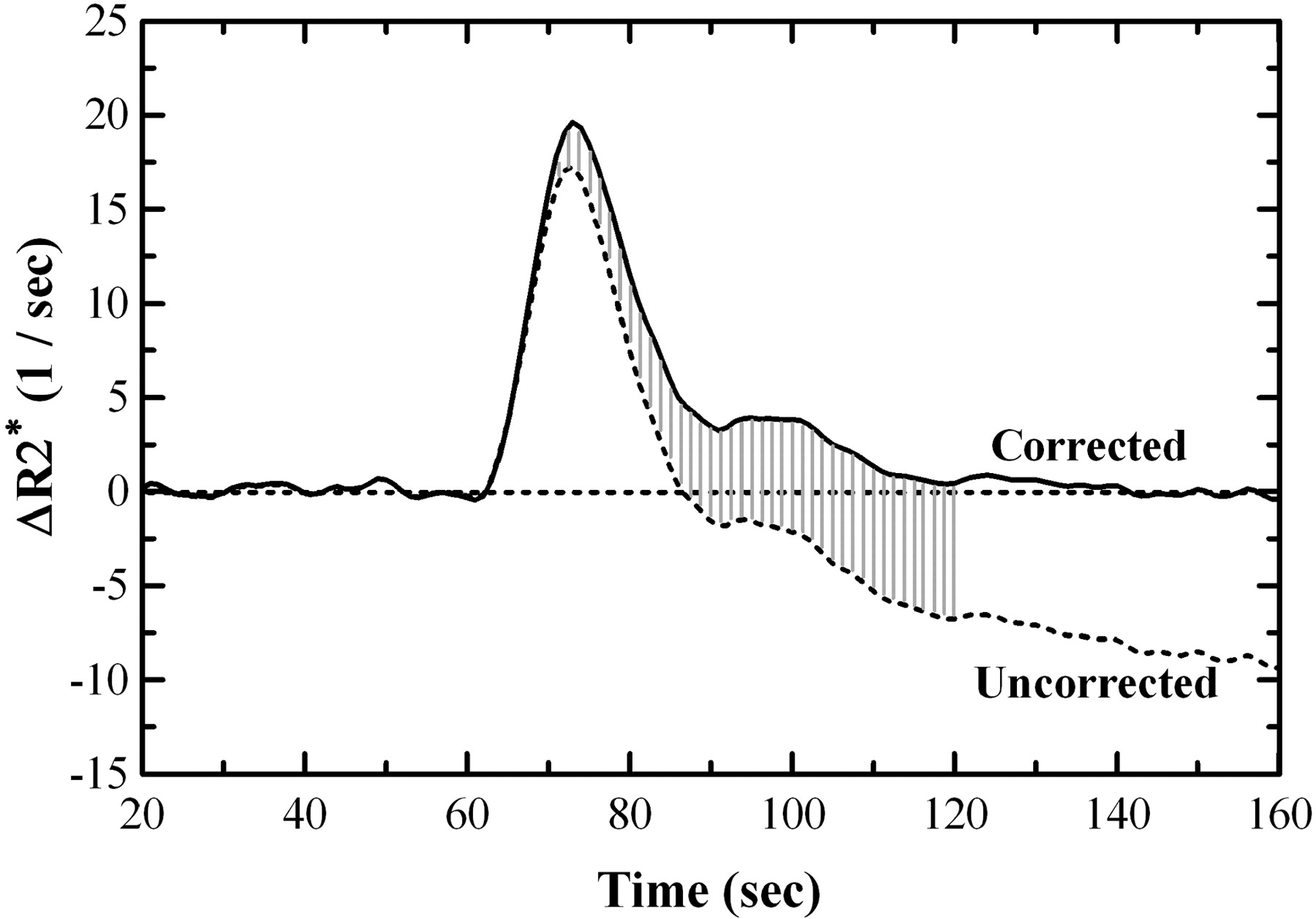
- Fig 1.
Typical corrected and uncorrected ΔR2* curves are shown. Note that even the first-pass curve is shifted to account for early leakage occurring during this segment. Without correction, the area under the curve (relative cerebral blood volume [rCBV]) is underestimated, even for integration techniques that stop at zero crossing or use gamma fitting.
- Fig 2.
Typical case (grade IV glioma) demonstrating post-Gd T1, uncorrected relative cerebral blood volume (rCBV) map with artificially low tumor blood volume, the K2 parameter map from our fitting algorithm showing areas of greatest correction, and the corrected rCBV map highlighting a focus of corrected very high blood volume. On the bottom are sample tumor and normal brain regions of interest based on the corrected rCBV map.
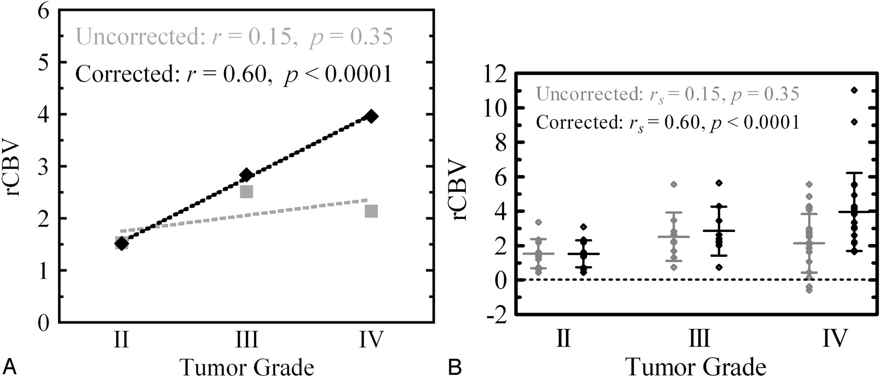
- Fig 3.
A, rCBV estimates corrected for contrast agent extravasation correlate significantly with glioma grade by using a Spearman rank correlation, whereas uncorrected relative cerebral blood volume (rCBV) does not. The disparity is due primarily to artificially low uncorrected blood-volume estimates in high-grade tumors that arises from the competing T1 effects of Gd leaking through disrupted blood-brain barrier.
B, Despite significant correlation, there exists moderate corrected rCBV variability within each grade and intergrade overlap. This supports the notion that glioma grading remains controversial and that multiple parameters, including vascular permeability and vessel size distribution, will probably be required in aggregate for accurate prognostication.
- Fig 4.
Percentage difference between corrected and uncorrected relative cerebral blood volume (rCBV) for each of the 43 gliomas separated by grade. For all 3 grades, there were several cases in which correction made no difference (shown in white), probably because of a combination of low vascular permeability (possibly from steroids) and the Gd preload. Most of the high-grade tumors, however, had >10% difference, with 6 cases differing by at least a factor of 2.


Tables
Patient histology and imaging results, grouped by tumor grade
Patient No./Age (y)/Sex Pathologic Diagnosis Corrected rCBV Uncorrected rCBV Grade II 1/80/M Ependymoma 2.18 2.18 2/50/M Low-grade glioma 1.56 1.56 3/59/M Oligodendroglioma 1.46 1.46 4/39/M Oligodendroglioma 0.72 0.72 5/50/M Astrocytoma 1.39 1.20 6/53/M Astrocytoma 1.35 1.35 7/66/F Astrocytoma 3.35 3.08 8/19/M Astrocytoma 0.44 0.44 9/48/F Astrocytoma 1.61 1.61 10/23/F Central neutrocytoma 2.33 2.33 11/30/M Mixed glioma 0.62 0.62 Grade III 1/40/F Anaplastic astrocytoma 3.44 3.47 2/32/F Anaplastic astrocytoma 0.74 0.74 3/37/F Anaplastic astrocytoma 5.63 5.55 4/47/M Anaplastic astrocytoma 4.28 1.31 5/77/M Anaplastic astrocytoma 2.42 2.82 6/50/M Anaplastic astrocytoma 2.02 1.68 7/41/M Anaplastic astrocytoma 2.18 2.18 8/55/M Anaplastic oligodendroglioma 2.25 2.25 9/41/M Anaplastic oligodendroglioma 2.63 2.63 Grade IV 1/50/M GBM 1.65 1.65 2/52/F GBM 3.35 1.06 3/73/M GBM 4.92 4.85 4/69/M GBM 11.03 −0.41 5/48/M GBM 9.18 0.15 6/68/M GBM 5.56 5.56 7/50/F GBM 4.16 2.09 8/42/M GBM 3.33 1.87 9/74/M GBM 3.91 0.44 10/49/F GBM 2.98 2.98 11/71/F GBM 4.23 4.21 12/30/M GBM 2.97 2.86 13/41/M GBM 2.59 −.39 14/65/F GBM 3.82 2.73 15/42/M GBM 2.59 2.51 16/66/M GBM 4.10 4.08 17/56/M GBM 1.70 1.06 18/77/M GBM 4.28 4.28 19/54/F GBM 2.13 1.63 20/55/M GBM 5.51 1.85 21/45/M GBM 1.71 −0.59 22/45/F Anaplastic oliogodendroglioma 2.22 2.17 23/66/M Mixed GBM + low-grade astrocytoma 3.07 2.59 Note.— rCBV indicates relative cerebral blood volume; GBM, glioblastoma multiforme.
In this issue


Jump to section
Related Articles
Cited By...
- Perfusion Showdown: Comparison of Multiple MRI Perfusion Techniques in the Grading of Pediatric Brain Tumors
- Multisite Benchmark Study for Standardized Relative CBV in Untreated Brain Metastases Using the DSC-MRI Consensus Acquisition Protocol
- "Synthetic" DSC Perfusion MRI with Adjustable Acquisition Parameters in Brain Tumors Using Dynamic Spin-and-Gradient-Echo Echoplanar Imaging
- Comparison of Arterial Spin-Labeling and DSC Perfusion MR Imaging in Pediatric Brain Tumors: A Systematic Review and Meta-Analysis
- Identification of a Single-Dose, Low-Flip-Angle-Based CBV Threshold for Fractional Tumor Burden Mapping in Recurrent Glioblastoma
- Whole-Brain Vascular Architecture Mapping Identifies Region-Specific Microvascular Profiles In Vivo
- Differentiating Low-Grade from High-Grade Intracranial Ependymomas: Comparison of Dynamic Contrast-Enhanced MRI and Diffusion-Weighted Imaging
- Blood-Brain Barrier Permeability and Kinetics of Inflammatory Markers in Acute Stroke Patients Treated With Thrombectomy
- Scanning ultrasound-mediated memory and functional improvements do not require amyloid-{beta} reduction
- Diffuse Large B-Cell Epstein-Barr Virus-Positive Primary CNS Lymphoma in Non-AIDS Patients: High Diagnostic Accuracy of DSC Perfusion Metrics
- Cytostatic hypothermia and its impact on glioblastoma and survival
- Simultaneous Mapping of Vasculature, Hypoxia, and Proliferation Using Dynamic Susceptibility Contrast MRI, 18F-FMISO PET, and 18F-FLT PET in Relation to Contrast Enhancement in Newly Diagnosed Glioblastoma
- Lack of benefit of temozolomide for MGMT methylated patients with high vascular glioblastoma: a confirmatory study
- MTT and Blood-Brain Barrier Disruption within Asymptomatic Vascular WM Lesions
- Vessel Type Determined by Vessel Architectural Imaging Improves Differentiation between Early Tumor Progression and Pseudoprogression in Glioblastoma
- Estimating Local Cellular Density in Glioma Using MR Imaging Data
- Uncertainty Quantification in Radiogenomics: EGFR Amplification in Glioblastoma
- Advanced ADC Histogram, Perfusion, and Permeability Metrics Show an Association with Survival and Pseudoprogression in Newly Diagnosed Diffuse Intrinsic Pontine Glioma: A Report from the Pediatric Brain Tumor Consortium
- Discrimination between Glioblastoma and Solitary Brain Metastasis: Comparison of Inflow-Based Vascular-Space-Occupancy and Dynamic Susceptibility Contrast MR Imaging
- Performance of Standardized Relative CBV for Quantifying Regional Histologic Tumor Burden in Recurrent High-Grade Glioma: Comparison against Normalized Relative CBV Using Image-Localized Stereotactic Biopsies
- Effects of Susceptibility Artifacts on Perfusion MRI in Patients with Primary Brain Tumor: A Comparison of Arterial Spin-Labeling versus DSC
- Sex-specific differences in white matter microvascular integrity after ischaemic stroke
- Prognostic Predictions for Patients with Glioblastoma after Standard Treatment: Application of Contrast Leakage Information from DSC-MRI within Nonenhancing FLAIR High-Signal-Intensity Lesions
- Utility of Percentage Signal Recovery and Baseline Signal in DSC-MRI Optimized for Relative CBV Measurement for Differentiating Glioblastoma, Lymphoma, Metastasis, and Meningioma
- A multi-scale sub-voxel perfusion model to estimate diffusive capillary wall conductivity in multiple sclerosis lesions from perfusion MRI data
- Moving Toward a Consensus DSC-MRI Protocol: Validation of a Low-Flip Angle Single-Dose Option as a Reference Standard for Brain Tumors
- Accurate Patient-Specific Machine Learning Models of Glioblastoma Invasion Using Transfer Learning
- Brain DSC MR Perfusion in Children: A Clinical Feasibility Study Using Different Technical Standards of Contrast Administration
- Optimization of Acquisition and Analysis Methods for Clinical Dynamic Susceptibility Contrast MRI Using a Population-Based Digital Reference Object
- Clinical Value of Vascular Permeability Estimates Using Dynamic Susceptibility Contrast MRI: Improved Diagnostic Performance in Distinguishing Hypervascular Primary CNS Lymphoma from Glioblastoma
- Multisite Concordance of DSC-MRI Analysis for Brain Tumors: Results of a National Cancer Institute Quantitative Imaging Network Collaborative Project
- Discrimination between Glioma Grades II and III Using Dynamic Susceptibility Perfusion MRI: A Meta-Analysis
- Effects of MRI Protocol Parameters, Preload Injection Dose, Fractionation Strategies, and Leakage Correction Algorithms on the Fidelity of Dynamic-Susceptibility Contrast MRI Estimates of Relative Cerebral Blood Volume in Gliomas
- Multiparametric Magnetic Resonance Imaging for Prediction of Parenchymal Hemorrhage in Acute Ischemic Stroke After Reperfusion Therapy
- A Simplified Model for Intravoxel Incoherent Motion Perfusion Imaging of the Brain
- Correlation of Tumor Immunohistochemistry with Dynamic Contrast-Enhanced and DSC-MRI Parameters in Patients with Gliomas
- Dynamic Susceptibility Contrast-Enhanced MR Perfusion Imaging in Assessing Recurrent Glioblastoma Response to Superselective Intra-Arterial Bevacizumab Therapy
- Childhood Cerebral Adrenoleukodystrophy: MR Perfusion Measurements and Their Use in Predicting Clinical Outcome after Hematopoietic Stem Cell Transplantation
- Improved Leakage Correction for Single-Echo Dynamic Susceptibility Contrast Perfusion MRI Estimates of Relative Cerebral Blood Volume in High-Grade Gliomas by Accounting for Bidirectional Contrast Agent Exchange
- Pretreatment blood-brain barrier disruption and post-endovascular intracranial hemorrhage
- Improving Perfusion Measurement in DSC-MR Imaging with Multiecho Information for Arterial Input Function Determination
- On the Use of DSC-MRI for Measuring Vascular Permeability
- Texture Feature Ratios from Relative CBV Maps of Perfusion MRI Are Associated with Patient Survival in Glioblastoma
- Impact of Software Modeling on the Accuracy of Perfusion MRI in Glioma
- The Added Prognostic Value of Preoperative Dynamic Contrast-Enhanced MRI Histogram Analysis in Patients with Glioblastoma: Analysis of Overall and Progression-Free Survival
- Comparison of the Diagnostic Accuracy of DSC- and Dynamic Contrast-Enhanced MRI in the Preoperative Grading of Astrocytomas
- Repeatability of Standardized and Normalized Relative CBV in Patients with Newly Diagnosed Glioblastoma
- ASFNR Recommendations for Clinical Performance of MR Dynamic Susceptibility Contrast Perfusion Imaging of the Brain
- Pixel-by-Pixel Comparison of Volume Transfer Constant and Estimates of Cerebral Blood Volume from Dynamic Contrast-Enhanced and Dynamic Susceptibility Contrast-Enhanced MR Imaging in High-Grade Gliomas
- Preoperative Prognostic Value of Dynamic Contrast-Enhanced MRI-Derived Contrast Transfer Coefficient and Plasma Volume in Patients with Cerebral Gliomas
- Advanced Magnetic Resonance Imaging of the Physical Processes in Human Glioblastoma
- Evaluation of Microvascular Permeability with Dynamic Contrast-Enhanced MRI for the Differentiation of Primary CNS Lymphoma and Glioblastoma: Radiologic-Pathologic Correlation
- Differentiation of Tumor Progression from Pseudoprogression in Patients with Posttreatment Glioblastoma Using Multiparametric Histogram Analysis
- Pretreatment Blood-Brain Barrier Damage and Post-Treatment Intracranial Hemorrhage in Patients Receiving Intravenous Tissue-Type Plasminogen Activator
- Assessment of Angiographic Vascularity of Meningiomas with Dynamic Susceptibility Contrast-Enhanced Perfusion-Weighted Imaging and Diffusion Tensor Imaging
- The Effect of Pulse Sequence Parameters and Contrast Agent Dose on Percentage Signal Recovery in DSC-MRI: Implications for Clinical Applications
- Differentiation of Primary Central Nervous System Lymphomas and Glioblastomas: Comparisons of Diagnostic Performance of Dynamic Susceptibility Contrast-Enhanced Perfusion MR Imaging without and with Contrast-Leakage Correction
- The Role of Preload and Leakage Correction in Gadolinium-Based Cerebral Blood Volume Estimation Determined by Comparison with MION as a Criterion Standard
- The Added Value of Apparent Diffusion Coefficient to Cerebral Blood Volume in the Preoperative Grading of Diffuse Gliomas
- Correlations between Perfusion MR Imaging Cerebral Blood Volume, Microvessel Quantification, and Clinical Outcome Using Stereotactic Analysis in Recurrent High-Grade Glioma
- Perfusion CT Imaging of Brain Tumors: An Overview
- Multimodality Assessment of Brain Tumors and Tumor Recurrence
- Percentage Signal Recovery Derived from MR Dynamic Susceptibility Contrast Imaging Is Useful to Differentiate Common Enhancing Malignant Lesions of the Brain
- O-(2-18F-Fluoroethyl)-L-Tyrosine PET Predicts Failure of Antiangiogenic Treatment in Patients with Recurrent High-Grade Glioma
- Reply to S. Heiland et al
- Perfusion Magnetic Resonance Imaging for Parametric Response Maps in Tumors: Is It Really That Easy?
- Differentiation among Glioblastoma Multiforme, Solitary Metastatic Tumor, and Lymphoma Using Whole-Tumor Histogram Analysis of the Normalized Cerebral Blood Volume in Enhancing and Perienhancing Lesions
- Optimized Preload Leakage-Correction Methods to Improve the Diagnostic Accuracy of Dynamic Susceptibility-Weighted Contrast-Enhanced Perfusion MR Imaging in Posttreatment Gliomas
- An Automatic Procedure for Normalization of Cerebral Blood Volume Maps in Dynamic Susceptibility Contrast-Based Glioma Imaging
- A "Vascular Normalization Index" as Potential Mechanistic Biomarker to Predict Survival after a Single Dose of Cediranib in Recurrent Glioblastoma Patients
- Relative Cerebral Blood Volume Values to Differentiate High-Grade Glioma Recurrence from Posttreatment Radiation Effect: Direct Correlation between Image-Guided Tissue Histopathology and Localized Dynamic Susceptibility-Weighted Contrast-Enhanced Perfusion MR Imaging Measurements
- Spin-Echo Echo-Planar Perfusion MR Imaging in the Differential Diagnosis of Solitary Enhancing Brain Lesions: Distinguishing Solitary Metastases from Primary Glioma