
Article Figures & Data
Figures
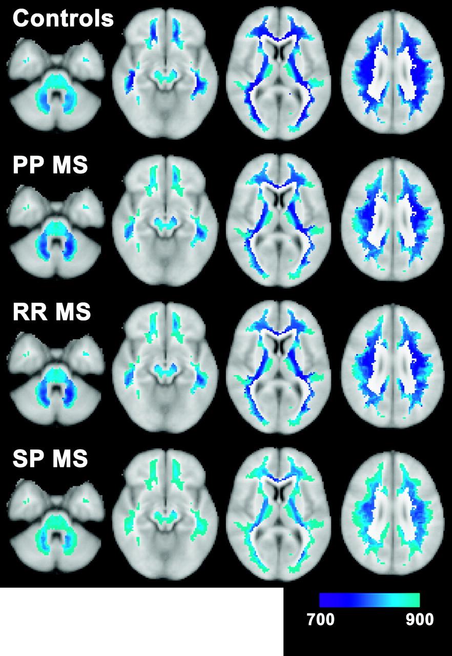
- Fig 2.
Mean T1 values in the 4 subject groups as fitted by the general linear model illustrate how T1 relaxation times increase throughout the normal-appearing white matter (NAWM) when going from control subjects (top row) to primary progressive multiple sclerosis (MS) (second row) to relapsing-remitting MS (third row) to secondary progressive MS (bottom row). The color range represents the T1 range of 700 to 900 ms, as indicated by the color bar.
- Fig 3.
Results of pairwise contrasts between multiple sclerosis (MS) groups (primary progressive [PP], relapsing-remitting [RR], and secondary progressive [SP]) and control subjects (C) are displayed as Z scores for PP MS > control subjects (top), RR MS > control subjects (middle), and SP MS > control subjects (bottom). The color range represents a Z score range of 3.1 to 8.0, as indicated by the color bar. Statistically significant T1 increases involve large fractions of normal-appearing white matter (NAWM) in RR and SP MS. The spatial extent of statistically significant T1 increases is small in PP MS.
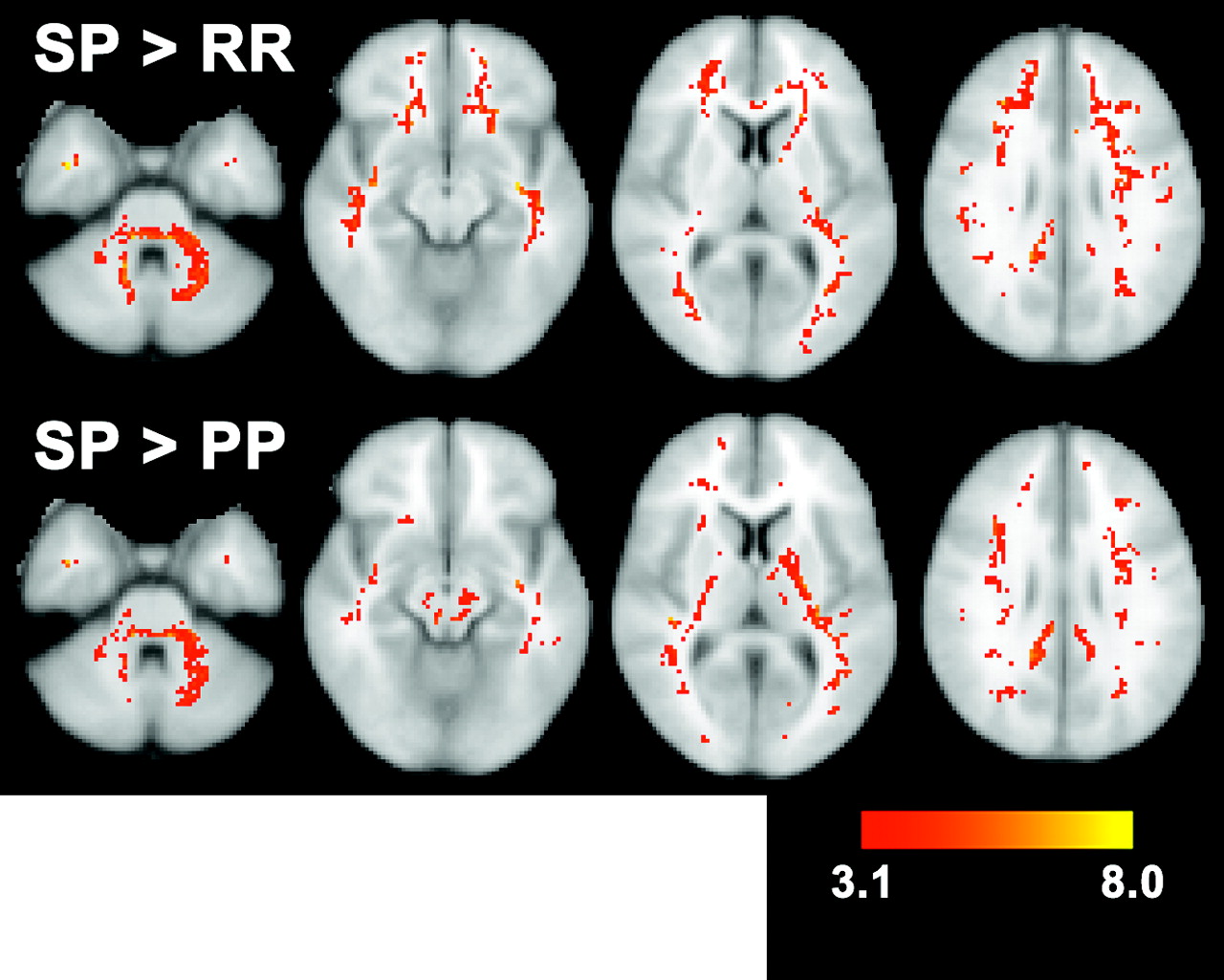
- Fig 4.
Results of pairwise contrasts of secondary progressive (SP) multiple sclerosis (MS) with relapsing-remitting (RR) MS (top, SP>RR) and with primary progressive (PP) MS (bottom, SP>PP) are displayed as Z scores. The color range represents a Z score range of 3.1–8.0, as indicated by the color bar. The effects of disease progression in RR/SP MS are visible as large areas with increased T1 in SP compared with RR MS. SP MS also has significantly higher T1 than PP MS in voxels throughout normal-appearing white matter (NAWM).


Tables
Characteristics of subject groups
PP MS RR MS SP MS Controls No. of subjects (M/F) 13 (7/6) 36 (11/25) 18 (7/11) 23 (12/11) Age (y) 57.2 ± 6.3 39.0 ± 7.3 44.3 ± 10.5 30.6 ± 7.4 Supratentorial lesion load (range) (mL) 8.6 ± 8.7 (0.2–27.8) 8.8 ± 9.4 (0.3–41.2) 14.6 ± 13.1 (2.5–54.7) Infratentorial lesion load (range) (mL) 0.2 ± 0.3 (0.0–0.8) 0.2 ± 0.3 (0.0–0.9) 0.5 ± 1.0 (0.0–4.2) Normalized brain volume (mL) 1417 ± 48 1473 ± 64 1406 ± 52 1518 ± 42 Median EDSS score (range) 4.5 (3.0–6.5) 2.0 (1.0–4.5) 6.0 (2.5–8.0) MSFC score 0.32 ± 0.19 0.56 ± 0.31 −0.27 ± 0.72 Note:— PP MS, RR MS, and SP MS indicate, respectively, primary progressive, relapsing-remitting, and secondary progressive multiple sclerosis; EDSS, expanded disability status scale; MSFC, multiple sclerosis functional composite.


