Advertisement

Main menu

  • Home
  • Content
    • Current Issue
    • Accepted Manuscripts
    • Article Preview
    • Past Issue Archive
    • AJNR Case Collection
    • Case of the Week Archive
    • Classic Case Archive
    • Case of the Month Archive
  • Special Collections
    • Spinal CSF Leak Articles (Jan 2020-June 2024)
    • 2024 AJNR Journal Awards
    • Most Impactful AJNR Articles
  • Multimedia
    • AJNR Podcast
    • AJNR Scantastics
    • Video Articles
  • For Authors
    • Submit a Manuscript
    • Author Policies
    • Fast publishing of Accepted Manuscripts
    • Graphical Abstract Preparation
    • Manuscript Submission Guidelines
    • Imaging Protocol Submission
    • Submit a Case for the Case Collection
  • About Us
    • About AJNR
    • Editorial Board
  • More
    • Become a Reviewer/Academy of Reviewers
    • Subscribers
    • Permissions
    • Alerts
    • Feedback
    • Advertisers
    • ASNR Home
  • Other Publications
    • ajnr

User menu

  • Alerts
  • Log in

Search

  • Advanced search
American Journal of Neuroradiology

American Journal of Neuroradiology

ASHNR American Society of Functional Neuroradiology ASHNR American Society of Pediatric Neuroradiology ASSR
  • Alerts
  • Log in

Advanced Search

  • Home
  • Content
    • Current Issue
    • Accepted Manuscripts
    • Article Preview
    • Past Issue Archive
    • AJNR Case Collection
    • Case of the Week Archive
    • Classic Case Archive
    • Case of the Month Archive
  • Special Collections
    • Spinal CSF Leak Articles (Jan 2020-June 2024)
    • 2024 AJNR Journal Awards
    • Most Impactful AJNR Articles
  • Multimedia
    • AJNR Podcast
    • AJNR Scantastics
    • Video Articles
  • For Authors
    • Submit a Manuscript
    • Author Policies
    • Fast publishing of Accepted Manuscripts
    • Graphical Abstract Preparation
    • Manuscript Submission Guidelines
    • Imaging Protocol Submission
    • Submit a Case for the Case Collection
  • About Us
    • About AJNR
    • Editorial Board
  • More
    • Become a Reviewer/Academy of Reviewers
    • Subscribers
    • Permissions
    • Alerts
    • Feedback
    • Advertisers
    • ASNR Home
  • Follow AJNR on Twitter
  • Visit AJNR on Facebook
  • Follow AJNR on Instagram
  • Join AJNR on LinkedIn
  • RSS Feeds

Welcome to the new AJNR, Updated Hall of Fame, and more. Read the full announcements.


AJNR is seeking candidates for the position of Associate Section Editor, AJNR Case Collection. Read the full announcement.


OtherBRAIN

MR Imaging of Human Herpesvirus-6–Associated Encephalitis in 4 Patients with Anterograde Amnesia after Allogeneic Hematopoietic Stem-Cell Transplantation

R.J.T. Gorniak, G.S. Young, D.E. Wiese, F.M. Marty and R.B. Schwartz
American Journal of Neuroradiology April 2006, 27 (4) 887-891;
R.J.T. Gorniak
  • Find this author on Google Scholar
  • Find this author on PubMed
  • Search for this author on this site
G.S. Young
  • Find this author on Google Scholar
  • Find this author on PubMed
  • Search for this author on this site
D.E. Wiese
  • Find this author on Google Scholar
  • Find this author on PubMed
  • Search for this author on this site
F.M. Marty
  • Find this author on Google Scholar
  • Find this author on PubMed
  • Search for this author on this site
R.B. Schwartz
  • Find this author on Google Scholar
  • Find this author on PubMed
  • Search for this author on this site
  • Article
  • Figures & Data
  • Info & Metrics
  • Responses
  • References
  • PDF
Loading

Article Figures & Data

Figures

  • Fig 1.
    • Download figure
    • Open in new tab
    • Download powerpoint
    Fig 1.

    Patient 1. Axial fluid-attenuated inversion recovery (FLAIR) imaging (A), diffusion-weighted imaging (DWI) (B), and apparent diffusion coefficient (ADC) mapping (C) performed on admission show T2 prolongation in the medial temporal lobes with reduced diffusion on the right. Follow-up FLAIR images obtained at approximately 5 weeks (D) and 8 weeks (E) after admission show persistent T2 prolongation in the medial temporal lobes and mild volume loss more pronounced in the medial temporal lobes.
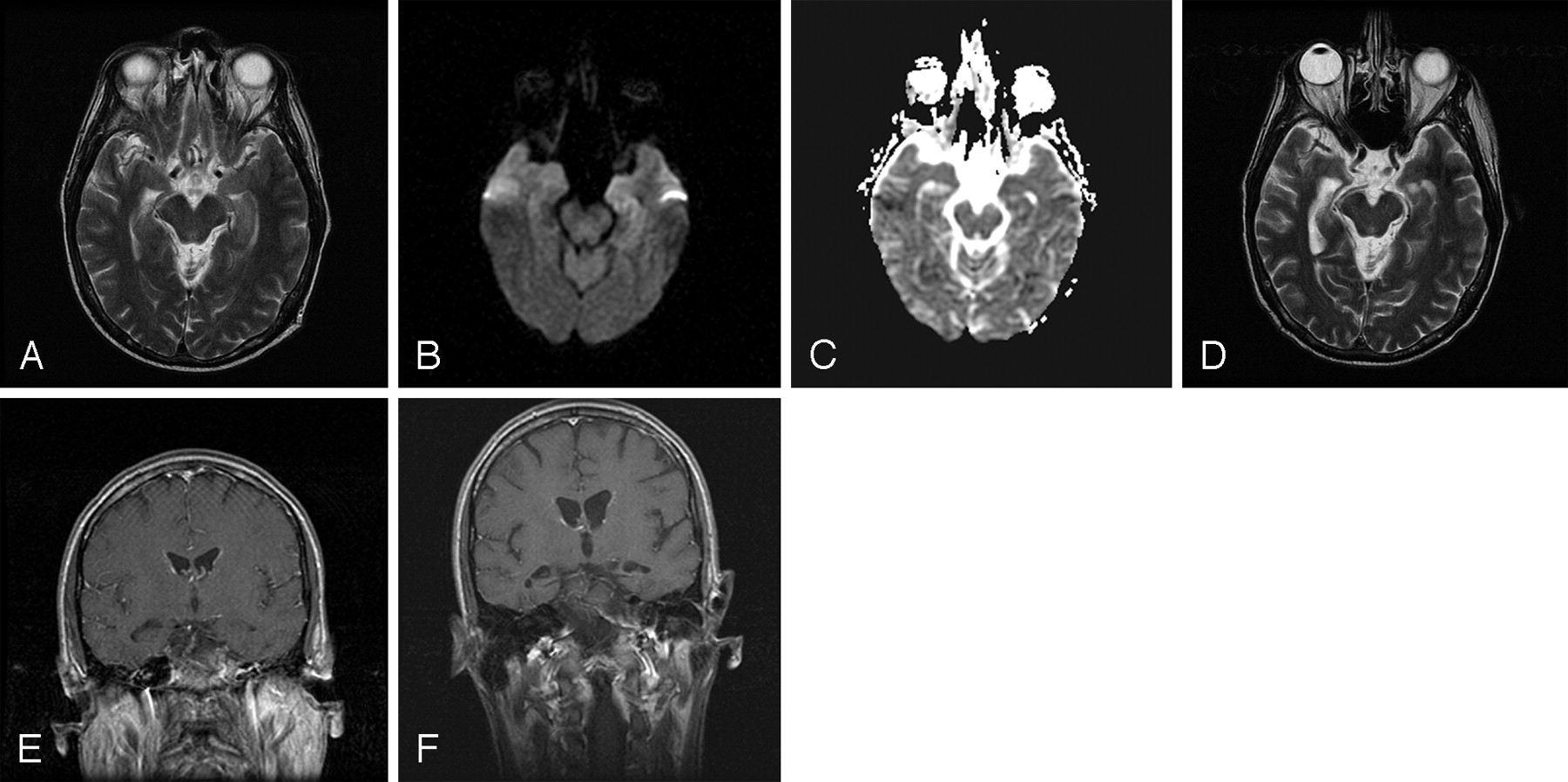
  • Fig 2.
    • Download figure
    • Open in new tab
    • Download powerpoint
    Fig 2.

    Patient 2. Axial T2-weighted image (A), diffusion-weighted imaging (DWI) (B), apparent diffusion coefficient (ADC) mapping (C), and coronal postcontrast T1-weighted (E) image obtained on admission show left medial temporal T2 prolongation on fast spin-echo T2-weighted image and DWI (“T2 shine-through” effect) as confirmed by ADC map demonstrating normal diffusivity. Axial T2-weighted and coronal T1 postcontrast images at the same level performed 3 months later (D) show resolution of abnormal T2 signal intensity with interval mild volume loss, most apparent in the medial temporal lobes (F).

  • Fig 3.
    • Download figure
    • Open in new tab
    • Download powerpoint
    Fig 3.

    Patient 3. Axial diffusion-weighted imaging (DWI) (A) and FLAIR imaging of the temporal lobes performed 2 days (B), 3 weeks (C), and 4 months (D) after admission demonstrate initial T2 prolongation in the medial temporal lobes with subsequent gradual normalization of signal intensity abnormality culminating in complete resolution by 4 months.
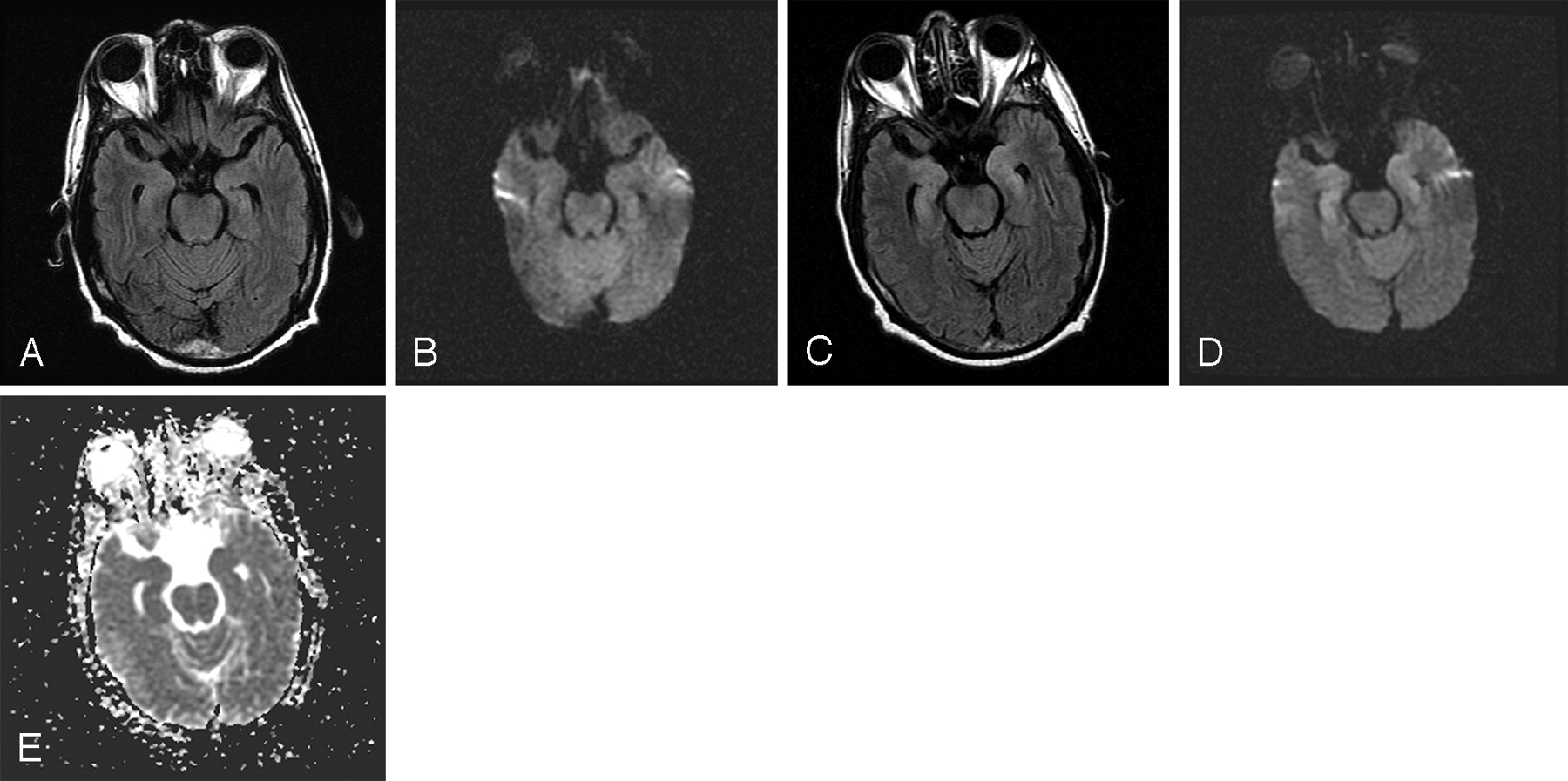
  • Fig 4.
    • Download figure
    • Open in new tab
    • Download powerpoint
    Fig 4.

    Patient 4. Axial FLAIR image (A) and diffusion-weighted imaging (DWI) (B) through the temporal lobes obtained on admission show no medial temporal lobe abnormality. Axial FLAIR image (C), DWI (D), and apparent diffusion coefficient (ADC) mapping (E) obtained 5 days later revealed new medial temporal T2 prolongation both on FLAIR and diffusion-weighted images. ADC map demonstrates normal diffusivity.

PreviousNext
Back to top

In this issue

American Journal of Neuroradiology: 27 (4)
American Journal of Neuroradiology
Vol. 27, Issue 4
April 2006
  • Table of Contents
  • Index by author
Advertisement
Print
Download PDF
Email Article

Thank you for your interest in spreading the word on American Journal of Neuroradiology.

NOTE: We only request your email address so that the person you are recommending the page to knows that you wanted them to see it, and that it is not junk mail. We do not capture any email address.

Enter multiple addresses on separate lines or separate them with commas.
MR Imaging of Human Herpesvirus-6–Associated Encephalitis in 4 Patients with Anterograde Amnesia after Allogeneic Hematopoietic Stem-Cell Transplantation
(Your Name) has sent you a message from American Journal of Neuroradiology
(Your Name) thought you would like to see the American Journal of Neuroradiology web site.
CAPTCHA
This question is for testing whether or not you are a human visitor and to prevent automated spam submissions.
Cite this article
R.J.T. Gorniak, G.S. Young, D.E. Wiese, F.M. Marty, R.B. Schwartz
MR Imaging of Human Herpesvirus-6–Associated Encephalitis in 4 Patients with Anterograde Amnesia after Allogeneic Hematopoietic Stem-Cell Transplantation
American Journal of Neuroradiology Apr 2006, 27 (4) 887-891;

Citation Manager Formats

  • BibTeX
  • Bookends
  • EasyBib
  • EndNote (tagged)
  • EndNote 8 (xml)
  • Medlars
  • Mendeley
  • Papers
  • RefWorks Tagged
  • Ref Manager
  • RIS
  • Zotero
0 Responses
Respond to this article
Share
Bookmark this article
MR Imaging of Human Herpesvirus-6–Associated Encephalitis in 4 Patients with Anterograde Amnesia after Allogeneic Hematopoietic Stem-Cell Transplantation
R.J.T. Gorniak, G.S. Young, D.E. Wiese, F.M. Marty, R.B. Schwartz
American Journal of Neuroradiology Apr 2006, 27 (4) 887-891;
del.icio.us logo Twitter logo Facebook logo Mendeley logo
  • Tweet Widget
  • Facebook Like
  • Google Plus One

Jump to section

  • Article
    • Abstract
    • Case Reports
    • Discussion
    • References
  • Figures & Data
  • Info & Metrics
  • Responses
  • References
  • PDF

Related Articles

  • No related articles found.
  • PubMed
  • Google Scholar

Cited By...

  • Human herpes 6 virus encephalitis complicating allogeneic hematopoietic stem cell transplantation
  • POST-TRANSPLANT ACUTE LIMBIC ENCEPHALITIS: CLINICAL FEATURES AND RELATIONSHIP TO HHV6
  • Post-transplant acute limbic encephalitis: Clinical features and relationship to HHV6
  • Crossref
  • Google Scholar

This article has not yet been cited by articles in journals that are participating in Crossref Cited-by Linking.

More in this TOC Section

  • Progression of Microstructural Damage in Spinocerebellar Ataxia Type 2: A Longitudinal DTI Study
  • Usefulness of Quantitative Susceptibility Mapping for the Diagnosis of Parkinson Disease
  • Evaluating the Effects of White Matter Multiple Sclerosis Lesions on the Volume Estimation of 6 Brain Tissue Segmentation Methods
Show more Brain

Similar Articles

Advertisement

Indexed Content

  • Current Issue
  • Accepted Manuscripts
  • Article Preview
  • Past Issues
  • Editorials
  • Editors Choice
  • Fellow Journal Club
  • Letters to the Editor

Cases

  • Case Collection
  • Archive - Case of the Week
  • Archive - Case of the Month
  • Archive - Classic Case

Special Collections

  • Special Collections

Resources

  • News and Updates
  • Turn around Times
  • Submit a Manuscript
  • Author Policies
  • Manuscript Submission Guidelines
  • Evidence-Based Medicine Level Guide
  • Publishing Checklists
  • Graphical Abstract Preparation
  • Imaging Protocol Submission
  • Submit a Case
  • Become a Reviewer/Academy of Reviewers
  • Get Peer Review Credit from Publons

Multimedia

  • AJNR Podcast
  • AJNR SCANtastic
  • Video Articles

About Us

  • About AJNR
  • Editorial Board
  • Not an AJNR Subscriber? Join Now
  • Alerts
  • Feedback
  • Advertise with us
  • Librarian Resources
  • Permissions
  • Terms and Conditions

American Society of Neuroradiology

  • Not an ASNR Member? Join Now

© 2025 by the American Society of Neuroradiology All rights, including for text and data mining, AI training, and similar technologies, are reserved.
Print ISSN: 0195-6108 Online ISSN: 1936-959X

Powered by HighWire