
Article Figures & Data
Figures
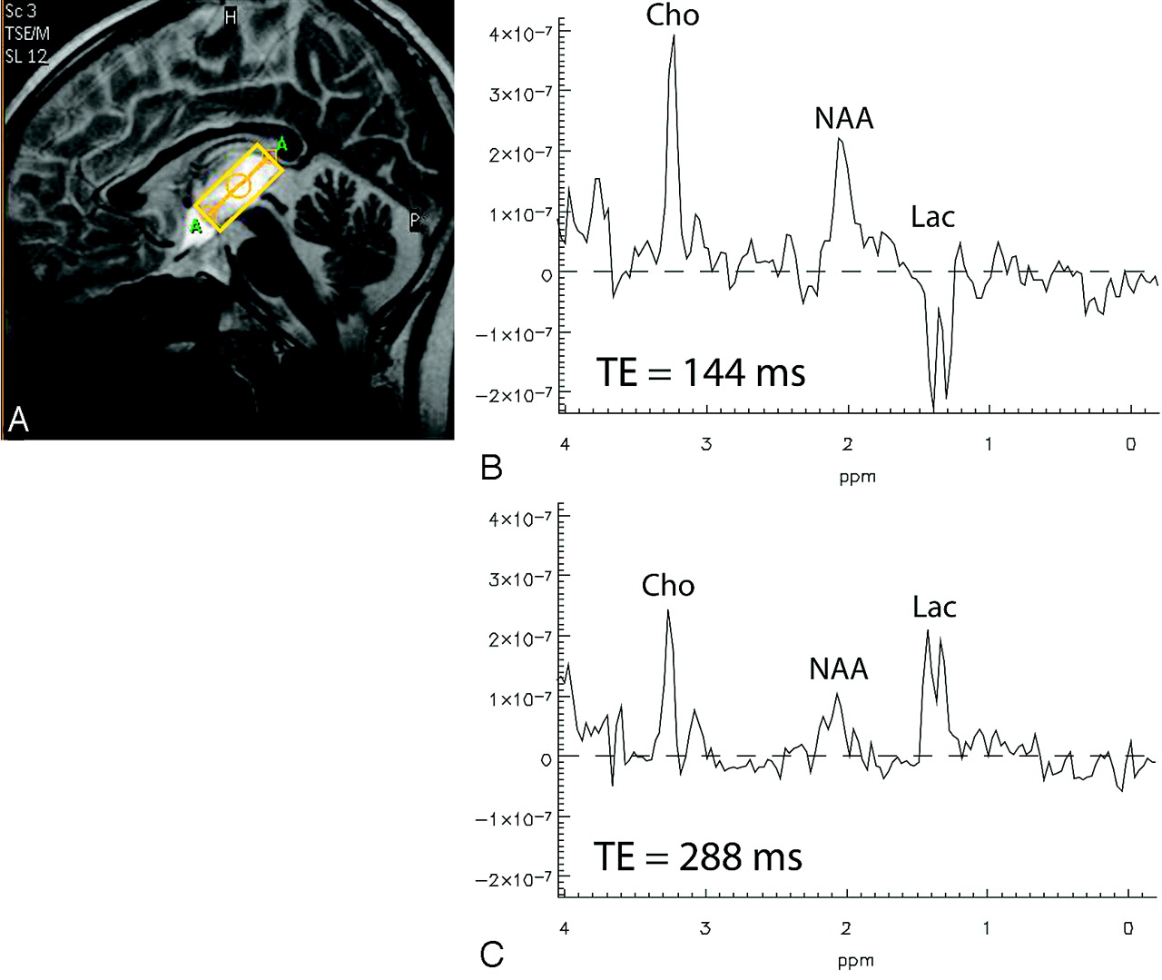
- Fig 1.
Single-voxel spectra acquired at 1.5T from the brain of a patient with a high-grade glioma by using PRESS localization. B, TE = 144 ms. C, TE = 288 ms. Cho indicates choline; Lac, lactate; NAA, N-acetylaspartate.
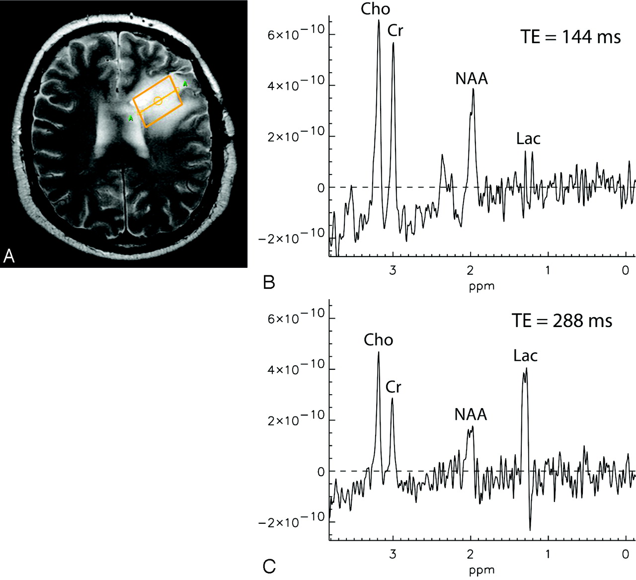
- Fig 2.
Single-voxel spectra acquired at 3T from the brain of a patient with a grade III glioma by using PRESS localization. B, TE = 144 ms. C, TE = 288 ms. Cr indicates creatine.
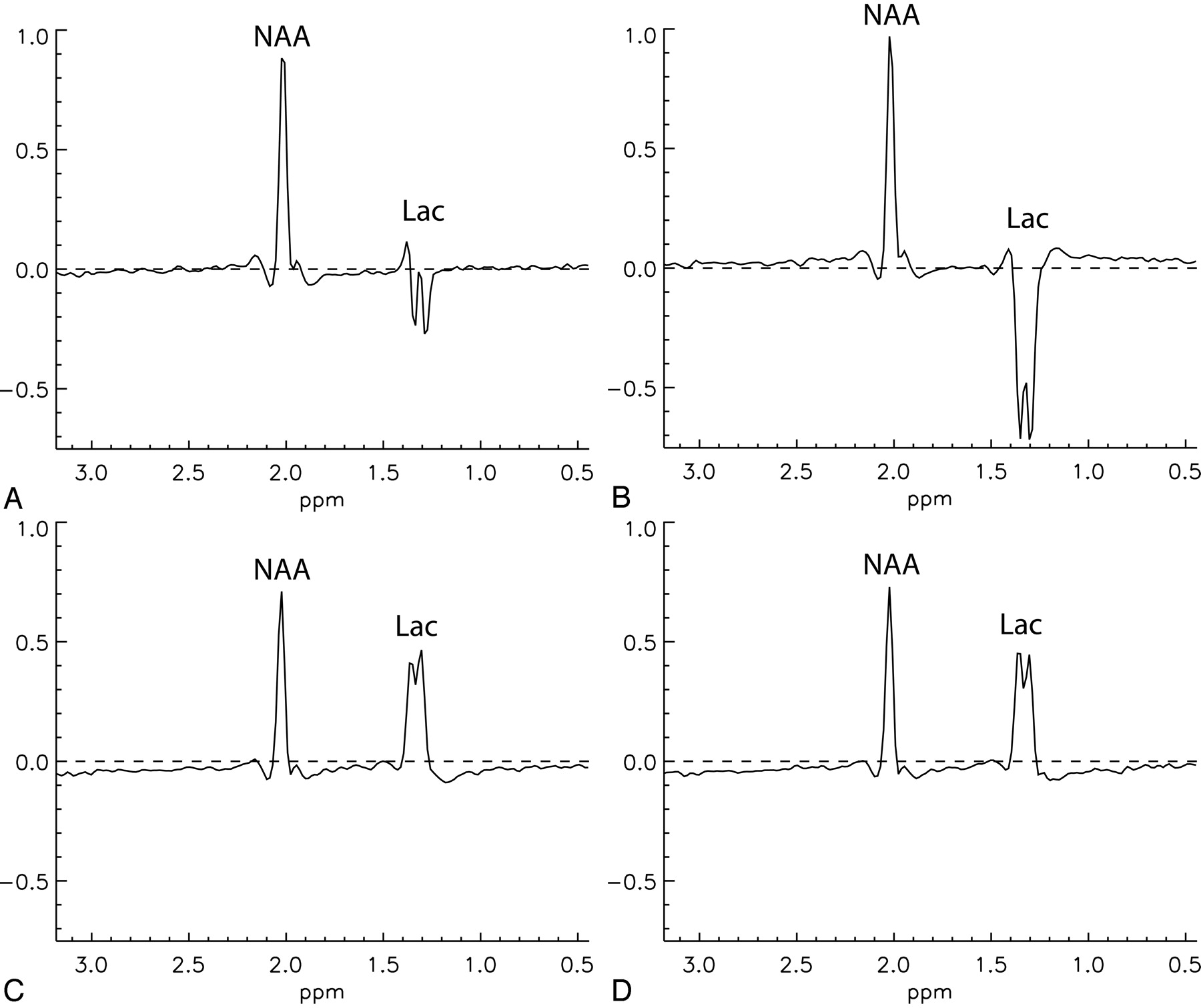
- Fig 3.
Multivoxel spectra acquired 1 hour apart at 1.5T and 3T from the same region in the brain of a patient with MELAS, by using standard PRESS localization with TE = 144 ms and TE = 288 ms. An inverted lactate doublet is clearly visible at 1.5T, but not at 3T (arrows). Upright lactate peaks at TE = 288 are seen equally well at both field strengths (arrows).
- Fig 4.
Proton spectra acquired from a standard brain metabolite phantom containing 5 mmol/L of lactate. The measurements were performed on 3 3T MR imaging scanners from 3 different vendors (Philips Medical Systems, GE Healthcare, and Siemens Medical Solutions). Single-voxel MR spectra (VOI size = 2 × 2 × 2 cm3, PRESS localization) were acquired from the same volume once with TE = 144 ms and once with TE = 288 ms. Radio-frequency pulse bandwidths for the selective refocusing pulses vary between vendors in the range of 874–2300 Hz.
- Fig 5.
Spectra from an MRSI imaging dataset acquired at 3T from a phantom containing 10 mmol/L of NAA and 20 mmol/L of lactate without PRESS localization. A, TE = 144 ms, with refocusing pulse gradient. B, TE = 144 ms, without refocusing pulse gradient. C, TE = 288 ms, with refocusing pulse gradient. D, TE = 288 ms, without refocusing pulse gradient.
- Fig 6.
Partial volumes and their coupling evolution for a single-voxel PRESS experiment: The 90° excitation pulse is applied with a gradient in the z direction, whereas the 2 refocusing pulses are applied with gradients in the x and y direction, respectively. The size of the partial volumes is determined by the chemical shift displacement. The signal phase of the magnetization is determined by TE, the scalar coupling constant (J) of lactate, and the time interval (t1) between the excitation pulse and the first refocusing pulse.


In this issue


Jump to section
Related Articles
Cited By...
- Band-selective IR PRESS for brain tumor spectroscopy allows robust detection of lactate
- Spatiospectral image processing workflow considerations for advanced MR spectroscopy of the brain
- Does Gadolinium Deposition Lead to Metabolite Alteration in the Dentate Nucleus? An MRS Study in Patients with MS
- Lower Lactate Levels and Lower Intracellular pH in Patients with IDH-Mutant versus Wild-Type Gliomas
- Expression Changes in Lactate and Glucose Metabolism and Associated Transporters in Basal Ganglia following Hypoxic-Ischemic Reperfusion Injury in Piglets
- Utility of Proton MR Spectroscopy for Differentiating Typical and Atypical Primary Central Nervous System Lymphomas from Tumefactive Demyelinating Lesions
- Focal MR spectroscopy of hippocampal CA-1 lesions in transient global amnesia