Advertisement

Main menu

  • Home
  • Content
    • Current Issue
    • Accepted Manuscripts
    • Article Preview
    • Past Issue Archive
    • AJNR Case Collection
    • Case of the Week Archive
    • Classic Case Archive
    • Case of the Month Archive
  • Special Collections
    • Spinal CSF Leak Articles (Jan 2020-June 2024)
    • 2024 AJNR Journal Awards
    • Most Impactful AJNR Articles
  • Multimedia
    • AJNR Podcast
    • AJNR Scantastics
    • Video Articles
  • For Authors
    • Submit a Manuscript
    • Author Policies
    • Fast publishing of Accepted Manuscripts
    • Graphical Abstract Preparation
    • Manuscript Submission Guidelines
    • Imaging Protocol Submission
    • Submit a Case for the Case Collection
  • About Us
    • About AJNR
    • Editorial Board
  • More
    • Become a Reviewer/Academy of Reviewers
    • Subscribers
    • Permissions
    • Alerts
    • Feedback
    • Advertisers
    • ASNR Home
  • Other Publications
    • ajnr

User menu

  • Alerts
  • Log in

Search

  • Advanced search
American Journal of Neuroradiology

American Journal of Neuroradiology

ASHNR American Society of Functional Neuroradiology ASHNR American Society of Pediatric Neuroradiology ASSR
  • Alerts
  • Log in

Advanced Search

  • Home
  • Content
    • Current Issue
    • Accepted Manuscripts
    • Article Preview
    • Past Issue Archive
    • AJNR Case Collection
    • Case of the Week Archive
    • Classic Case Archive
    • Case of the Month Archive
  • Special Collections
    • Spinal CSF Leak Articles (Jan 2020-June 2024)
    • 2024 AJNR Journal Awards
    • Most Impactful AJNR Articles
  • Multimedia
    • AJNR Podcast
    • AJNR Scantastics
    • Video Articles
  • For Authors
    • Submit a Manuscript
    • Author Policies
    • Fast publishing of Accepted Manuscripts
    • Graphical Abstract Preparation
    • Manuscript Submission Guidelines
    • Imaging Protocol Submission
    • Submit a Case for the Case Collection
  • About Us
    • About AJNR
    • Editorial Board
  • More
    • Become a Reviewer/Academy of Reviewers
    • Subscribers
    • Permissions
    • Alerts
    • Feedback
    • Advertisers
    • ASNR Home
  • Follow AJNR on Twitter
  • Visit AJNR on Facebook
  • Follow AJNR on Instagram
  • Join AJNR on LinkedIn
  • RSS Feeds

Welcome to the new AJNR, Updated Hall of Fame, and more. Read the full announcements.


AJNR is seeking candidates for the position of Associate Section Editor, AJNR Case Collection. Read the full announcement.


Research ArticleBRAIN

MR Characteristics and Neuropathology in Adult-Onset Autosomal Dominant Leukodystrophy with Autonomic Symptoms

A. Melberg, L. Hallberg, H. Kalimo and R. Raininko
American Journal of Neuroradiology April 2006, 27 (4) 904-911;
A. Melberg
  • Find this author on Google Scholar
  • Find this author on PubMed
  • Search for this author on this site
L. Hallberg
  • Find this author on Google Scholar
  • Find this author on PubMed
  • Search for this author on this site
H. Kalimo
  • Find this author on Google Scholar
  • Find this author on PubMed
  • Search for this author on this site
R. Raininko
  • Find this author on Google Scholar
  • Find this author on PubMed
  • Search for this author on this site
  • Article
  • Figures & Data
  • Info & Metrics
  • Responses
  • References
  • PDF
Loading

Article Figures & Data

Figures

  • Fig 1.
    • Download figure
    • Open in new tab
    • Download powerpoint
    Fig 1.

    T2 signal-intensity alterations in a 37-year-old asymptomatic family member. Subtle high-signal-intensity lesions in the corticospinal tracts under the motor cortex (A), in the posterior limb of the internal capsule (B, arrow), mesencephalon (C, arrow), pons (D, arrow), and medulla oblongata (E, arrow). Only a tiny signal-intensity abnormality is seen in the right corticospinal tract at the level of the pons. Note the lesions in the upper cerebellar peduncles (C, arrowhead). Fluid-attenuated inversion recovery (FLAIR), 10,000/140/2500.

  • Fig 2.
    • Download figure
    • Open in new tab
    • Download powerpoint
    Fig 2.

    More extensive signal-intensity alterations in a 34-year-old asymptomatic subject.

    A, Frontoparietal changes are less severe in the periventricular region.

    B, Increased signal intensity in the internal capsules, in the anterior and posterior corpus callosum and in the vicinity of the atria (arrow).

    C, Changes in the middle cerebellar peduncles and in the pontine nuclei. Turbo spin-echo (TSE), 5000/90.

  • Fig 3.
    • Download figure
    • Open in new tab
    • Download powerpoint
    Fig 3.

    A 48-year-old woman with a history of urinary bladder dysfunction for the past 2 years. Frontoparietal white matter alteration does not involve the periventricular area (A and B). Increased signal intensity in the corticospinal tracts in the internal capsules (B, arrow).

    A, Fluid-attenuated inversion recovery (FLAIR), 10,000/140/2500.

    B, Turbo spin-echo (TSE), 8116/100.
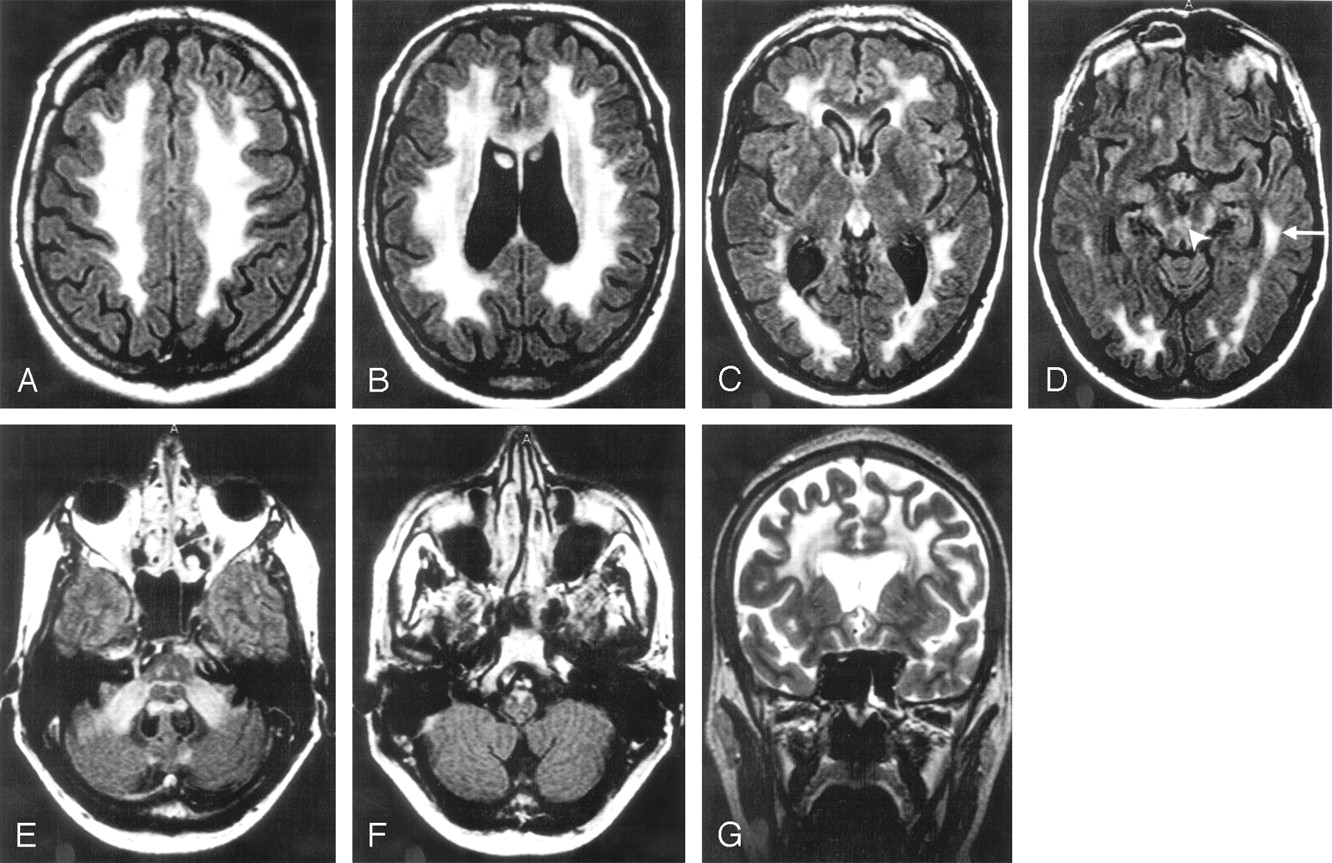
  • Fig 4.
    • Download figure
    • Open in new tab
    • Download powerpoint
    Fig 4.

    A 55-year-old man with a history of impotence, urinary bladder dysfunction, and obstipation for the past 6 years. Most of the frontoparietal and occipital white matter is affected (A–D and G). The periventricular white matter looks less pathologic (B, C, G). The circular periventricular layers are best seen in a coronal plane (G). The leukodystrophic area continues in the temporal lobes (D, arrow). The corticospinal tracts show a high signal intensity in the internal capsules (C), mesencephalon (D), pons (E), and medulla oblongata (F). The upper (arrowhead) and middle cerebellar peduncles are affected (D and E). High signal intensities are seen in the corpus callosum (B). Its posterior part (B) is very thin.

    A–F, Fluid-attenuated inversion recovery (FLAIR), 10,000/140/2500.

    G, Turbo spin-echo (TSE), 8116/100.
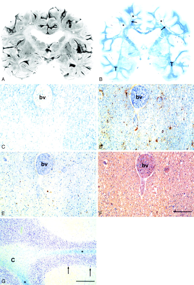
  • Fig 5.
    • Download figure
    • Open in new tab
    • Download powerpoint
    Fig 5.

    A, Coronal formalin-fixed section at the level of the basal ganglia of a 69-year-old man. White matter is reduced and there are irregularly shaped miscolored areas of demyelination (asterisk).

    B, Whole-brain microscopic section from the same level. Widespread diffuse loss of myelin (pallor of the blue color) in the centrum semiovale bilaterally (asterisk, arrows) with sparing of the U fibers (arrows). The myelin is better preserved in the temporal lobes (T). The Sylvian fissures are widened and the lateral ventricles moderately dilated, which indicates mild brain atrophy.

    C–F, Consecutive sections from the parietal white matter of a 56-year-old woman. C, Diffuse loss of myelin. Note the less severe pallor around the blood vessel (bv), which indicates that the myelin loss is not perivascular as in multiple sclerosis. Luxol fast blue-cresyl violet (LFB-CV) staining. D, There is only minor astrogliosis in the demyelinated area. The astrocytes (brown cells) are rather plump with irregular processes. Anti-glial fibrillar acidic protein (GFAP) + hematoxylin counterstain. E, No lymphocyte infiltrates are present, but scattered macrophages (brown cells) are seen in the demyelinated area. Anti-CD68 + hematoxylin counterstain. The number of oligodendroglial nuclei (blue) in panels D and E appears the same irrespective of the intensity of the myelin stain (C). F, The density of neurofilament positive axons did not appear to be affected by demyelination. Anti-neurofilament + hematoxylin counterstain. Scale bar, 100 μm.

    G, Cerebellum of the 69-year-old man. Severe diffuse loss of myelin in the cerebellar white matter, more marked centrally (C) than in the folia (asterisk). The number of Purkinje cells (2 preserved cells marked with arrows) is greatly decreased, but there is only slightly increased number of reactive Bergmann astrocytes. LFB-CV. Scale bar, 100 μm.

PreviousNext
Back to top

In this issue

American Journal of Neuroradiology: 27 (4)
American Journal of Neuroradiology
Vol. 27, Issue 4
April 2006
  • Table of Contents
  • Index by author
Advertisement
Print
Download PDF
Email Article

Thank you for your interest in spreading the word on American Journal of Neuroradiology.

NOTE: We only request your email address so that the person you are recommending the page to knows that you wanted them to see it, and that it is not junk mail. We do not capture any email address.

Enter multiple addresses on separate lines or separate them with commas.
MR Characteristics and Neuropathology in Adult-Onset Autosomal Dominant Leukodystrophy with Autonomic Symptoms
(Your Name) has sent you a message from American Journal of Neuroradiology
(Your Name) thought you would like to see the American Journal of Neuroradiology web site.
CAPTCHA
This question is for testing whether or not you are a human visitor and to prevent automated spam submissions.
Cite this article
A. Melberg, L. Hallberg, H. Kalimo, R. Raininko
MR Characteristics and Neuropathology in Adult-Onset Autosomal Dominant Leukodystrophy with Autonomic Symptoms
American Journal of Neuroradiology Apr 2006, 27 (4) 904-911;

Citation Manager Formats

  • BibTeX
  • Bookends
  • EasyBib
  • EndNote (tagged)
  • EndNote 8 (xml)
  • Medlars
  • Mendeley
  • Papers
  • RefWorks Tagged
  • Ref Manager
  • RIS
  • Zotero
0 Responses
Respond to this article
Share
Bookmark this article
MR Characteristics and Neuropathology in Adult-Onset Autosomal Dominant Leukodystrophy with Autonomic Symptoms
A. Melberg, L. Hallberg, H. Kalimo, R. Raininko
American Journal of Neuroradiology Apr 2006, 27 (4) 904-911;
del.icio.us logo Twitter logo Facebook logo Mendeley logo
  • Tweet Widget
  • Facebook Like
  • Google Plus One

Jump to section

  • Article
    • Abstract
    • Materials and Methods
    • Results
    • Discussion
    • Conclusion
    • Footnotes
    • References
  • Figures & Data
  • Info & Metrics
  • Responses
  • References
  • PDF

Related Articles

  • No related articles found.
  • PubMed
  • Google Scholar

Cited By...

  • An oligodendrocyte silencer element underlies the pathogenic impact of lamin B1 structural variants
  • Imaging Patterns Characterizing Mitochondrial Leukodystrophies
  • Duplication and deletion upstream of LMNB1 in autosomal dominant adult-onset leukodystrophy
  • Defects of Lipid Synthesis Are Linked to the Age-Dependent Demyelination Caused by Lamin B1 Overexpression
  • Nuclear Lamins and Neurobiology
  • A practical approach to diagnosing adult onset leukodystrophies
  • Adult-onset autosomal dominant leukodystrophy presenting with REM sleep behavior disorder
  • Nuclear lamins: Functions and clinical implications
  • Adult-Onset Autosomal Dominant Leukodystrophy: Linking Nuclear Envelope to Myelin
  • Invited Article: An MRI-based approach to the diagnosis of white matter disorders
  • MR Imaging Characteristics and Neuropathology of the Spinal Cord in Adult-Onset Autosomal Dominant Leukodystrophy with Autonomic Symptoms
  • Crossref
  • Google Scholar

This article has not yet been cited by articles in journals that are participating in Crossref Cited-by Linking.

More in this TOC Section

  • Multimodal CT Provides Improved Performance for Lacunar Infarct Detection
  • Statin Therapy Does Not Affect the Radiographic and Clinical Profile of Patients with TIA and Minor Stroke
  • Optimal MRI Sequence for Identifying Occlusion Location in Acute Stroke: Which Value of Time-Resolved Contrast-Enhanced MRA?
Show more Brain

Similar Articles

Advertisement

Indexed Content

  • Current Issue
  • Accepted Manuscripts
  • Article Preview
  • Past Issues
  • Editorials
  • Editors Choice
  • Fellow Journal Club
  • Letters to the Editor

Cases

  • Case Collection
  • Archive - Case of the Week
  • Archive - Case of the Month
  • Archive - Classic Case

Special Collections

  • Special Collections

Resources

  • News and Updates
  • Turn around Times
  • Submit a Manuscript
  • Author Policies
  • Manuscript Submission Guidelines
  • Evidence-Based Medicine Level Guide
  • Publishing Checklists
  • Graphical Abstract Preparation
  • Imaging Protocol Submission
  • Submit a Case
  • Become a Reviewer/Academy of Reviewers
  • Get Peer Review Credit from Publons

Multimedia

  • AJNR Podcast
  • AJNR SCANtastic
  • Video Articles

About Us

  • About AJNR
  • Editorial Board
  • Not an AJNR Subscriber? Join Now
  • Alerts
  • Feedback
  • Advertise with us
  • Librarian Resources
  • Permissions
  • Terms and Conditions

American Society of Neuroradiology

  • Not an ASNR Member? Join Now

© 2025 by the American Society of Neuroradiology All rights, including for text and data mining, AI training, and similar technologies, are reserved.
Print ISSN: 0195-6108 Online ISSN: 1936-959X

Powered by HighWire