
Article Figures & Data
Figures
- Fig 1.
Examples of region of interest placement for mean diffusivity (MD) and fractional anisotropy (FA) measurements. FA images show regions of interest of medulla oblongata (A), cerebral peduncle (B), internal capsule (C), optic radiation (D), splenium and genu of corpus callosum (E), and putamen and thalamus (F).
- Fig 2.
Average mean diffusivity (mm2/s) histograms of the brain tissue from patients with relapsing neuromyelitis optica (RNMO) (solid line) and control subjects (dotted line).
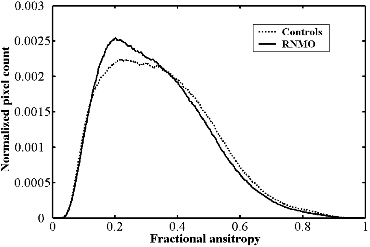
- Fig 3.
Average fractional anisotropy histograms of the brain tissue from patients with relapsing neuromyelitis optica (RNMO) (solid line) and control subjects (dotted line).
- Fig 4.
Average mean diffusivity (mm2/s) histograms of the brain white matter from patients with relapsing neuromyelitis optica (RNMO) (solid line) and control subjects (dotted line).
- Fig 5.
Average mean diffusivity (mm2/s) histograms of the brain gray matter from patients with relapsing neuromyelitis optica (RNMO) (solid line) and control subjects (dotted line).
- Fig 6.
Average fractional anisotropy histograms of the brain white matter from patients with relapsing neuromyelitis optica (RNMO) (solid line) and control subjects (dotted line).


Tables
- Table 1:
MD and FA histogram-derived measures of brain tissue, gray matter, and white matter from patients with RNMO and control subjects
RNMO Controls P* (95% CI) Brain tissue Average MD (×10−3 mm2 s−1) 0.946 ± 0.022 0.909 ± 0.031 <.001 (0.934–0.958) (0.893–0.925) (0.018 to 0.056) Mean MD peak height (‰) 10.326 ± 0.657 11.224 ± 1.260 .017 (9.975–10.676) (10.552–11.895) (−1.624 to −0.172) Mean MD peak location (×10−3 mm2 s−1) 0.777 ± 0.022 0.764 ± 0.025 .136 (0.765–0.789) (0.751–0.778) (−0.004 to 0.030) Average FA 0.250 ± 0.008 0.253 ± 0.009 .356 (0.246–0.254) (0.248–0.258) (−0.009 to 0.003) Mean FA peak height (‰) 4.109 ± 0.213 4.090 ± 0.272 .828 (3.996–4.222) (3.945–4.235) (−0.157 to 0.195) Mean FA peak location 0.127 ± 0.009 0.125 ± 0.010 .577 (0.123–0.132) (0.120–0.131) (−0.005 to 0.009) White matter Average MD (×10−3 mm2 s−1) 0.822 ± 0.016 0.803 ± 0.019 .004 (0.813–0.830) (0.793–0.812) (0.007 to 0.032) Mean MD peak height (‰) 15.856 ± 0.889 16.310 ± 1.402 .283 (15.382–16.329) (15.563–17.057) (−1.301 to 0.393) Mean MD peak location (×10−3 mm2 s−1) 0.760 ± 0.022 0.744 ± 0.019 .042 (0.748–0.771) (0.734–0.754) (0.001 to 0.030) Average FA 0.333 ± 0.012 0.345 ± 0.016 .020 (0.327–0.339) (0.336–0.353) (−0.022 to −0.002) Mean FA peak height (‰) 2.614 ± 0.179 2.383 ± 0.180 .001 (2.519–2.709) (2.288–2.480) (0.101 to 0.360) Mean FA peak location 0.216 ± 0.034 0.254 ± 0.063 .043 (0.199–0.234) (0.221–0.288) (−0.074 to −0.001) Gray matter Average MD (×10−3 mm2 s−1) 1.057 ± 0.037 0.992 ± 0.053 <.001 (1.037–1.077) (0.964–1.020) (0.032 to 0.098) Mean MD peak height (‰) 7.368 ± 0.889 8.913 ± 1.707 .003 (6.894–7.842) (8.003–9.822) (−2.527 to −0.562) Mean MD peak location (×10−3 mm2 s−1) 0.833 ± 0.016 0.809 ± 0.017 <.001 (0.825–0.842) (0.800–0.818) (0.013 to 0.036) Note:—RNMO indicates relapsing neuromyelitis optica; MD, mean diffusivity; FA, fractional anisotropy. Values are expressed as means ± SD (95% confidence interval).
* Statistical analysis: Student t test for independent samples; P < .005 is considered significant.
ROI Average MD (×10−3 mm2 s−1) Average FA RNMO Controls P* (95% CI) RNMO Controls P* (95% CI) Medulla oblongata 1.271 ± 0.111 1.111 ± 0.102 <.001 0.266 ± 0.014 0.287 ± 0.026 .005 (1.212–1.330) (1.057–1.165) (0.084 to 0.237) (0.259–0.274) (0.275–0.300) (−0.035 to −0.007) Cerebral peduncle 1.007 ± 0.111 0.872 ± 0.053 <.001 0.489 ± 0.035 0.535 ± 0.024 <.001 (0.948–1.066) (0.843–0.900) (0.072 to 0.198) (0.471–0.508) (0.522–0.548) (−0.067 to −0.024) Internal capsule 0.735 ± 0.010 0.718 ± 0.011 <.001 0.661 ± 0.019 0.693 ± 0.022 <.001 (0.730–0.741) (0.712–0.723) (0.010 to 0.065) (0.651–0.671) (0.681–0.705) (−0.047 to −0.017) Optic radiation 0.876 ± 0.033 0.824 ± 0.031 <.001 0.527 ± 0.039 0.585 ± 0.030 <.001 (0.859–0.894) (0.808–0.840) (−0.076 to −0.029) (0.506–0.547) (0.569–0.601) (−0.083 to −0.033) Splenium of corpus callosum 0.759 ± 0.034 0.726 ± 0.033 .009 0.815 ± 0.034 0.817 ± 0.033 .846 (0.741–0.777) (0.708–0.743) (0.009 to 0.057) (0.796–0.833) (0.799–0834) (−0.027 to 0.022) Genu of corpus callosum 0.765 ± 0.049 0.731 ± 0.038 .033 0.832 ± 0.033 0.829 ± 0.020 .741 (0.739–0.794) (0.711–0.751) (0.003 to 0.066) (0.814–0.849) (0.818–0.839) (−0.016 to 0.023) Putamen 0.729 ± 0.012 0.721 ± 0.011 .068 (0.722–0.735) (0.715–0.727) (−0.001 to 0.016) Thalamus 0.775 ± 0.022 0.760 ± .025 .066 (0.763–0.786) (0.746–0.772) (−0.001 to 0.032) Frontal gray matter 0.674 ± 0.042 0.634 ± 0.050 .023 (0.651–0.696) (0.608–0.661) (0.006 to 0.073) Parietal gray matter 0.807 ± 0.014 0.779 ± 0.016 <.001 (0.800–0.814) (0.771–0.788) (0.017 to 0.038) Temporal gray matter 0.838 ± 0.020 0.823 ± 0.011 .016 (0.827–0.849 (0.818–0.829) (0.003 to 0.026) Occipital gray matter 0.780 ± 0.012 0.758 ± 0.016 .007 (0.770–0.790) (0.746–0.770) (0.006 to 0.036) Note:—RNMO indicates relapsing neuromyelitis optica; MD, mean diffusivity; FA, fractional anisotropy; ROI, region of interest. Values are expressed as mean ± SD (95% confidence interval).
* Statistical analysis: Student t test for independent samples; P < .005 is considered significant.
In this issue


Jump to section
Related Articles
Cited By...
- Attack-related damage of thalamic nuclei in neuromyelitis optica spectrum disorders
- Gray Matter Volume Reduction Is Associated with Cognitive Impairment in Neuromyelitis Optica
- Nonconventional MRI biomarkers for in vivo monitoring of pathogenesis in multiple sclerosis
- Diameter, Length, Speed, and Conduction Delay of Callosal Axons in Macaque Monkeys and Humans: Comparing Data from Histology and Magnetic Resonance Imaging Diffusion Tractography
- No MRI evidence of cortical lesions in neuromyelitis optica
- Serum and CSF N-acetyl aspartate levels differ in multiple sclerosis and neuromyelitis optica
- Imaging evaluation of demyelinating processes of the central nervous system
- Neuromyelitis optica: an overview