
Article Figures & Data
Figures
- Fig 1.
Anteroposterior views of rotational angiogram, magnitude image, and 2D velocity vector field traversing M1 segment and two M2 segments of the right middle cerebral artery (MCA).
A, A surface rendering image obtained from rotational digital subtraction angiogram of the right internal carotid artery reveals that the blood flow moves through the M1 segment of right middle cerebral artery (MCA) (white arrow) and then diverges into the two M2 segments (anterior trunk of MCA [white small arrow] and posterior trunk of MCA [white arrowhead]). There is an aneurysm in this bifurcation of MCA.
B, A magnitude image traversing M1 segment and two M2 segments of the right MCA. White arrow, M1 segment; white small arrow, anterior trunk of MCA; white arrowhead, posterior trunk of MCA.
C, 2D velocity vector field traversing the M1 segment and two M2 segments of right MCA shows counter-clockwise vortex flow in the aneurysm. It demonstrates flow velocities near the vascular wall. 2D velocity vector fields can be displayed as cine images like a movie on a computer display. Numbers and colors correspond with flow rate in the legend at the right side in the figure regarding 2D velocity vector fields. Units in meters per second. Flow rate in the aneurysm is less than 15 cm/s.
- Fig 2.
Left anterior oblique views of a rotational angiogram and 3D streamlines of the silicon middle cerebral artery (MCA) aneurysm. B is stereoscopic 3D streamlines corresponding the systolic phase. C shows hemodynamics of B by arrows.
A, A left anterior oblique view of surface rendering of rotational digital subtraction angiogram of the right internal carotid artery shows M1 segment of right MCA (white arrow) and two M2 segment (anterior trunk of MCA [white small arrow] and posterior trunk of MCA [white arrowhead]) very clearly. Black arrows indicate the bleb.
B, Blood flow from the M1 segment of MCA strikes the posterior wall of the intracranial aneurysm. Blood flow flows along the wall of the intracranial aneurysm (the highest flow rate, 15 cm/s) and diverges into the two M2 segments near the inlet of the aneurysm. A helical flow at the left aspect of the aneurysm is seen along the aneurysmal wall. Where the helical flow reverses direction is coincident with the bleb (shown by arrows in Fig 2A). Flow rate of the bleb is low (less than 5 cm/s). Numbers and colors correspond with flow rate in the legend at the right side of the figure regarding 3D streamlines. Units are meters per second.
C, Arrows indicate flow schematically.
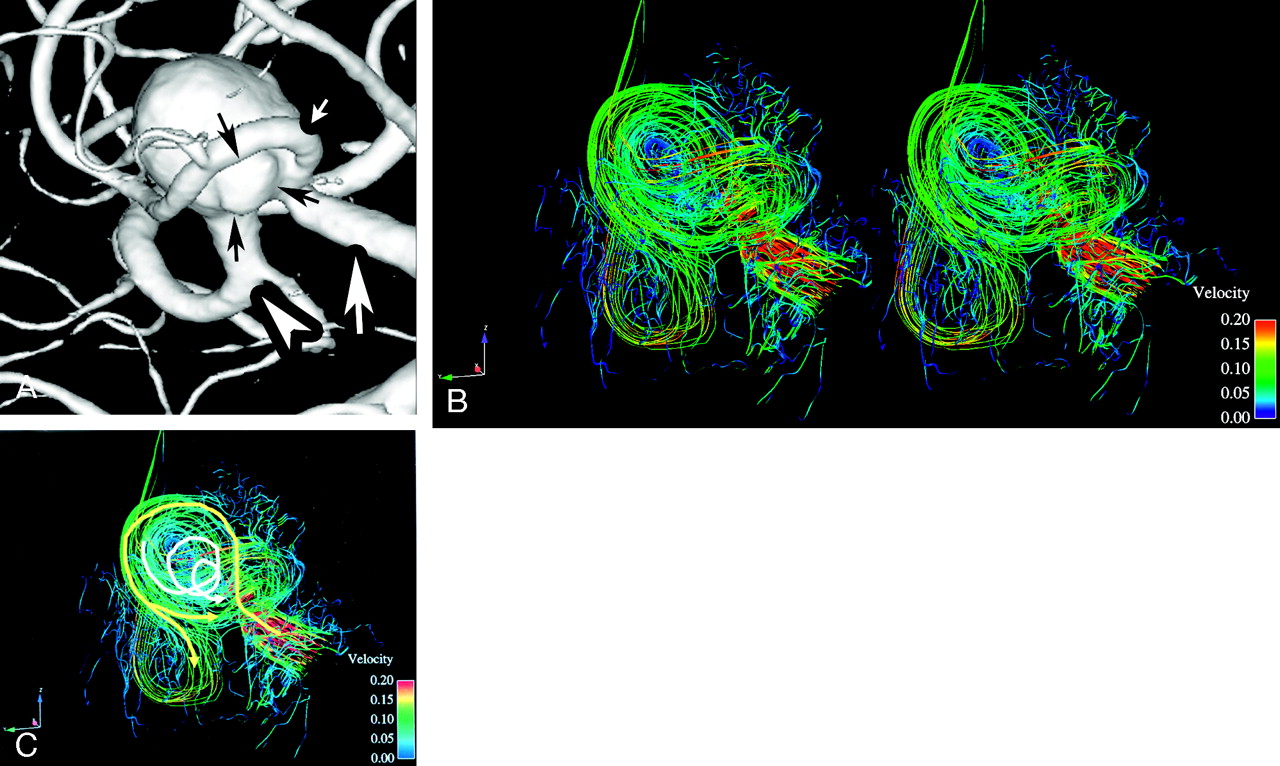
- Fig 3.
Posterior-anterior views of a rotational angiogram and 3D streamlines of the silicon middle cerebral artery (MCA) aneurysm. B is stereoscopic 3D streamlines corresponding with the systolic phase. C shows hemodynamics of B by arrows.
A, A posterior-anterior view of surface rendering of rotational digital subtraction angiogram of the right internal carotid artery shows M1 segment of right MCA (white arrow) and two M2 segment (anterior trunk of MCA [white small arrow] and posterior trunk of MCA [white arrowhead]) very clearly. Black arrows indicate irregular surface of the aneurysm.
B, Blood flow from the M1 segment of MCA strikes the posterior wall of the intracranial aneurysm. Blood flow flows along the wall of the intracranial aneurysm and 2 main flows diverge into the two M2 segments near the inlet of the aneurysm. Another weak flow flows out from the anterior trunk of MCA (white small arrow in Fig 3A) from behind the central incoming jet of the M1 segment. This slow flow (yellow arrow in Fig 3C, around 5 cm/s) corresponds with the irregular surface of the aneurysmal wall (shown by black arrows in Fig 3A).
C, Arrows indicate flow schematically.


