
Article Figures & Data
Figures
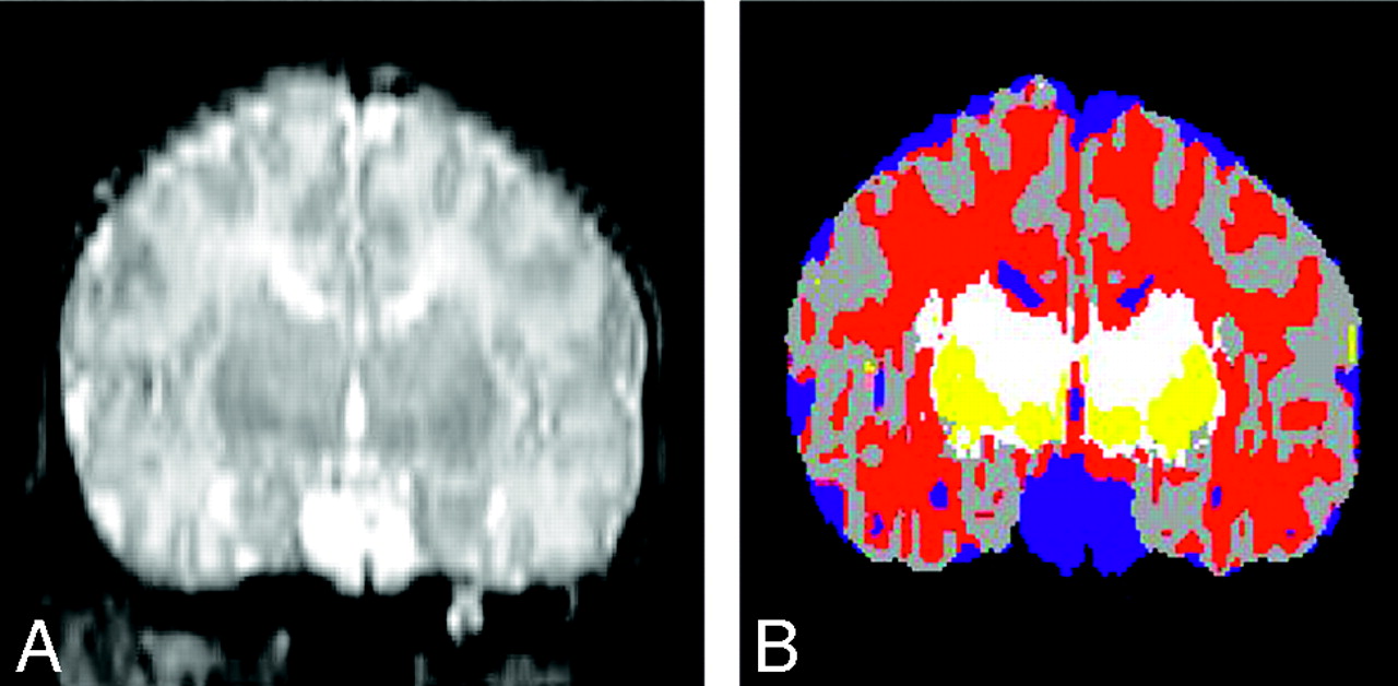
- Fig 1.
Segmentation. Reconstructed coronal T2-weighted image on the left compared with the segmented image (on the right) by using the Slicer software. On the segmented image, gray matter is gray, CSF is blue, the BG are yellow, unmyelinated white matter is red, and myelinated white matter is white.
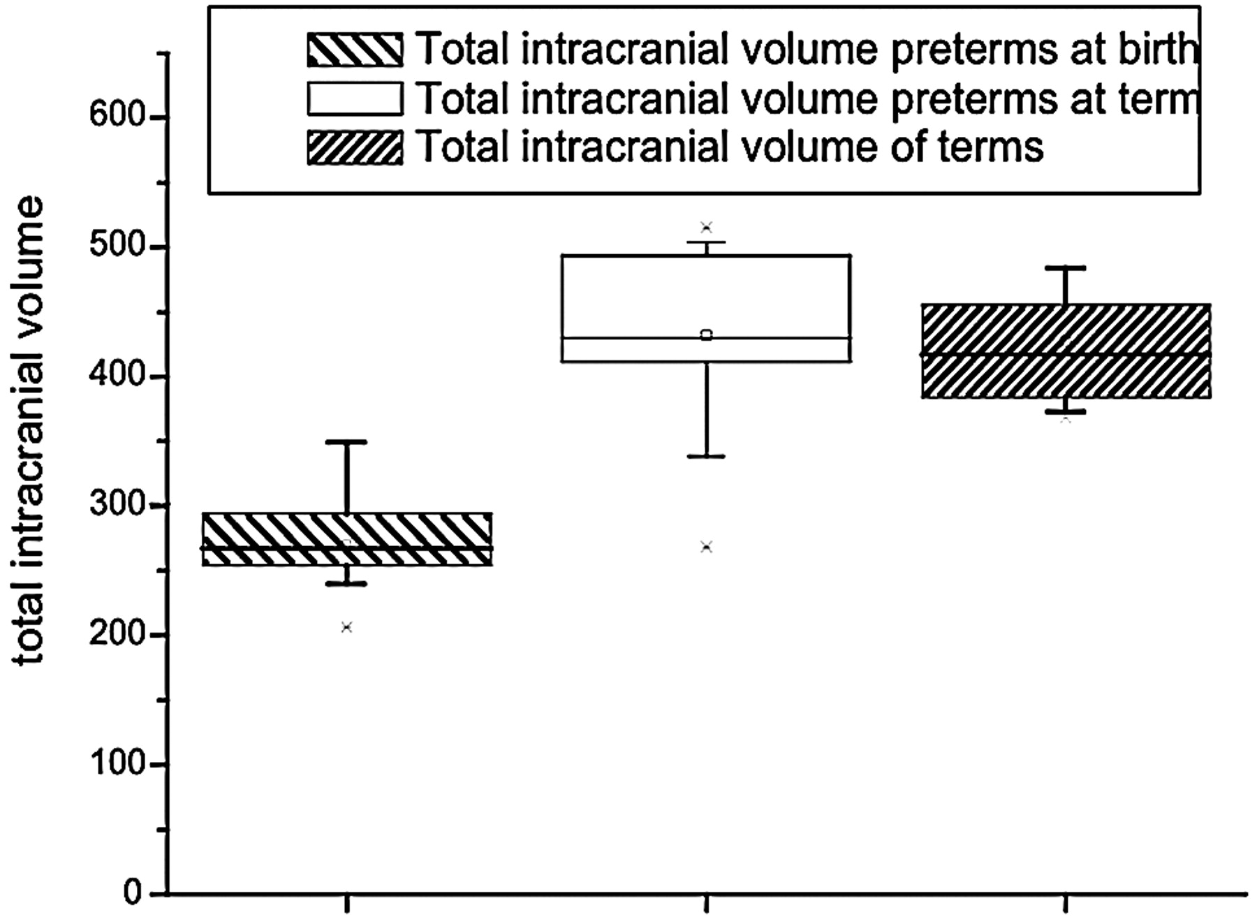
- Fig 2.
Total intracranial volume. The preterm newborns at birth had a lower intracranial volume, but at term, they had a volume similar to that of the term neonates.
- Fig 3.
Volume of cortical gray matter. The preterm infants at birth had a lower volume of cortical gray matter, but at term, they had a volume similar to that of the term neonates.
- Fig 4.
Volume of unmyelinated white matter. The preterm neonates at birth had a lower volume of unmyelinated white matter, but at term, they had a volume similar to that of the term neonates.
- Fig 5.
The relative volume of myelinated white matter showed no difference between preterms, preterms at term, and full-term infants
- Fig 6.
The relative volume of BG was highest in preterms. Preterm infants at term and full-term infants had similar relative volumes of basal ganglia.


Tables
Results in newborns and premature babies
PT (n = 11) PTT (n = 13) FT (n = 12) P (ANOVA, PT vs PTT vs FT) P (ANOVA, PT vs PTT) Absolute Units (cm3) Relative to ICV (%) Absolute Units (cm3) Relative to ICV (%) Absolute Units (cm3) Relative to ICV (%) P Absolute Units P Relative to ICV P Absolute Units P Relative to ICV ICV 269.8 ± 36.5 431.7 ± 70.0 427.4 ± 53.8 F = 31.0P < .001 F = 47.6 P < .001 Total brain volume 246.5 ± 32.3 391 ± 66.1 395 ± 49.2 F = 30.2 P < .001 F = 43.5 P < .001 CGM 83.5 ± 22.2 30.6 ± 5.1 179 ± 41.5 41.0 ± 5.4 181.4 ± 29.3 42.4 ± 4.1 F = 33.6 P < .001 F = 23.1 P < .001 F = 46.6 P < .001 F = 23.1 P < .001 UMWM 142.4 ± 15.0 53.1 ± 4.8 185.3 ± 30.8 43.2 ± 5.1 183.4 ± 27.4 43.0 ± 4.4 F = 10.1 P < .001 F = 16.8 P < .001 F = 17.6 P < .001 F = 23.7 P < .001 MWM 6.1 ± 1.8 2.2 ± 0.5 10.7 ± 3.0 2.5 ± 0.8 10.72 ± 4.63 2.5 ± 1.0 F = 7.0 P = .003 F = 0.4 P > .1 F = 18.8 P < .001 F = 1.0 P > .1 BG 14.2 ± 4.2 5.4 ± 1.7 15.7 ± 5.7 3.7 ± 1.1 17.14 ± 4.39 4.0 ± 0.9 F = 1.0 P > .1 F = 5.8 P = .007 F = 0.5 P > .1 F = 8.6 P = .008 CSF 23.3 ± 9.6 8.6 ± 3.0 40.7 ± 21.2 9.5 ± 4.3 32.4 ± 11.0 7.5 ± 2.3 F = 3.8 P = .03 F = 1.0 P > .1 F = 6.2 P = .02 F = 0.4 P > .1 Note:—Fifteen premature babies had 2 MRIs, one after birth (PT) and one at 40 wks (PTT); 12 newborns had an MRI at 40 wks (FT). ICV indicates intracranial volume; CGM, cortical gray matter; UMWM, ummyelinated white matter; MWM, myelinated white matter; BG, basal ganglia; CSF, cerebrospinal fluid.


