
Article Figures & Data
Figures
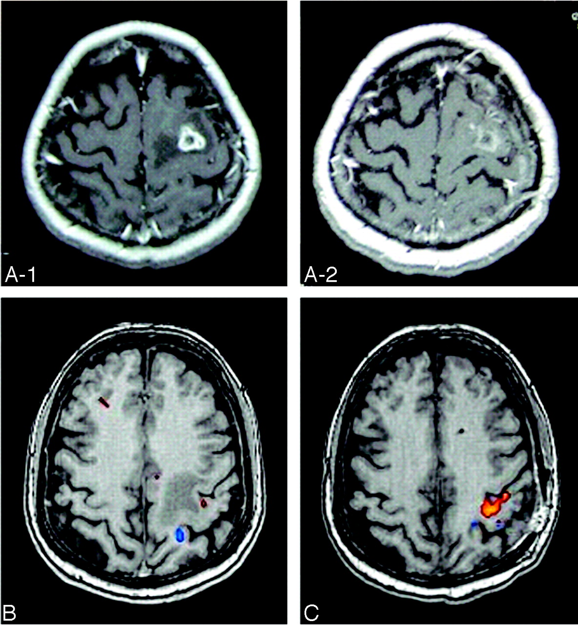
- Fig 1.
Case 1. A 57-year-old woman with metastatic brain tumor located inside the left-sided M1.
A-1, Preoperative MR imaging displays heterogeneously enhancing tumor in the M1.
A-2, Postoperative MR imaging shows ring enhancing lesion, but there was no residual tumor in the M1 according to the intraoperative observation for tumor cavity.
B, fMRI images activated by right hand clenching (B, orange) and right elbow flexion (B, blue) before surgery.
C, fMRI images activated by right hand clenching (orange) and right elbow flexion (blue) after surgery. A relatively small area of the contralateral M1 was activated by right hand clenching before surgery, whereas a large area of the contralateral M1 was activated by right hand clenching after surgery.
- Fig 2.
Case 2. A 71-year-old man with metastatic brain tumor located inside the right-sided S1.
A-1, Preoperative MR imaging displays heterogeneously enhancing tumor in the S1.
A-2, Postoperative MR imaging shows no residual tumor in the S1.
B, fMRI images activated by left hand clenching (B, orange) and right hand clenching (B, blue) before surgery.
C, fMRI images activated by left hand clenching (orange) and right hand clenching (blue) after surgery. Left hand clenching (orange area in B) activated the contralateral M1 and S1 areas separately before surgery and (orange area in C) activated the large area of the contralateral M1 and S1, and contralateral supplementary motor area after surgery.
- Fig 3.
Case 3. A 49-year-old man with metastatic brain tumor located inside the left-sided premotor area.
A-1, Preoperative MR imaging displays heterogeneously enhancing tumor in the premotor area.
A-2, Postoperative MR imaging shows no residual tumor in the premotor area.
B, fMRI images activated by right hand clenching (B-1, B-3; orange) and left hand clenching (B-2, B-4; blue) before surgery,
C, fMRI images activated by right hand clenching (orange) and left hand clenching (blue) after surgery. The ipsilateral and contralateral M1 and the anterior temporal lobe was activated by right hand clenching before surgery, whereas the contralateral M1 was activated by right hand clenching after surgery. The area in the anterior temporal lobe activated by right hand clenching before surgery (B-1) was not activated by right hand clenching after surgery (C-1).
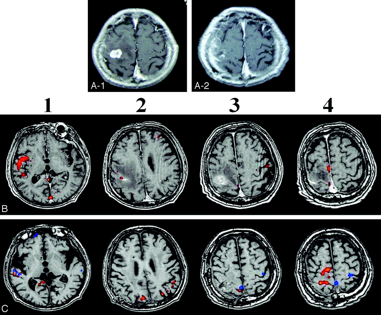
- Fig 4.
Case 4. A 71-year-old woman with metastatic brain tumor located inside the right-sided premotor area.
A-1, Preoperative MR imaging displays heterogeneously enhancing tumor in the premotor area.
A-2, Postoperative MR imaging shows no residual tumor in the premotor area.
B, fMRI images activated by right hand clenching (blue) and left hand clenching (red) before surgery,
C, fMRI images activated by right hand clenching (blue) and left hand clenching (orange) after surgery. Both left and right hand clenching activated the right temporal and frontal lobes before surgery. After surgery, left and right hand clenching activated the anterior temporal lobe (C-1,-2,-3), and left hand clenching activated a relatively small area of contralateral M1 (C-6). Because both preoperative and postoperative fMRI show similar atypical results such as no activation in the left hand M1 during right hand clenching, it seems that this result was not artifactual. It is not clear why there is no activation in the left M1 during right hand clenching, but the right hemisphere might be mainly activated by both right and left hand clenching in the case of severe paresis, possibly because of the preferential processing of spatial information in the right hemisphere.
- Fig 5.
Case 5. 76-year-old man with metastatic brain tumor located inside the right-sided M1.
A-1, Preoperative MR imaging displays heterogeneously enhancing tumor in the M1.
A-2, Postoperative MR imaging shows no residual tumor in the M1.
B, fMRI images activated by left hand clenching (orange) before surgery. fMRI by right hand clenching was not performed before surgery.
C, fMRI images activated by left hand clenching (orange) and right hand clenching (blue) after surgery. A relatively small area of the ipsilateral (B-3) and contralateral (B-2) M1 was activated by left hand clenching before surgery. A large portion of the right premotor and M1 were activated by left hand clenching after surgery (C-4, orange). The anterior temporal area was activated by left hand clenching before surgery (B-1) and by right or left hand clenching after surgery (C-1). The right medial M1 was activated by right or left hand clenching (C-3).
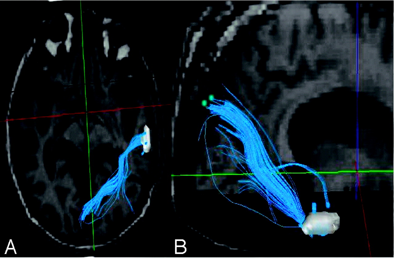
- Fig 6.
Case 5. 3D reconstruction of fMRI and DTI images constructed during left hand (affected side) clenching after surgery. Fibers (blue) connecting the premotor area (yellow) and the M1 (white) were detected.
- Fig 7.
Case 5. 3D reconstruction of fMRI and DTI images constructed during left and right hand clenching after surgery. Fibers (blue), possibly the ILF, arise from the anterior temporal area activated by left or right hand clenching and connect between temporal and occipital lobes. At the current state of the art, the contribution of Figs. 6 and 7 is only demonstrative, not scientific.


Tables
- Table 1:
Tumor location, main activated area in pre- and postoperative fMRI, and brain mapping
Patient No. Tumor Location Main Activated Area of Preoperative fMRI Main Activated Area of Brain Mapping Main Activated Area of Postoperative fMRI 1 L M1 L M1 (small) L M1 L M1 (large) 2 R S1 R M1 (small), R S1 R M1 R M1 (large), R S1 3 L PMA R and L M1, R T L M1 L M1 4 R PMA R F and T (large) R M1 R F and T (small), R M1 5 R M1 R T (large), R M1 (small) R PMA R T (small), R PMA and M1 (large) Note:—L indicates left; R, right; M1, primary motor area; S1, primary sensory area; PMA, premotor area; F, frontal; T, temporal lobe; small, relatively small area was activated; large, relatively large area was activated.
In this issue


Jump to section
Related Articles
Cited By...
- No citing articles found.