
Article Figures & Data
Figures
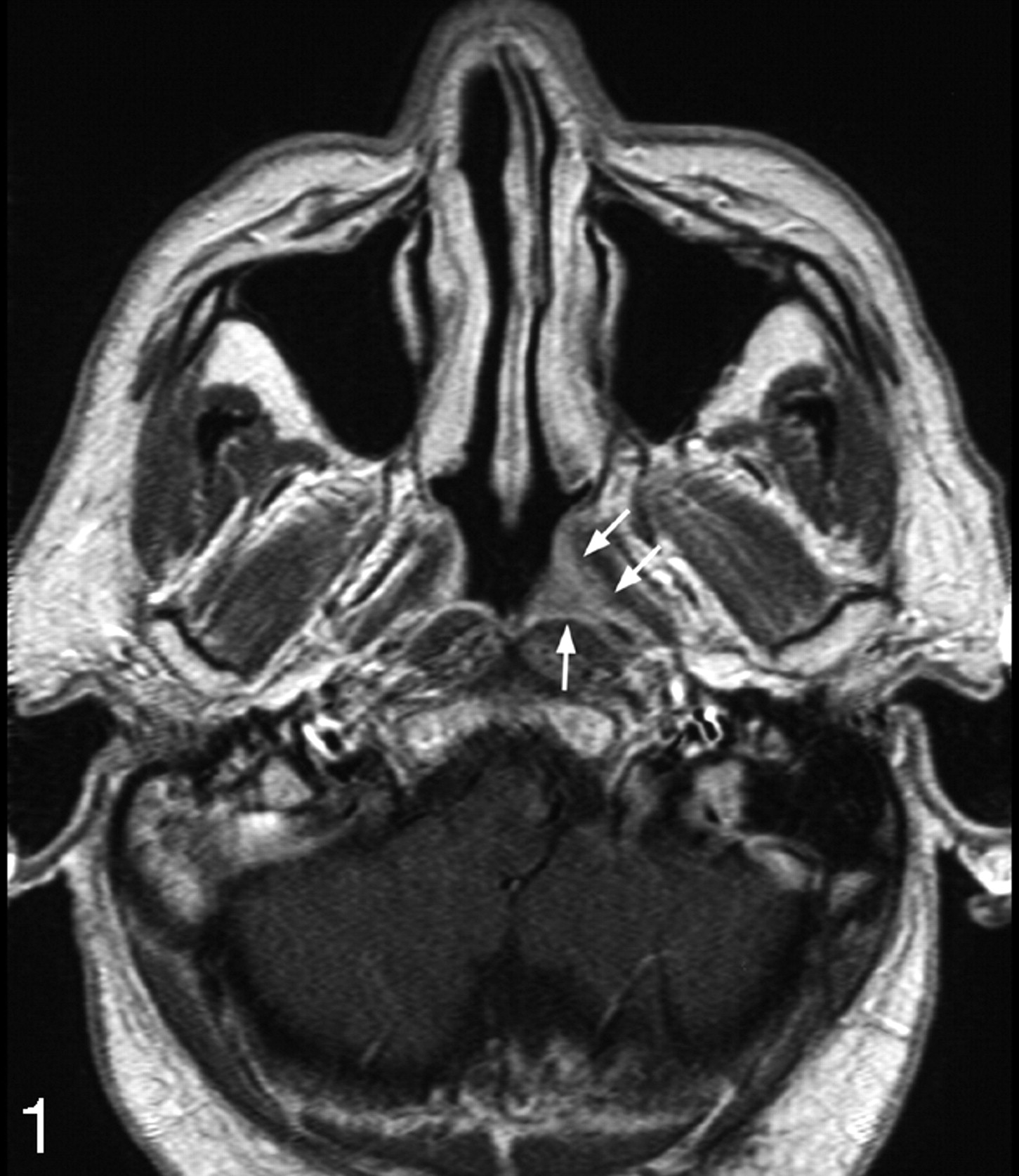
- Fig 1.
Axial T1-weighted MR image postcontrast of the nasopharynx of a patient with proved NPC (group 1) undergoing staging with a small cancer confined to the left side of the nasopharynx (arrows) (stage T1).
- Fig 2.
Axial T1-weighted contrast-enhanced MR image of a patient with suspected NPC (group 2) with a normal nasopharynx (arrows show normal enhancing mucosa).
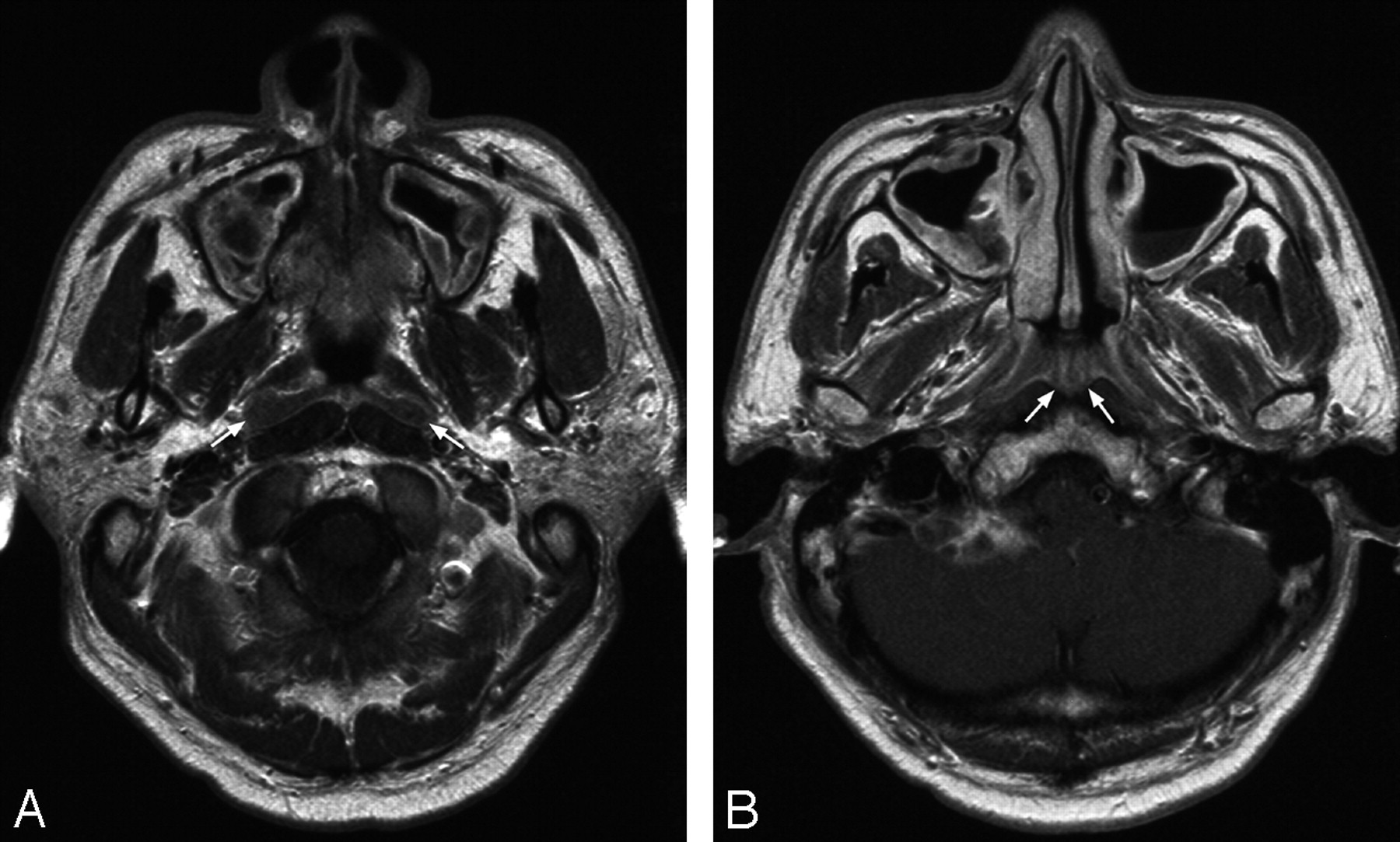
- Fig 3.
Axial T1-weighted contrast-enhanced MR image of the nasopharynx in 2 patients with suspected NPC (group 2) where cancer was initially missed by endoscopy and biopsy but identified by MR imaging. Patient with a small cancer in the left fossa of Rosenmuller (arrows) (stage T1) (A) and patient with a small cancer over the torus tubarius (arrows) (stage T1) (B).
- Fig 4.
Axial T1-weighted contrast-enhanced MR image of the nasopharynx in a patient with suspected NPC (group 2) where MR imaging incorrectly diagnosed cancer that was later shown by biopsy to be lymphoid hyperplasia. A, Section at the level of the fossa of Rosenmuller shows mucosal abnormality in the fossa bilaterally (arrows), giving the false-positive result on MR imaging for cancer.
B, Section at the level of the roof shows the “striped” appearance of normal lymphoid tissue in the adenoids (arrows).


Tables
Stage of Primary Tumor Extent of Tumor T1 Tumor confined to the nasopharynx T2a Tumor extends to the oropharynx and/or nasal fossa without parapharyngeal extension T2b Tumor extends to the oropharynx and/or nasal fossa with parapharyngeal extension T3 Tumor invades bony structures and/or paranasal sinuses T4 Tumor with intracranial extension and/or involvement of cranial nerves, infratemporal fossa, hypopharynx, or orbit Score MRI Findings No. of Patients % of Patients 1 Normal nasopharynx 46 60 2 Normal nasopharynx with lymphoid tissue in the adenoids 15 19 3 Mild diffuse mucosal thickening with the signal intensity of mucosa 9 12 4 Focal mucosal thickening with the signal intensity of NPC 2 3 5 Definite NPC 5 6 Note:—NPC indicates nasopharyngeal carcinoma.
T-stage No. of Patients % of Patients T1 118 26 T2 65 14 T3 163 36 T4 110 24 T1–T4 456 100 Biopsy Positive for NPC Biopsy Negative for NPC MRI positive for NPC 3 4 MRI negative for NPC 0 70 Note:—NPC indicates nasopharyngeal carcinoma.
In this issue


Jump to section
Related Articles
Cited By...
- Early Detection of Cancer: Evaluation of MR Imaging Grading Systems in Patients with Suspected Nasopharyngeal Carcinoma
- Optimal Mass Transport Kinetic Modeling for Head and Neck DCE-MRI: Initial Analysis
- MR Imaging Criteria for the Detection of Nasopharyngeal Carcinoma: Discrimination of Early-Stage Primary Tumors from Benign Hyperplasia
- Detection of Nasopharyngeal Carcinoma by MR Imaging: Diagnostic Accuracy of MRI Compared with Endoscopy and Endoscopic Biopsy Based on Long-Term Follow-Up
- Diagnostic Accuracy of Sonography Versus Magnetic Resonance Imaging for Primary Nasopharyngeal Carcinoma
- Imaging of the pharynx and larynx
- Imaging of the pharynx and larynx