Advertisement

Main menu

  • Home
  • Content
    • Current Issue
    • Accepted Manuscripts
    • Article Preview
    • Past Issue Archive
    • AJNR Case Collection
    • Case of the Week Archive
    • Classic Case Archive
    • Case of the Month Archive
  • Special Collections
    • Spinal CSF Leak Articles (Jan 2020-June 2024)
    • 2024 AJNR Journal Awards
    • Most Impactful AJNR Articles
  • Multimedia
    • AJNR Podcast
    • AJNR Scantastics
    • Video Articles
  • For Authors
    • Submit a Manuscript
    • Author Policies
    • Fast publishing of Accepted Manuscripts
    • Graphical Abstract Preparation
    • Manuscript Submission Guidelines
    • Imaging Protocol Submission
    • Submit a Case for the Case Collection
  • About Us
    • About AJNR
    • Editorial Board
  • More
    • Become a Reviewer/Academy of Reviewers
    • Subscribers
    • Permissions
    • Alerts
    • Feedback
    • Advertisers
    • ASNR Home
  • Other Publications
    • ajnr

User menu

  • Alerts
  • Log in

Search

  • Advanced search
American Journal of Neuroradiology

American Journal of Neuroradiology

ASHNR American Society of Functional Neuroradiology ASHNR American Society of Pediatric Neuroradiology ASSR
  • Alerts
  • Log in

Advanced Search

  • Home
  • Content
    • Current Issue
    • Accepted Manuscripts
    • Article Preview
    • Past Issue Archive
    • AJNR Case Collection
    • Case of the Week Archive
    • Classic Case Archive
    • Case of the Month Archive
  • Special Collections
    • Spinal CSF Leak Articles (Jan 2020-June 2024)
    • 2024 AJNR Journal Awards
    • Most Impactful AJNR Articles
  • Multimedia
    • AJNR Podcast
    • AJNR Scantastics
    • Video Articles
  • For Authors
    • Submit a Manuscript
    • Author Policies
    • Fast publishing of Accepted Manuscripts
    • Graphical Abstract Preparation
    • Manuscript Submission Guidelines
    • Imaging Protocol Submission
    • Submit a Case for the Case Collection
  • About Us
    • About AJNR
    • Editorial Board
  • More
    • Become a Reviewer/Academy of Reviewers
    • Subscribers
    • Permissions
    • Alerts
    • Feedback
    • Advertisers
    • ASNR Home
  • Follow AJNR on Twitter
  • Visit AJNR on Facebook
  • Follow AJNR on Instagram
  • Join AJNR on LinkedIn
  • RSS Feeds

Welcome to the new AJNR, Updated Hall of Fame, and more. Read the full announcements.


AJNR is seeking candidates for the position of Associate Section Editor, AJNR Case Collection. Read the full announcement.


OtherHEAD AND NECK

A Case of Horner Syndrome with Intermittent Mydriasis in a Patient with Hypoplasia of the Internal Carotid Artery

M. Ibrahim, H.M. Branson, J.R. Buncic and M.M. Shroff
American Journal of Neuroradiology June 2006, 27 (6) 1318-1320;
M. Ibrahim
  • Find this author on Google Scholar
  • Find this author on PubMed
  • Search for this author on this site
H.M. Branson
  • Find this author on Google Scholar
  • Find this author on PubMed
  • Search for this author on this site
J.R. Buncic
  • Find this author on Google Scholar
  • Find this author on PubMed
  • Search for this author on this site
M.M. Shroff
  • Find this author on Google Scholar
  • Find this author on PubMed
  • Search for this author on this site
  • Article
  • Figures & Data
  • Info & Metrics
  • Responses
  • References
  • PDF
Loading

Abstract

SUMMARY: We report a rare case of hypoplasia of the right internal carotid artery (ICA) with ipsilateral congenital Horner syndrome. The etiology and pathogenesis of hypoplasia of the ICA is not well understood. Multiple types of collateral flow have been reported to develop to maintain blood supply to the ipsilateral cerebral hemisphere. Although collateral flow may allow these patients to remain asymptomatic, we postulate that the enlarged posterior communicating artery (PcomA) in our patient caused mass effect on the cisternal segment of cranial nerve III causing intermittent mydriasis apart from Horner syndrome.

Case Report

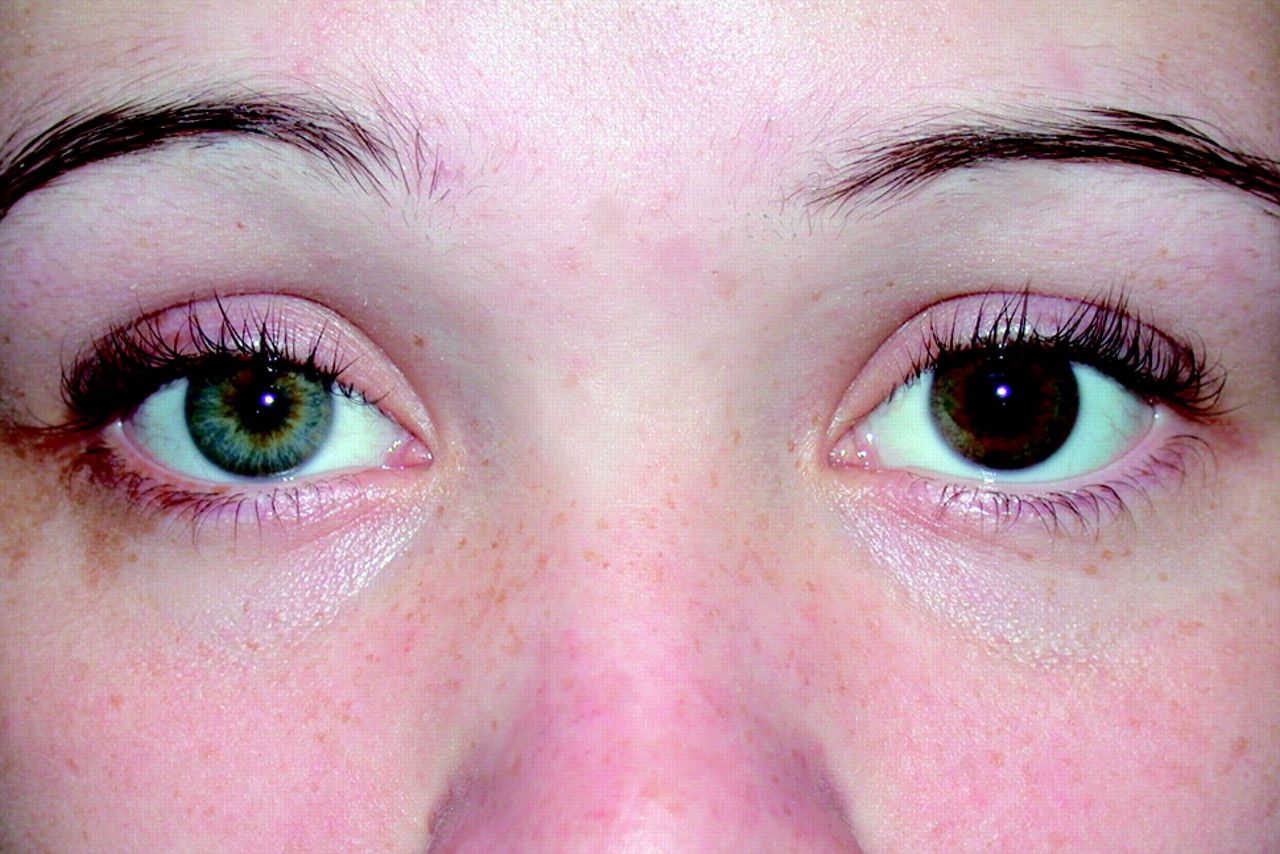
A 15-year-old girl was referred to the ophthalmology clinic for assessment of right iris heterochromia and miosis, with intermittent episodes of asymmetric dilation of the right pupil. Since her birth, the parents had noticed the right pupil was smaller and the right iris lighter in color than the left eye (Fig 1).

Fig. 1.
  • Download figure
  • Open in new tab
  • Download powerpoint
Fig. 1.

Clinical photograph demonstrating right Horner syndrome with ptosis, miosis, and iris heterochromia.

Recently the patient complained of recurrent episodes of pupil dilation, which lasted approximately 10 minutes once a day for 2–3 months and then decreased to once a month. These episodes of pupil dilation were accompanied with tingling in the right cheek. In addition, the patient experienced intermittent blurring of the vision in the right eye with aching around her eyes, especially on the right. Examination of the patient demonstrated uncorrected visual acuity of 20/20 in both eyes. There was slight (1–2 mm) right-sided ptosis. The right pupil was smaller than the left, and the right iris was lightly pigmented compared with the left. The right iris was asymmetrically hypoplastic; the inferior sphincter cuff was hypoplastic, whereas the upper portion was normal. Failure of the right pupil to dilate after administration of 4% cocaine eye drops verifiGed the diagnosis of unilateral right Horner syndrome. The rest of the ocular and visual examination was normal.

Time-of-flight MR angiography (MRA) of the circle of Willis showed absent flow in the right internal carotid artery (ICA) with collateral flow to the right cerebral hemisphere via an enlarged PcomA (Fig 2A, -B). Figure 3 demonstrates compression of the right cranial nerve III by the enlarged right PcomA. Time-of-flight MRA of the neck reveals absence of the right cervical ICA (Fig. 4). High-resolution CT through the skull base demonstrated an atretic right bony carotid canal, confirming presence of the hypoplastic ICA before skull base development (Fig 5).

Fig. 2.
  • Download figure
  • Open in new tab
  • Download powerpoint
Fig. 2.

A, Source images from axial time-of-flight MRA demonstrates absence of the right ICA in the right parasellar region. The left ICA and basilar arteries are clearly seen.

B, Axial source MRA image superior to the prior image demonstrates an enlarged right PcomA.

Fig. 3.
  • Download figure
  • Open in new tab
  • Download powerpoint
Fig. 3.

Parasagittal T1-weighted sequence to the right of the midline demonstrates the enlarged right PcomA coursing along the superior aspect of the right third nerve.

Fig. 4.
  • Download figure
  • Open in new tab
  • Download powerpoint
Fig. 4.

Axial time-of-flight MRA of the neck with multiplanar intensity projection reformats demonstrating absent flow in the cervical right ICA.

Fig. 5.
  • Download figure
  • Open in new tab
  • Download powerpoint
Fig. 5.

Axial CT through the skull base demonstrates an atretic right carotid canal on the right. Note the normal left-sided bony carotid canal.

Discussion

Hypoplasia of the ICA is an uncommon congenital disease. Slightly more than 100 cases have been reported in the literature,1 though only a few cases have been reported in association with congenital Horner syndrome.2 To the best of our knowledge, the association of Horner syndrome with intermittent mydriasis in the setting of hypoplasia of the ICA has not been reported.

Most reported cases of aplasia and hypoplasia of the ICA are in asymptomatic patients. Arterial insufficiency is prevented via collateral pathways that include the circle of Willis, persistence of embryonic vessels, and transcranial collaterals via the external carotid artery.3,4 These collateral pathways include fetal-type hypertrophied anterior communicating artery (AcomA) and PcomA supplying the anterior cerebral artery (ACA) and middle cerebral artery (MCA) respectively, adult type (hypertrophied AcomA supplying both of the ACA and MCA), and transcranial anastomosis arising from the contralateral ICA or from primitive vessels. Lie 5 described 6 pathways of collateral circulation in association with absence of the ICA. In types A, B, and C, unilateral (types A and B) or bilateral (type C) absence of the ICA is associated with hypertrophied AcomA and PcomA (type A), patent AcomA (type B), or carotid-vertebrobasilar anastomoses (type C). Type D represents intercavernous communication to the ipsilateral carotid siphon from the contralateral cavernous ICA in the setting of unilateral agenesis of the cervical segment of the ICA. In type E the ACAs are supplied by hypoplastic ICAs, whereas in type F transcranial anastomoses from the internal maxillary branches of the external carotid artery (ECA) system form supply to the distal ICA, the so-called rete mirabile. Our patient had a fetal type of collateral supply (Lie type A), with hypoplastic ICA and hypertrophied PcomA.

Changes in pupil size are governed by pupillary sphincter (parasympathetic) and the pupillary dilator muscle (sympathetic) tone in response to ambient light, adrenergic and cholinergic tone, and local pharmacologic or pathophysiologic conditions. The parasympathetic (pupilloconstrictor) pathway originates in the Edinger-Westphal nucleus in the dorsal midbrain and exits with the motor fibers of the third cranial nerve (CN III) to the ciliary ganglion via the cavernous sinus. Postganglionic fibers then travel to the sphincter muscle of the iris. Differential diagnosis of mydriasis includes cranial nerve dysfunction, Adie tonic pupil, CN III dysfunction, and pharmacologic blockade. Dysfunction of CN III can occur in a wide variety of settings and is found in association with uncal herniation, PcomA aneurysm, ophthalmoplegic migraine, peripheral neuropathies, Lyme disease, and alcoholism.6 We postulate the underlying cause of mydriasis in our patient is related to hypertrophied PcomA intermittently compressing the cisternal segment of CN III and causing transient dysfunction of the sphincter muscle.

Horner syndrome results from an interruption of the sympathetic nerve supply to the eye. It may be congenital, acquired, or hereditary (autosomal dominant).7 Birth trauma is a frequent cause of Horner syndrome.7 Acquired Horner syndrome with no antecedent surgery is usually approached as being associated with potentially serious underlying disease. The interruption of the sympathetic fibers may occur centrally (ie, between the hypothalamus and the fibers’ point of exit from the spinal cord [C8–T2]) or peripherally (ie, cervical sympathetic chain, superior cervical ganglion, or along the carotid artery). It is characterized by classic triad of miosis (constricted pupil), partial ptosis, and loss of hemifacial sweating (anhydrosis). Anhydrosis occurs with hyperaemia and warmth when oculosympathetic denervation of the sweat and vasoconstrictor fibers distributed through branches of the ECA occurs. Iris heterochromia is seen in congenital Horner syndrome, Horner syndrome that occurs in children younger than 2 years of age and in long-standing Horner syndrome. The association of Horner syndrome with congenital hypoplasia of the ICA is rare, and only a few case reports have been published.2,8

In conclusion, the differential diagnosis of Horner syndrome is broad, with birth trauma the most common cause of congenital Horner syndrome. The rare association with congenital hypoplasia of the ipsilateral ICA is reported. Multiple collateral flow pathways have been described to establish blood flow to the ipsilateral cerebral hemisphere. Such compensatory flow is essential but as seen in our case may cause further abnormalities.

References

  1. ↵
    Claros P, Bandos R, Gilea I, et al. Case report: major congenital anomalies of the internal carotid artery: agenesis, aplasia, and hypoplasia. Int J Pediatr Otorhinolaryngol 1999;49:69–76
    CrossRefPubMed
  2. ↵
    Dinç H, Alioglu Z, Erdöl Z, et al. Agenesis of the internal carotid artery associated with aortic arch anomaly in a patient with congenital Horner’s syndrome. AJNR Am J Neuroradiol 2002;23:929–31
    Abstract/FREE Full Text
  3. ↵
    Midkiff RB, Boykin MW, McFarland DR, et al. Agenesis of the internal carotid artery with intercavernous anastomosis. AJNR Am J Neuroradiol 1995;16:1356–59
    Abstract
  4. ↵
    Chen CJ, Chen ST, Hsieh FY, et al. Hypoplasia of the internal carotid artery with intercavernous anastomosis. Neuroradiology 1998;40:252–54
    CrossRefPubMed
  5. ↵
    Lie TA. Congenital anomalies of the carotid arteries. Amsterdam: Excerpta Medica;1968;35–51
  6. ↵
    Lee AG, Taber KH, Hayman LA, et al. A guide to the isolated dilated pupil. Arch Fam Med 1997;4:385–88
  7. ↵
    Jeffrey AR, Ellis FJ, Repka MX, et al. Pediatric Horner syndrome. J Am Assoc Ped Ophthalmol 1998;2:159–67
  8. ↵
    Given CA II, Huang-Hellinger F, Baker MD, et al. Congenital absence of the internal carotid artery: case reports and review of the collateral circulation. AJNR Am J Neuroradiol 2001;22:1953–59
    Abstract/FREE Full Text
  • Received June 17, 2005.
  • Accepted after revision August 10, 2005.
  • Copyright © American Society of Neuroradiology
View Abstract
PreviousNext
Back to top

In this issue

American Journal of Neuroradiology: 27 (6)
American Journal of Neuroradiology
Vol. 27, Issue 6
June 2006
  • Table of Contents
  • Index by author
Advertisement
Print
Download PDF
Email Article

Thank you for your interest in spreading the word on American Journal of Neuroradiology.

NOTE: We only request your email address so that the person you are recommending the page to knows that you wanted them to see it, and that it is not junk mail. We do not capture any email address.

Enter multiple addresses on separate lines or separate them with commas.
A Case of Horner Syndrome with Intermittent Mydriasis in a Patient with Hypoplasia of the Internal Carotid Artery
(Your Name) has sent you a message from American Journal of Neuroradiology
(Your Name) thought you would like to see the American Journal of Neuroradiology web site.
CAPTCHA
This question is for testing whether or not you are a human visitor and to prevent automated spam submissions.
Cite this article
M. Ibrahim, H.M. Branson, J.R. Buncic, M.M. Shroff
A Case of Horner Syndrome with Intermittent Mydriasis in a Patient with Hypoplasia of the Internal Carotid Artery
American Journal of Neuroradiology Jun 2006, 27 (6) 1318-1320;

Citation Manager Formats

  • BibTeX
  • Bookends
  • EasyBib
  • EndNote (tagged)
  • EndNote 8 (xml)
  • Medlars
  • Mendeley
  • Papers
  • RefWorks Tagged
  • Ref Manager
  • RIS
  • Zotero
0 Responses
Respond to this article
Share
Bookmark this article
A Case of Horner Syndrome with Intermittent Mydriasis in a Patient with Hypoplasia of the Internal Carotid Artery
M. Ibrahim, H.M. Branson, J.R. Buncic, M.M. Shroff
American Journal of Neuroradiology Jun 2006, 27 (6) 1318-1320;
del.icio.us logo Twitter logo Facebook logo Mendeley logo
  • Tweet Widget
  • Facebook Like
  • Google Plus One

Jump to section

  • Article
    • Abstract
    • Case Report
    • Discussion
    • References
  • Figures & Data
  • Info & Metrics
  • Responses
  • References
  • PDF

Related Articles

  • No related articles found.
  • PubMed
  • Google Scholar

Cited By...

  • Carotid Near-Occlusion: A Comprehensive Review, Part 2--Prognosis and Treatment, Pathophysiology, Confusions, and Areas for Improvement
  • Crossref
  • Google Scholar

This article has not yet been cited by articles in journals that are participating in Crossref Cited-by Linking.

More in this TOC Section

  • Correlation of Apparent Diffusion Coefficient at 3T with Prognostic Parameters of Retinoblastoma
  • Parathyroid Lesions: Characterization with Dual-Phase Arterial and Venous Enhanced CT of the Neck
  • MR Evaluation of Sinonasal Angiomatous Polyp
Show more Head and Neck

Similar Articles

Advertisement

Indexed Content

  • Current Issue
  • Accepted Manuscripts
  • Article Preview
  • Past Issues
  • Editorials
  • Editors Choice
  • Fellow Journal Club
  • Letters to the Editor

Cases

  • Case Collection
  • Archive - Case of the Week
  • Archive - Case of the Month
  • Archive - Classic Case

Special Collections

  • Special Collections

Resources

  • News and Updates
  • Turn around Times
  • Submit a Manuscript
  • Author Policies
  • Manuscript Submission Guidelines
  • Evidence-Based Medicine Level Guide
  • Publishing Checklists
  • Graphical Abstract Preparation
  • Imaging Protocol Submission
  • Submit a Case
  • Become a Reviewer/Academy of Reviewers
  • Get Peer Review Credit from Publons

Multimedia

  • AJNR Podcast
  • AJNR SCANtastic
  • Video Articles

About Us

  • About AJNR
  • Editorial Board
  • Not an AJNR Subscriber? Join Now
  • Alerts
  • Feedback
  • Advertise with us
  • Librarian Resources
  • Permissions
  • Terms and Conditions

American Society of Neuroradiology

  • Not an ASNR Member? Join Now

© 2025 by the American Society of Neuroradiology All rights, including for text and data mining, AI training, and similar technologies, are reserved.
Print ISSN: 0195-6108 Online ISSN: 1936-959X

Powered by HighWire