
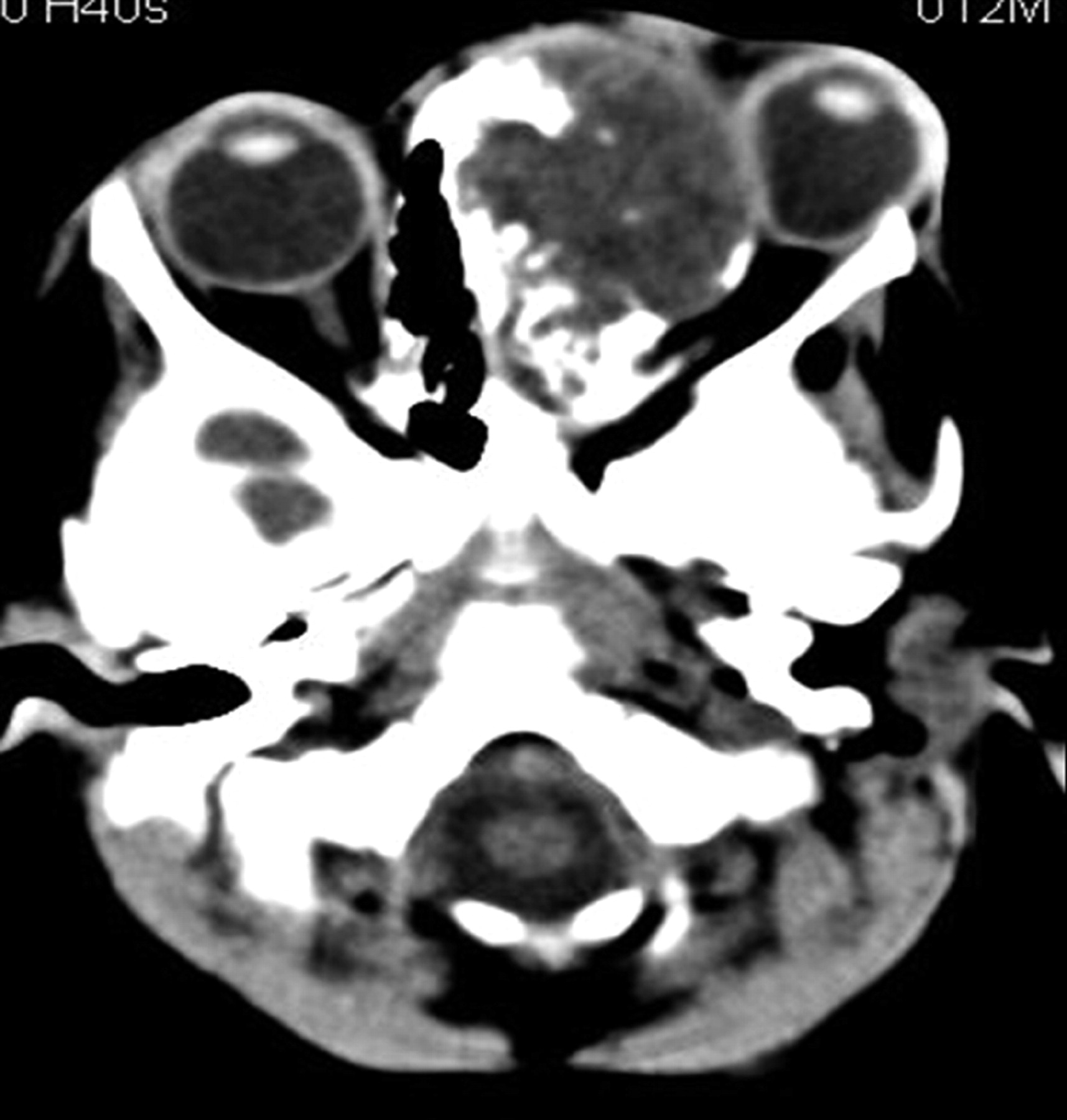
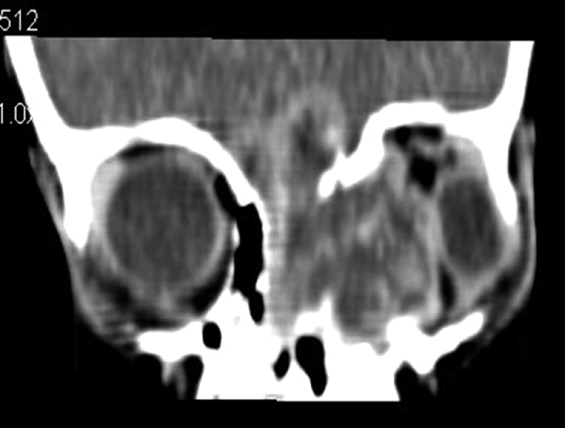
We report an unusual case of “nasal chondrosarcoma in an infant.” An 11-month-old girl presenting with swelling near the inner canthus of her left eye she had experienced since 2 months of age underwent axial CT to assess the suspected orbital mass (Fig 1). CT showed a large heterogeneously enhancing low-attenuation mass centered in the left ethmoid sinuses with extension into left orbit and left maxillary sinuses (Fig 2). The tumor also eroded the cribriform plate and floor of anterior cranial fossa with intracranial extension (Fig 2). Only subtle calcific foci were noted within the mass (Fig 1). Biopsy of the mass showed histologic findings consistent with myxoid chondrosarcoma. Her parents, unfortunately, refused treatment.
Chondrosarcomas are malignant cartilagenous tumors that constitute approximately 10%–20% of all primary malignant osseous neoplasms, of which only about 10% arise in the head and neck region.1 The highest incidence of craniofacial chondrosarcoma occurs in the 4th decade of life.
In the pediatric population, primary chondrosarcoma of head and neck is rare and usually occurs in the maxillary sinus or mandible. It is also typically low grade.2 In rare cases, it may arise from the nasal cavity and nasal septum. There are only a few studies in the literature. In a study done by Gadwal et al, 14 such cases between 3 and 18 years of age were reported, only 2 of which originated from the nasal cavity.2 The case discussed here is of nasal chondrosarcoma and is unusual because it was seen in infancy and showed myxoid degeneration, which is more common in higher grades of chondrosarcoma.
Chondrosarcomas are classified pathologically as conventional, mesenchymal, clear cell, and dedifferentiated types. Myxoid degeneration is commonly found, particularly in higher-grade conventional chondrosarcomas, but it is not a well-established entity. The myxoid variety has a more aggressive clinical course than the low-grade conventional chondrosarcoma.
Imaging diagnosis principally depends on CT, which shows characteristic chondroid type of calcification. The typical appearance of mineralized chondroid matrix is a ring-and-arc pattern of calcification that may coalesce to form a flocculent, flecklike pattern. This characteristic chondroid calcification is the most useful and dominant feature of a cartilagenous lesion.3 Higher-grade chondrosarcomas and myxoid variety, because of their aggressive clinical behavior, however, frequently show an aggressive type of bone lysis and less extensive calcifications.4 Because of high water content, it also shows low attenuation on CT and high signal intensity on T2-weighted images. The enhancement is commonly mild and septal to peripheral.4 MR imaging can depict the exact extent of the tumor and has a significant role in monitoring recurrence. CT is optimal to detect the matrix mineralization, particularly when it is subtle or when the lesion is located in anatomically complex areas.3
Complete surgical resection is the treatment of choice. Even with extensive local invasion, the long-term prognosis is excellent for pediatric patients with primary chondrosarcoma of the head and neck.2

Plain axial CT scan shows a large expansile low-attenuation mass originating in left nasal cavity with extension into left orbit. There are subtle chondroid matrix mineralizations within the mass.

Postcontrast reformatted coronal CT shows septal and peripheral enhancement of the mass with extension into left maxillary sinus and anterior cranial fossa.
- Copyright © American Society of Neuroradiology
In this issue


Jump to section
Related Articles
Cited By...
- No citing articles found.