
Article Figures & Data
Figures
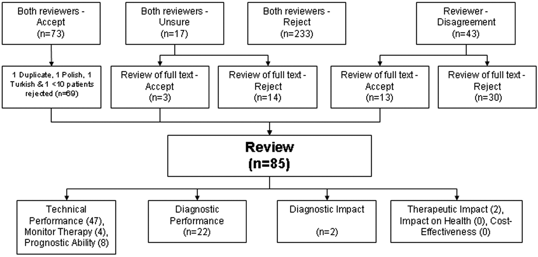
- Fig 1.
Studies identified by the systematic review.
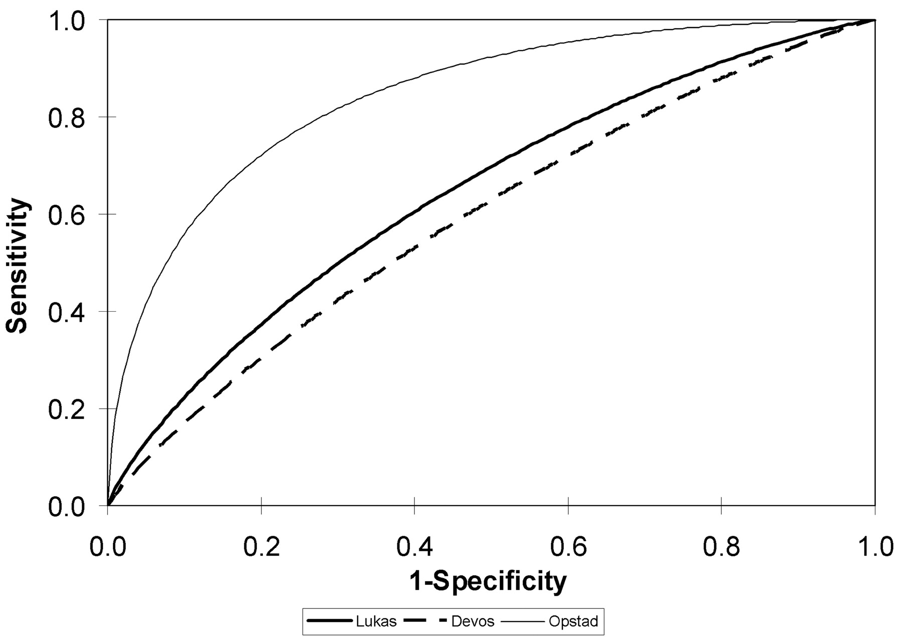
- Fig 2.
Receiver operating characteristic (ROC) curves measuring the sensitivity and specificity of 1H-MR spectroscopy for distinguishing metastases from high-grade astrocytomas. The ROC curves are back-calculated from the area-under-the-curve figures provided by the authors. They approximate, but are not perfect matches, for the ROC curves based on the individual patient data.
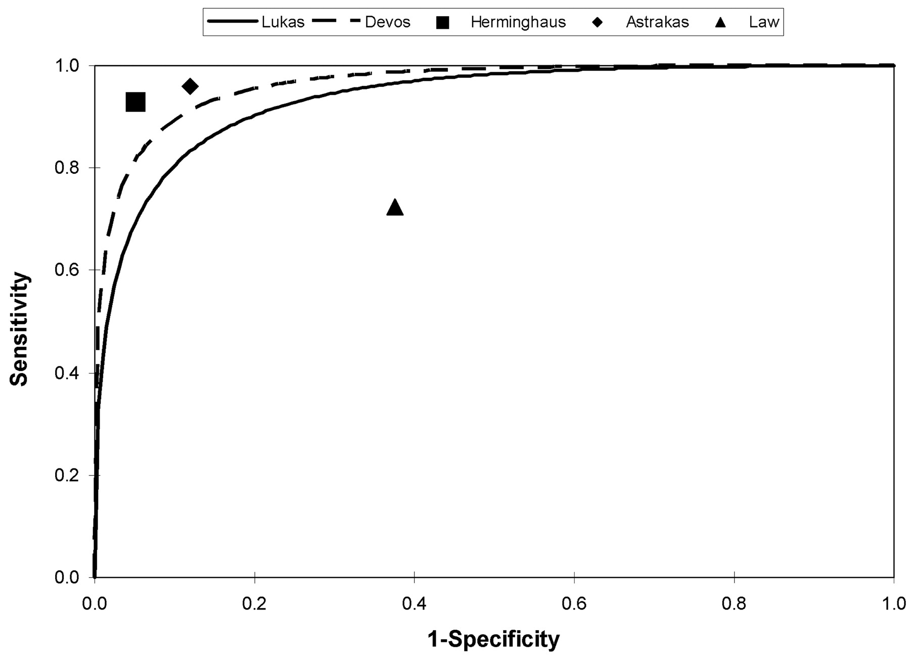
- Fig 3.
Receiver operating characteristic (ROC) curves and point estimates of sensitivity and specificity of 1H-MR spectroscopy for distinguishing high- and low-grade astrocytomas. The ROC curves are back-calculated from the area-under-the-curve figures provided by the authors. They approximate, but are not perfect matches, for the ROC curves based on the individual patient data.
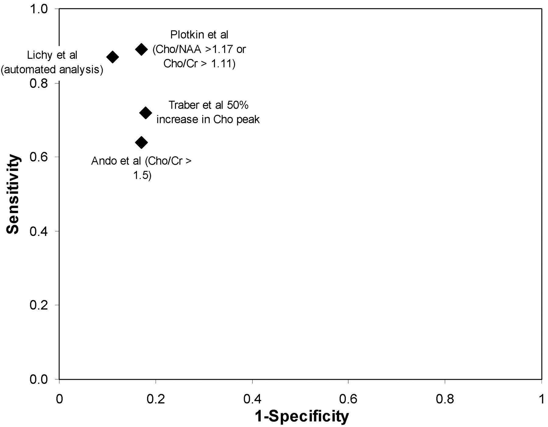
- Fig 4.
Sensitivity and specificity of 1H-MR spectroscopy for differentiating recurrent or residual tumor from treatment-related changes.


Tables
Study Clinical Subgroup Start/End Year No. of Patients Reference Standard(s) Metabolites Diagnostic Categorization* Voxels TR (ms) TE Pulse Sequence Ando et al, 200422 Residual-recurrent/necrosis Unclear 20 Biopsy/resection, clinical & radiologic follow-up Cho, Cr, NAA, Lac, Lip Quantitative Single 1500 270 Unclear Astrakas et al, 200428 Tumor grading Unclear 66 Biopsy/resection Cho, Cr, NAA, Lip, Lac Quantitative Multiple 1000 65 PRESS Devos et al, 20049 Tumor grading, primary/met Unclear 205 Biopsy/resection Complete spectrum Automated Single 1600, 2000, 2018, 2020 20, 30, 31, 32 STEAM and PRESS Fountas et al, 200410 Tumor grading 2000/2001 71 Biopsy/resection Cho, Cr, NAA, Lac, Lip, mIns/Gly Quantitative Single 1600 135 PRESS Gajewicz et al, 200323 Tumor/tumorlike; tumor grading, primary/met Unclear 29 Biopsy/resection Cho, Cr, NAA, Lac, Lip, mIns/Gly, Glx, Ala Automated Single 2000 20, 136 STEAM and PRESS Herminghaus et al, 200311 Tumor grading Unclear 94 Biopsy/resection Cho, Cr, NAA, Lac, Lip Automated Single 1500 135 PRESS Huang et al, 200312 Tumor grading Unclear 41 + 11 (test set) Biopsy/resection Complete spectrum Automated Single 1600 135 PRESS Law et al, 200313 Tumor grading 1999/2002 160 Biopsy/resection Cho, Cr, NAA, Lac, Lip Quantitative Multiple 1500 144 PRESS Lichy et al, 200429 Residual-recurrent/necrosis Unclear 24 Clinical and radiologic follow-up Cho, Cr, NAA, Lac, Lip Quantitative Multiple 1500 135 PRESS Lukas et al, 200424 Tumor grading Unclear 183 Biopsy/resection Complete spectrum Automated Single 1500–2020 135 or 136 PRESS Majos et al, 200214 Tumor grading, primary/met Unclear 95 + 24 (test set) Biopsy/resection Cho, Cr, NAA, Lac, Lip, mIns/Gly, Glx, Ala Quantitative Single 2000 136 PRESS Majos et al, 200316 Tumor grading, primary/met Unclear 108 + 25 (test set) Biopsy/resection, clinical and radiologic follow-up Cho, Cr, NAA, Lac, Lip, mIns/Gly, Glx, Ala Quantitative Single 2000 136 PRESS Majos et al, 200315 Tumor grading, primary/met Unclear 130 Biopsy/resection, clinical and radiologic follow-up Cho, Cr, NAA, Lac, Lip, Glx, Ala Quantitative Single 2000 136 PRESS Majos et al, 200430 Tumor grading 1998/2003 151 Biopsy/resection, clinical and radiologic follow-up Cho, Cr, NAA, Lac, Lip, Glx, Ala, mIns/Gly Automated Single 2000 136 and 30 PRESS McKnight et al, 200217 Tumor extent Unclear 44 (100 biopsies) Biopsy/resection Cho, NAA Quantitative Multiple 1000 144 PRESS Mishra et al, 200418 Tumor/tumorlike Unclear 52 Biopsy/resection Cho, Cr, NAA, Lac, Lip, mIns/Gly, Ala, Suc, Ace Qualitative Single 3000 144 Unclear Moller-Hartmann et al, 200219 Tumor/tumorlike; tumor grading, primary/met Unclear 176 Biopsy/resection, clinical and radiologic follow-up, CSF and laboratory tests Cho, Cr, NAA, Lac, Lip Qualitative Single 1500 135 PRESS Nafe et al, 200320 Tumor grading Unclear 46 Biopsy/resection Cho, Cr, NAA, Lac, Lip Automated Single 1500 135 PRESS Opstad et al, 200425 Primary/met Unclear 47 Biopsy/resection Cho, Cr, NAA, Lac, Lip, mIns/Gly, Glx, Others Quantitative Single 2000 30 STEAM or PRESS Plotkin et al, 200426 Residual-recurrent/necrosis Unclear 25 Clinical and radiologic follow-up Cho, Cr, NAA Quantitative Single 6000 30 PRESS Tate et al, 200321 Tumor grading, primary/met Unclear 144 Biopsy/resection Complete spectrum Automated Single 2000, 1600 30 or 20 STEAM or PRESS Traber et al, 200227 Residual-recurrent/necrosis Unclear 54 Biopsy/resection, clinical and radiologic follow-up Cho, Cr, NAA, Lac Quantitative Multiple 2000 272 Unclear Note:—TR indicates repetition time; TE, echo time; Ala, alanine; Cho, choline; Cr, creatine; Gly, glycine; Glx, glutamate and glutamine; Lac, lactate; Lip, lipids; mIns, myo-inositol; NAA, N-acetylaspartate; Suc, succinate; PRESS, point-resolved spectroscopy sequence; STEAM, stimulated echo acquisition mode; primary/met, metastasis versus high grade tumor; Ace, acetate.
* Authors who made diagnostic classifications based on visualizing the spectra are categorized as “qualitative”; authors who present specific ratios or threshold values for distinguishing lesions are categorized as “quantitative”; authors who used statistical modeling, such as linear discriminant analysis, are categorized as “automated”.
Quality item %* Is the reference standard likely to correctly classify the target condition? 90 Did the whole sample or a random selection of the sample receive verification using a reference standard? 90 Did patients receive the same reference standard regardless of the index test result? 80 Were selection criteria clearly described? 76 Was the spectrum of patients representative of the patients who will receive the test in practice? 73 Were the MRS results interpreted without knowledge of the results of the reference standard? 71 Was the execution of MRS described in sufficient detail to permit replication of the test? 68 Was the reference standard independent of the MRS (ie, MRS did not contribute to the reference standard)? 66 Was the execution of the reference standard described in sufficient detail to permit its replication? 63 Were uninterpretable/intermediate test results reported? 59 Were the same clinical data available when test results were interpreted as would be available when the test is used in practice? 49 Were withdrawals from the study explained? 49 Were the reference test results interpreted without knowledge of the results of MRS? 41 Is the time period between MRS and the reference standard short enough to be reasonably sure that the target condition did not change between the 2 tests? 34 Was the reproducibility of (inter-radiologist or inter-technologist) MRS described? 12 Note:—MRS indicates magnetic resonance spectroscopy.
* Each of the quality items were assessed by 2 reviewers for English language articles and by one reviewer for the foreign language articles. Percentages represent the proportion of these assessments which judged the article to have met the quality criterion.
In this issue


Jump to section
Related Articles
Cited By...
- Imaging in low-grade glioma: a guide for neurologists
- Magnetic resonance spectroscopy of the brain
- Accurate Differentiation of Recurrent Gliomas from Radiation Injury by Kinetic Analysis of {alpha}-11C-Methyl-L-Tryptophan PET
- Clinical applications of imaging biomarkers. Part 1. The neuroradiologist's perspective
- Distinguishing Recurrent Intra-Axial Metastatic Tumor from Radiation Necrosis Following Gamma Knife Radiosurgery Using Dynamic Susceptibility-Weighted Contrast-Enhanced Perfusion MR Imaging
- Brain SPECT by 99mTc-Tetrofosmin for the Differentiation of Tumor Recurrence from Radiation Injury