
Article Figures & Data
Figures
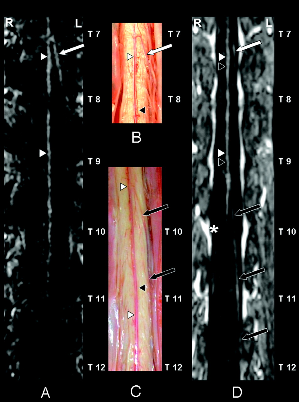
- Fig 1.
Coronal curved multiplanar reformation of the first dynamic phase of the CE-MRA examination (A), postmortem spinal cord specimens (B and C), and the second dynamic phase (D) of the anterior spinal cord surface. In the first (ie, arterial) dynamic phase (A) only the anterior spinal artery (ASA) (white arrowheads) and the AKA (white arrow) are depicted. The AKA derived from the left 8th thoracic segmental artery (SA) according to the CE-MRA (A) in agreement with the postmortem specimen (B). On the anterior cord surface of the postmortem specimens (B and C) both the ASA (white arrowheads) and anterior median vein (AMV) (black arrowheads) are visualized. Furthermore, the anterior radiculomedullary vein (black arrows) could be identified. This vein entered the epidural space at the left 12th thoracic vertebral level (C). Note the close anatomical relation (B and C) between the ASA (white arrowheads) and the AMV (black arrowheads), which explains that these vessels are not spatially resolved in the second-phase image (D). The second dynamic phase (D) shows a diminished signal intensity of the AKA (white arrow). In contrast to the first-phase angiogram (A), the anterior radiculomedullary vein (black arrows) is visualized and localized in the second-phase angiogram (D) and was in agreement with the postmortem specimen (C). In the midline the combination of the ASA (white arrowheads) and AMV (black arrowheads) are visualized. Note the strong enhancement of the epidural venous plexus (asterisk) in the second phase (D).
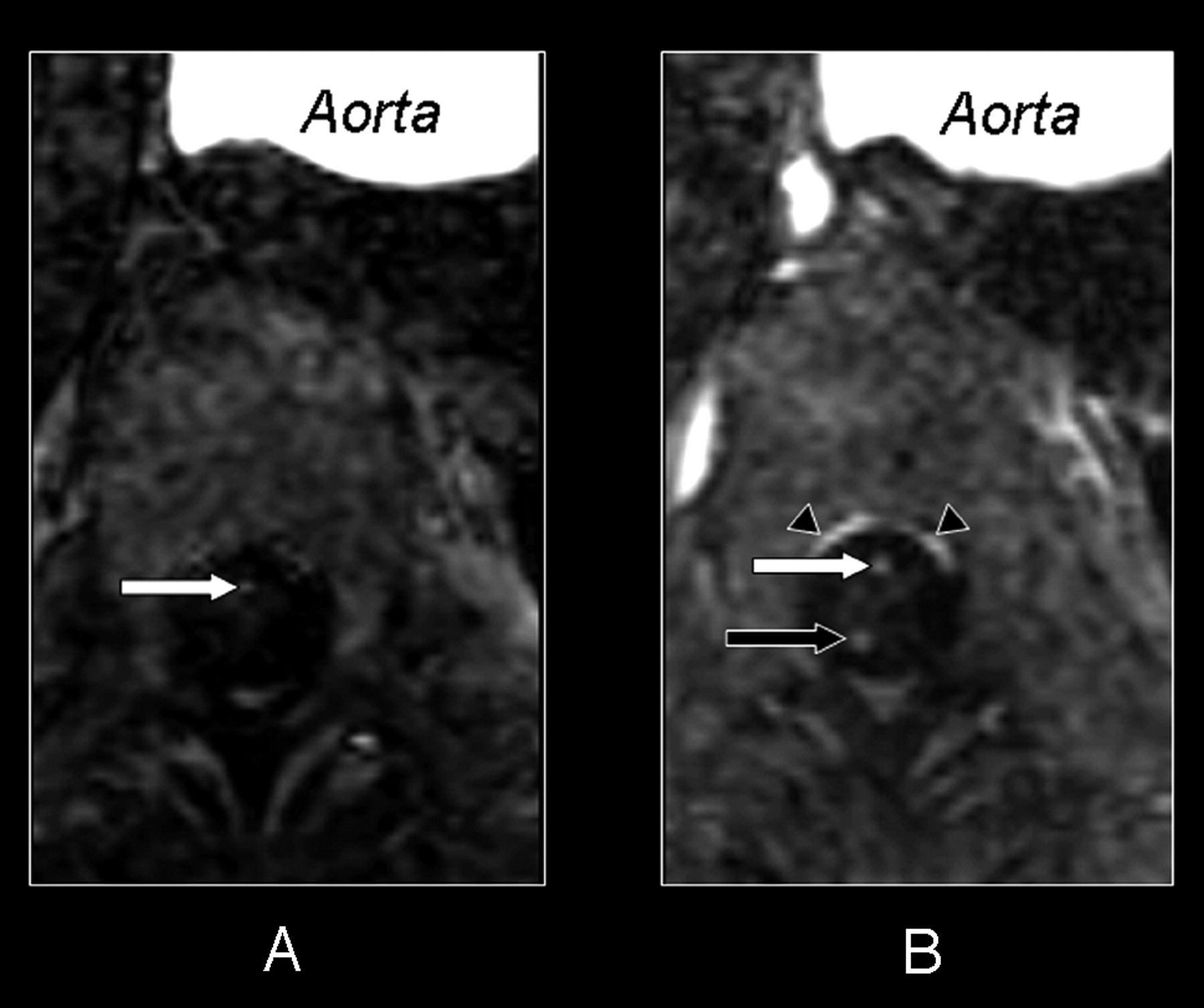
- Fig 2.
Axial section of the first (A) and second (B) dynamic phase of the CE-MRA examination. In both phases a cross-section at the 10th thoracic vertebral level shows the aorta and the intra- and extradural vessels. In the first phase (A) there is only enhancement on the midline of the anterior side of the spinal cord. This enhancement corresponds with the cross-section of the ASA (white arrow). The second phase (B) shows enhancement in the midline of both the anterior (white arrow) and posterior surface (black arrow) of the spinal cord. As there usually is only one vessel running along the midline of the posterior surface, the posterior median vein (black arrow), there must also be venous enhancement on the anterior surface. Therefore both the ASA and AMV are displayed on the midline of the anterior surface of the spinal cord in the second phase (B). Because of their close anatomic relation, no separation can be achieved between the ASA and AMV in the second phase (B). Again note the strong enhancement of the epidural venous plexus (black arrowheads) in the second phase (B).
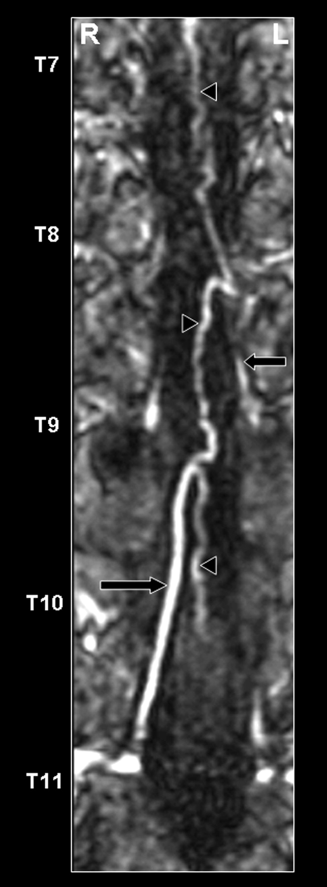
- Fig 3.
Coronal curved multiplanar reformation of the second dynamic phase of the CE-MRA examination of the posterior spinal cord surface. The posterior median vein (black arrowheads), as well as 2 draining posterior radiculomedullary veins (thoracic vertebral levels 9 and 11), are displayed (black arrows). The great radiculomedullary vein, which is defined as the largest of the radiculomedullary veins, entered the epidural space at the level of the 11th thoracic segmental vein on the right side.


In this issue


Jump to section
Related Articles
Cited By...
- No citing articles found.