
Article Figures & Data
Figures
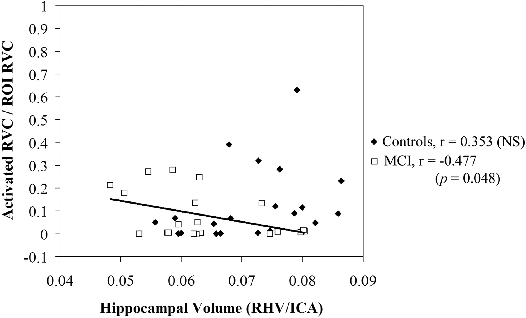
- Fig 2.
Correlation of right hippocampal activation in a template-space-based ROI with right hippocampal volume in subjects with MCI and control subjects during retrieval. Hippocampal volumes have been normalized by single-section intracranial area and hippocampal activation has been adjusted for subject age. Units of activation are represented as proportion of ROI activated. The line demonstrates a significant negative correlation between hippocampal volume and activation in the subjects with MCI.
- Fig 3.
Correlation of right hippocampal activation in a native-space-based manual ROI with right hippocampal volume in subjects with MCI and control subjects during retrieval. Hippocampal volumes have been normalized by single-section intracranial area and hippocampal activation has been adjusted for subject age. Units of activation are represented as proportion of ROI activated.


Tables
Characteristics Mild Cognitive Impairment (N = 20) Control (N = 20) Age, y (SD) 75.0 (7.6) 71.2 (4.5) Age range, y 55.5–83.3 63.3–80.5 Men/women 12/8 9/11 Education, y 15.0 (2.2) 15.9 (2.9) CVLT (SD) 5.0 (2.5) 11.0 2.6)* MMSE (SD) 26.7 (1.5) 28.4 (1.4)* LHV (as % of ICA) (SD) 6.19 (1.16) 7.29 (1.01)* RHV (as % of ICA) (SD) 6.40 (1.00) 7.16 (0.91)* Note:—Values shown are means (SD) unless otherwise noted. CVLT indicates delayed recall score on the California Verbal Learning Test-II; MMSE, Mini Mental State Examination; LHV, left hippocampal volume; RHV, right hippocampal volume; ICA, intracranial area.
* P < .05.
- Table 2:
Results from ANCOVA (with P values for independent variables included in the model)
ANCOVA Model Template-Based Analysis Native Subject Space with Manual ROI LH, encoding Group .699 .617 Age .616 .017 LHV/ICA .428 .490 Group X LHV/ICA .789 .700 LH, retrieval Group .591 .542 Age .271 .181 LHV/ICA .496 .619 Group X LHV/ICA .620 .593 RH, encoding Group .909 .724 Age .148 .070 RHV/ICA .731 .943 Group X RHV/ICA .108 .897 RH, retrieval Group .030* .694 Age .707 .828 RHV/ICA .812 .121 Group X RHV/ICA .020 .712 Note:—Dependent variable is proportion of activated voxels in hippocampal region of interest.
LH indicates left hippocampal activation; LHV/ICA, left hippocampal volume divided by single-section intracranial volume; RH, right hippocampal activation; RHV/ICA, right hippocampal volume divided by single-section intracranial volume; Group X LHV/ICA and Group X RHV/ICA, interaction term between group and hippocampal volume. Significant values of P are indicated in boldface type.
* Group term becomes nonsignificant (P = .314) when the interaction term, Group X RHV/ICA, is removed from the ANCOVA model.


