Advertisement

Main menu

  • Home
  • Content
    • Current Issue
    • Accepted Manuscripts
    • Article Preview
    • Past Issue Archive
    • Video Articles
    • AJNR Case Collection
    • Case of the Week Archive
    • Case of the Month Archive
    • Classic Case Archive
  • Special Collections
    • AJNR Awards
    • Low-Field MRI
    • Alzheimer Disease
    • ASNR Foundation Special Collection
    • Photon-Counting CT
    • View All
  • Multimedia
    • AJNR Podcasts
    • AJNR SCANtastic
    • Trainee Corner
    • MRI Safety Corner
    • Imaging Protocols
  • For Authors
    • Submit a Manuscript
    • Submit a Video Article
    • Submit an eLetter to the Editor/Response
    • Manuscript Submission Guidelines
    • Statistical Tips
    • Fast Publishing of Accepted Manuscripts
    • Graphical Abstract Preparation
    • Imaging Protocol Submission
    • Author Policies
  • About Us
    • About AJNR
    • Editorial Board
    • Editorial Board Alumni
  • More
    • Become a Reviewer/Academy of Reviewers
    • Subscribers
    • Permissions
    • Alerts
    • Feedback
    • Advertisers
    • ASNR Home

User menu

  • Alerts
  • Log in

Search

  • Advanced search
American Journal of Neuroradiology

American Journal of Neuroradiology

ASHNR American Society of Functional Neuroradiology ASHNR American Society of Pediatric Neuroradiology ASSR
  • Alerts
  • Log in

Advanced Search

  • Home
  • Content
    • Current Issue
    • Accepted Manuscripts
    • Article Preview
    • Past Issue Archive
    • Video Articles
    • AJNR Case Collection
    • Case of the Week Archive
    • Case of the Month Archive
    • Classic Case Archive
  • Special Collections
    • AJNR Awards
    • Low-Field MRI
    • Alzheimer Disease
    • ASNR Foundation Special Collection
    • Photon-Counting CT
    • View All
  • Multimedia
    • AJNR Podcasts
    • AJNR SCANtastic
    • Trainee Corner
    • MRI Safety Corner
    • Imaging Protocols
  • For Authors
    • Submit a Manuscript
    • Submit a Video Article
    • Submit an eLetter to the Editor/Response
    • Manuscript Submission Guidelines
    • Statistical Tips
    • Fast Publishing of Accepted Manuscripts
    • Graphical Abstract Preparation
    • Imaging Protocol Submission
    • Author Policies
  • About Us
    • About AJNR
    • Editorial Board
    • Editorial Board Alumni
  • More
    • Become a Reviewer/Academy of Reviewers
    • Subscribers
    • Permissions
    • Alerts
    • Feedback
    • Advertisers
    • ASNR Home
  • Follow AJNR on Twitter
  • Visit AJNR on Facebook
  • Follow AJNR on Instagram
  • Join AJNR on LinkedIn
  • RSS Feeds

AJNR Awards, New Junior Editors, and more. Read the latest AJNR updates

OtherHEAD & NECK

Temporal Giant Cell Reparative Granuloma: A Reappraisal of Pathology and Imaging Features

C. Reis, J.M. Lopes, E. Carneiro, A. Vilarinho, R. Portugal, F. Duarte and J. Fonseca
American Journal of Neuroradiology September 2006, 27 (8) 1660-1662;
C. Reis
  • Find this author on Google Scholar
  • Find this author on PubMed
  • Search for this author on this site
J.M. Lopes
  • Find this author on Google Scholar
  • Find this author on PubMed
  • Search for this author on this site
E. Carneiro
  • Find this author on Google Scholar
  • Find this author on PubMed
  • Search for this author on this site
A. Vilarinho
  • Find this author on Google Scholar
  • Find this author on PubMed
  • Search for this author on this site
R. Portugal
  • Find this author on Google Scholar
  • Find this author on PubMed
  • Search for this author on this site
F. Duarte
  • Find this author on Google Scholar
  • Find this author on PubMed
  • Search for this author on this site
J. Fonseca
  • Find this author on Google Scholar
  • Find this author on PubMed
  • Search for this author on this site
  • Article
  • Figures & Data
  • Info & Metrics
  • Responses
  • References
  • PDF
Loading

Abstract

SUMMARY: We report a case of large temporal giant cell reparative granuloma in a 72-year-old man. MR imaging depicted a right temporal expansile multiloculated lesion, with hyper- and hypointense signal areas on T2-weighted images, heterogeneously enhancing after gadolinium administration. Cortical thinning and bone remodeling of the temporal squamous portion were better seen on CT. The patient underwent surgery, and the diagnosis was achieved by the correlation of imaging, histologic, and laboratory findings.

The term “giant cell reparative granuloma” (GCRG) was introduced by Jaffe in 1953 as a benign lesion originating from an inflammatory response due to intraosseous hemorrhage following trauma.1,2 It is a non-neoplastic fibro-osseous lesion,1,3 mostly found in the mandible and maxilla.4–6 Temporal bone location is less frequent and was first described in 1974 by Hirschl and Katz,7 with only 21 cases reported since then in the literature, according to a Medline search by using the key words “GCRG” and “temporal.” We present a case of temporal GCRG and also review the imaging and histologic correlation, according to other reports.

Case Report

A 72-year-old man presented with a 1-year right progressive hypoacusia, dizziness, vertigo, and right temporal pain. He had previous episodes of repeated middle ear infections, mainly in the right side. There was no head trauma history.

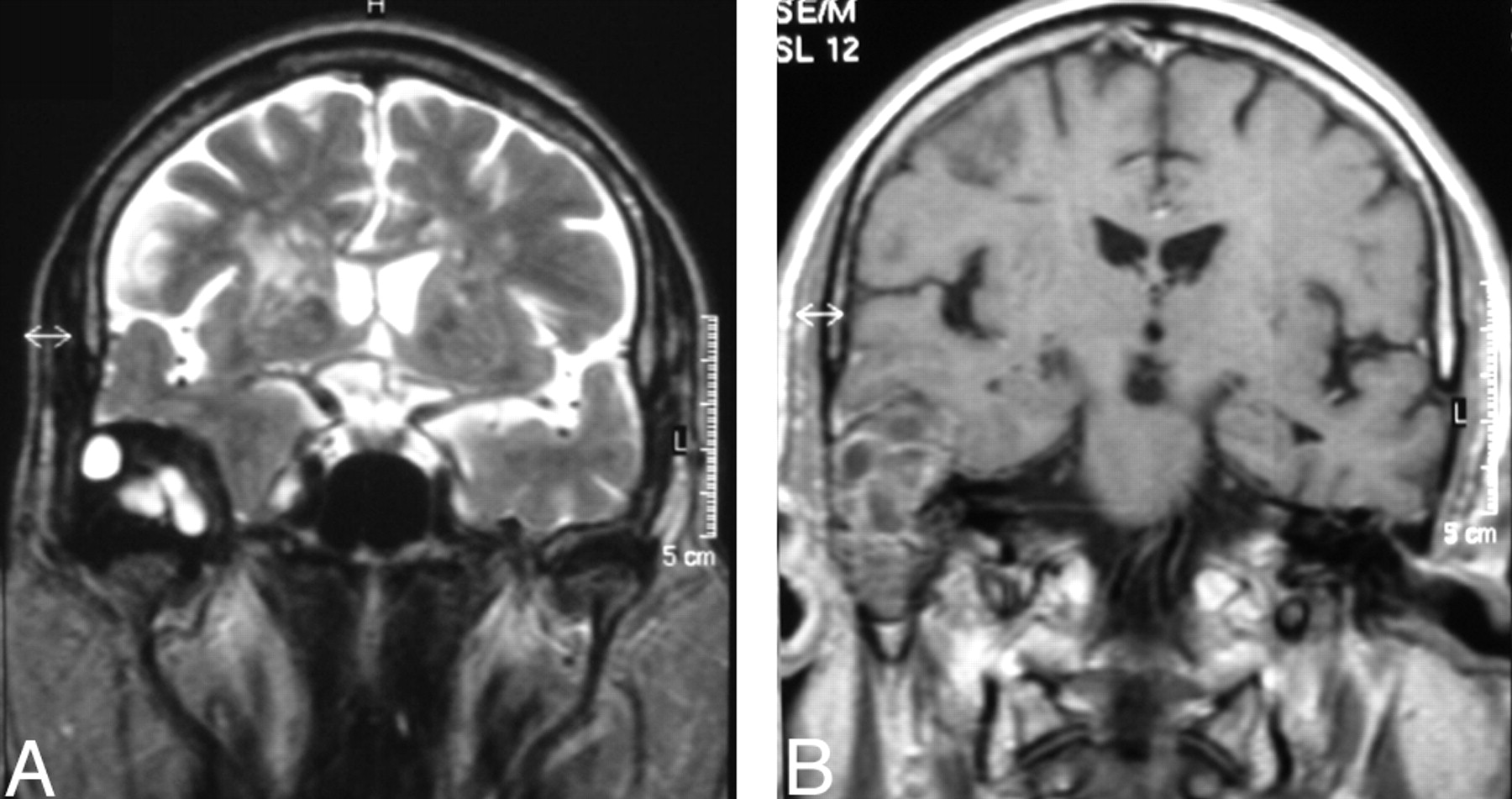
Head CT showed a large heterogeneous right temporal bone lesion (3.2 × 3.8 × 4.9 cm in greatest diameters), with cortical thinning and bone remodeling of the temporal squamous portion, along with lytic involvement of tympanic cavity, mastoid cells, and posterior wall of glenoid fossa; the lesion had a major intracranial component (Fig 1A, -B). Brain MR imaging disclosed a well-circumscribed round multiloculated right temporal extra-axial lesion. On T2-weighted sequences, it was mostly hyperintense with hypointense septations; after gadolinium administration, there was heterogeneous enhancement of the lesion and adjacent temporal dura. The lesion expanded mostly to the temporal fossa, with a slight mass effect on the temporal parenchyma but no edema signs, and, to a much lesser extent, to the glenoid fossa, with no evidence of condylar destruction (Fig 2A, -B).

Fig. 1.
  • Download figure
  • Open in new tab
  • Download powerpoint
Fig. 1.

A, Axial non-enhanced CT scan (soft-tissue algorithm) shows a heterogeneous right temporal bone lesion, with cortical thinning and bone remodeling of the temporal squamous portion and lytic destruction of middle ear (B, axial CT scan, bone algorithm).

Fig. 2.
  • Download figure
  • Open in new tab
  • Download powerpoint
Fig. 2.

A, Coronal T2-weighted image shows a right temporal extra-axial lesion, mostly hyperintense but with surrounding and septal areas of low signal intensity. Note a mild mass effect on the temporal parenchyma. There is no evidence of condyle destruction of the mandible. B, Contrast-enhanced coronal T1-weighted image shows the lesion occupying the right middle ear, which enhances heterogeneously after gadolinium. There is enhancement in the adjacent temporal dura.

These findings were consistent with a slow-growing expansive osseous lesion, with a myriad of possible diagnoses, including: benign epithelial and nonepithelial lesions, infectious and inflammatory lesions, or histiocytosis.8 Cholesterol granuloma was ruled out because of an absence of hyperintense areas on T1-weighted images.8 Sarcoma or metastasis was less likely because of the lack of rapid-growth imaging signs.

In August 2004, the patient was referred to the neurosurgery department for right temporal craniotomy and lesion excision (total removal, macroscopically) along with radical mastoidectomy. At surgery, the lesion was grayish, infiltrative, partially cystic, and calcified. There was dural (but no cortex) invasion, being replaced by Neuropatch (B. Braun Melsungen, Melsungen, Germany). After surgery, the patient had ipsilateral facial nerve palsy.

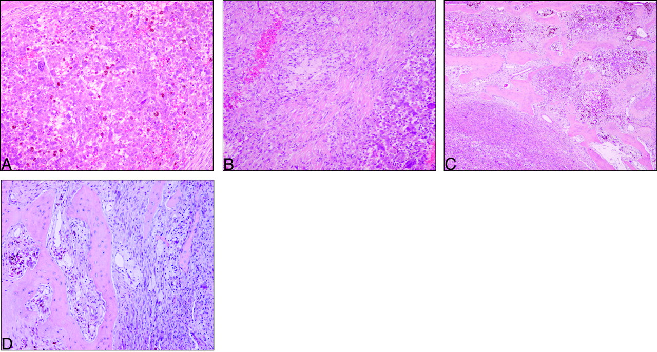
The entire surgical specimen was submitted for pathologic evaluation. Microscopically, the lesion was heterogeneous (Fig 3A–D) and composed of multinucleated osteoclast-like giant cells and spindle-shaped fibroblastic and polygonal xanthomatous cells in variable numbers. The mitotic index was low, necrosis was absent, and the lesion contained extensive areas of old hemorrhage and focal areas of neoformed osseous trabeculae at the advancing edge of the lesion. Tumor cells expressed vimentin and CD68 without immunoreactivity for CAM 5.2, AE1/3, CD31, S-100 protein, and HMB 45.

Fig. 3.
  • Download figure
  • Open in new tab
  • Download powerpoint
Fig. 3.

GCRG light microscopy discloses heterogeneous areas with multinucleated cells (A), alternating with spindle and xanthomatous cells (B). The lesion infiltrates the adjacent bone (C), both depicting similar heterogeneous features (D). Note signs of recent (B) and old hemorrhage (A, C, D).

Subsequent MR imaging revealed a small tumor remaining around the right condyle.

The patient had temporal pain relief after lesion removal and participated in an outpatient rehabilitation program, resulting in progressive recovery of facial motor deficits.

Discussion

The pathologic features of the lesion narrowed down the several diagnostic possibilities to the following: tenosynovial giant cell tumor of temporomandibular joint, brown tumor associated with hyperparathyroidism, GCRG, and giant cell tumor (GCT). Tenosynovial GCT of the temporomandibular joint was excluded by lesion topography on MR images: The lesion abutted the joint focally instead of originating from it. Brown tumor associated with hyperparathyroidism was excluded by laboratory studies disclosing normal serum calcium, phosphorous, and parathormone levels. Paget disease, frequently associated with GCT, was less likely because of the absence of a diffuse bone thickening appearance with mixed densities.8,9

The most challenging differential diagnosis encompasses GCT versus GCRG, due to their histologic resemblances.4 Nonetheless, the presence of giant cell osteoclast-like low-mitotic-rate signs of osteoid and new bone production and extensive hemorrhage with hemosiderin deposits favored GCRG.10

GCRG is a rare lesion,1,5,11 thought to be related to previous trauma or inflammation,12 but its pathogenesis remains unknown.1,13 GCRG mostly affects children, young adults, and women.4,11 However, according to GCRG cases reported in temporal locations, sex and age distributions generally differ from those in other locations and trauma is less frequent (Table).

Author(s)Age (y)/SexTraumaInitial Symptoms
Hirschl and Katz, 1974736/FYesHearing loss
Colclasure et al, 19812210/MNRHearing loss
22/MNRHearing loss and mass
Tesluk et al, 19892356/MNoMass, pain
Ciappetta et al, 19902425/MYesDysphagia, pain, protrusion
Cohen and Granada-Ricart, 1993254m/FNoMass
Lewis et al, 19941932/FYesHearing loss
Nemoto et al, 19951536/MNoHearing loss, tinnitus
28/MNoHearing loss, tinnitus
Maruno et al, 1997143/FNoFacial weakness, hearing loss
Ung et al, 19982636/FNoHearing loss
Liu et al, 20011044/MNoHearing loss, tinnitus
72/MNoHearing loss
Khodaei et al, 20011736/MNRPain, tinnitus, vertigo
Sharma et al, 20021812/MYesFacial weakness, hearing loss, mass, tinnitus, vertigo
Matsui et al, 20022741/MNRMass
Yoshimura et al, 20021638/MNRNR
Boedeker et al, 2003117/FNoHearing loss
Mohammed et al, 2001638/MNRHearing loss, bilateral headache
Montero et al, 2003460/MNRHearing loss
Kim et al, 2003550/MNoHearing loss, tinnitus
  • Note:—NR indicates not reported.

  • Temporal GCRG case reports

    In the Table, 10 of the 21 cases (47.5%) were not related to trauma. Only 4 patients had positive trauma history. Ages ranged from 4 months to 72 years, with a mean of 32 years. There was a predominance of 15 men versus 6 women. The most frequent associated symptom was hearing loss.

    In the available MR imaging findings of the reported cases mentioned in the Table,1,5,6,10,14–19 the lesions always had a hypointense component in long repetition time (TR) sequences and heterogeneous enhancement, although a multiloculated appearance similar to that in our case report was only described by Mohammed et al,6 Nemoto et al,15 and Sharma et al.18 The hypointense areas on long TR sequences probably reflected hemosiderin and bone formation.

    The first temporal GRCG case was described by Hirschl and Katz7; they reviewed 23 cases initially diagnosed as GCT and concluded that 18 were GCRG. GCRG is considered a benign lesion6 that presents as a mass with cortical thinning20 and potential local aggressiveness.1 When localized in the temporal region, associated symptoms include hearing loss, tinnitus, pain, vertigo, and facial palsy.1

    Some authors regard GCRG as the last phase of aneurysmal bone cyst and consider theses lesions as solid aneurysmal bone cyst variants,21 because of the overlap of clinical and histologic features shared by GCRG and aneurysmal bone cyst.21 Despite these histologic similarities, GCRG has no large blood-filled spaces, a finding that is at variance with aneurysmal bone cyst.21

    After complete removal of GCRG, prognosis is good, with a 10%–15% rate of recurrence.1 If the removal is partial, the recurrence rate is higher.1 Radiation therapy could be an alternative treatment, but the risk of sarcomatous degeneration makes radiation therapy controversial in this setting.1,5

    Conclusion

    GCRG is rare, especially in the temporal location. The differential diagnosis with GCT is of the utmost importance to establish a suitable follow-up and treatment plan. There are no specific imaging features for the diagnosis of GCRG; thus, it relies on close correlation with pathology and clinical and laboratory data. Additionally, we emphasize the absence of trauma history in our patient, in accordance to recent reports of GCRG.

    References

    1. ↵
      Boedeker CC, Kayser G, Ridder GJ, et al. Giant-cell reparative granuloma of the temporal bone: a case report and review of the literature. Ear Nose Throat J 2003;82:926–29, 33–34, 36–37
      PubMed
    2. ↵
      Arda HN, Karakus MF, Ozcan M, et al. Giant cell reparative granuloma originating from the ethmoid sinus. Int J Pediatr Otorhinolaryngol 2003;67:83–87
      PubMed
    3. ↵
      Morris JM, Lane JI, Witte RJ, et al. Giant cell reparative granuloma of the nasal cavity. AJNR Am J Neuroradiol 2004;25:1263–65
      Abstract/FREE Full Text
    4. ↵
      Montero EH, Navarro JS, Pueyo JL, et al. Giant-cell reparative granuloma in temporal bone. Am J Otolaryngol 2003;24:191–93
      PubMed
    5. ↵
      Kim HJ, Lee HK, Suh DC, et al. Giant cell reparative granuloma of the temporal bone: MR findings with pathologic correlation. AJNR Am J Neuroradiol 2003;24:1136–38
      Abstract/FREE Full Text
    6. ↵
      Mohammed TL, Brummett DP, Hahn FJ, et al. Intracranial giant cell reparative granuloma arising from the temporal bone. AJNR Am J Neuroradiol 2001;22:873–75
      Abstract/FREE Full Text
    7. ↵
      Hirschl S, Katz A. Giant cell reparative granuloma outside the jaw bone: diagnostic criteria and review of the literature with the first case described in the temporal bone. Hum Pathol 1974;5:171–81
      CrossRefPubMed
    8. ↵
      Sakai D, Curtin HD, Hasso AN, et al. Otosclerosis and dysplasias of the temporal bone. In: Som PM, Curtin HD, eds. Head and Neck Imaging. 4th ed. St. Louis: Mosby;2003 :1256–59, 1339
    9. ↵
      Swartz JD, Harnsberger HR. The otic capsule and otodystrophies.In: Imaging of the Temporal Bone. 3rd ed. New York: Thieme Medical Publications;1998 :288–90
    10. ↵
      Liu J, Zhong D-R, Liu L-F, et al. Giant cell reparative granuloma of the temporal bone. Acta Otolaryngol 2001;121:523–28
      PubMed
    11. ↵
      Uchino A, Kato A, Yonemitsu N, et al. Giant cell reparative granuloma of the cranial vault. AJNR Am J Neuroradiol 1996;17:1791–93
      Abstract
    12. ↵
      Ustundag E, Iseri M, Keskin G, et al. Central giant cell granuloma. Int J Pediatr Otorhinolaryngol 2002;65:143–46
      PubMed
    13. ↵
      Rosai J. Central giant cell granuloma and other giant cell-containing lesions. In: Rosai and Ackerman’s Surgical Pathology. 9th ed. Toronto, Ontario: Mosby;2004 :281–82
    14. ↵
      Maruno M, Yoshimine T, Kubo T, et al. A case of giant cell reparative granuloma of the petrous bone: demonstration of the proliferative component. Surg Neurol 1997;48:64–68
      PubMed
    15. ↵
      Nemoto Y, Inoue Y, Tashiro T, et al. Central giant cell granuloma of the temporal bone. AJNR Am J Neuroradiol 1995;16(4 suppl):982–85
    16. Yoshimura J, Onda K, Tanaka R, et al. Giant cell reparative granuloma of the temporal bone: neuroradiological and immunohistochemical findings. Neurol Med Chir (Tokyo) 2002;42:510–15
      PubMed
    17. Khodaei I, Rowley H, Farrell MA, et al. An unusual cause for tinnitus. Ir Med J 2001;94:312–13
      PubMed
    18. ↵
      Sharma RR, Verma A, Pawar SJ, et al. Pediatric giant cell granuloma of the temporal bone: a case report and brief review of the literature. J Clin Neurosci 2002;9:459–62
      CrossRefPubMed
    19. ↵
      Lewis ML, Weber AL, McKenna MJ. Reparative cell granuloma of the temporal bone. Ann Otol Rhinol Laryngol 1994;103:826–28
      FREE Full Text
    20. ↵
      Felsberg GJ, Tien RD, McLendon RE. Frontoethmoidal giant cell reparative granuloma. AJNR Am J Neuroradiol 1995;16:1551–54
      Abstract
    21. ↵
      Oda Y, Tsuneyoshi M, Shinohara N. “Solid” variant of aneurysmal bone cyst (extragnathic giant cell reparative granuloma) in the axial skeleton and long bones. Cancer 1992;70:2642–49
      CrossRefPubMed
    22. Colclasure JB, Shea MC Jr, Graham SS. Giant cell lesions of the temporal bone. Am J Otol 1981;2:188–92.
      CrossRefPubMed
    23. Tesluk H, Senders CW, Dublin AB. Case report 562: Giant cell reparative granuloma of temporal bone. Skeletal Radiol 1989;18:599–602
      CrossRefPubMed
    24. Ciappetta P, Salvati M, Bernardi C, et al. Giant cell reparative granuloma of the skull base mimicking an intracranial tumor. Case report and review of the literature. Surg Neurol 1990;33:52–56
      CrossRefPubMed
    25. Cohen D, Granda-Ricart MC. Giant cell reparative granuloma of the base of the skull in a 4-month-old infant—CT findings. Pediatr Radiol 1993;23:319–20
      CrossRefPubMed
    26. Ung F, Li KK, Keith DA, et al. Giant cell reparative granuloma of the temporal bone: case report and review of the literature. Otolaryngol Head Neck Surg 1998;118:525–29.
      CrossRefPubMed
    27. Matsui T, Iwamuro K, Ishikawa T, et al. Large giant cell reparative granuloma of the petrous bone—case report. Neurol Med Chir (Tokyo) 2002;42:232–36
      PubMed
    • Received July 19, 2005.
    • Accepted after revision September 30, 2005.
    • Copyright © American Society of Neuroradiology
    View Abstract
    PreviousNext
    Back to top

    In this issue

    American Journal of Neuroradiology: 27 (8)
    American Journal of Neuroradiology
    Vol. 27, Issue 8
    September 2006
    • Table of Contents
    • Index by author
    Advertisement
    Print
    Download PDF
    Email Article

    Thank you for your interest in spreading the word on American Journal of Neuroradiology.

    NOTE: We only request your email address so that the person you are recommending the page to knows that you wanted them to see it, and that it is not junk mail. We do not capture any email address.

    Enter multiple addresses on separate lines or separate them with commas.
    Temporal Giant Cell Reparative Granuloma: A Reappraisal of Pathology and Imaging Features
    (Your Name) has sent you a message from American Journal of Neuroradiology
    (Your Name) thought you would like to see the American Journal of Neuroradiology web site.
    CAPTCHA
    This question is for testing whether or not you are a human visitor and to prevent automated spam submissions.
    Cite this article
    C. Reis, J.M. Lopes, E. Carneiro, A. Vilarinho, R. Portugal, F. Duarte, J. Fonseca
    Temporal Giant Cell Reparative Granuloma: A Reappraisal of Pathology and Imaging Features
    American Journal of Neuroradiology Sep 2006, 27 (8) 1660-1662;

    Citation Manager Formats

    • BibTeX
    • Bookends
    • EasyBib
    • EndNote (tagged)
    • EndNote 8 (xml)
    • Medlars
    • Mendeley
    • Papers
    • RefWorks Tagged
    • Ref Manager
    • RIS
    • Zotero
    0 Responses
    Respond to this article
    Share
    Bookmark this article
    Temporal Giant Cell Reparative Granuloma: A Reappraisal of Pathology and Imaging Features
    C. Reis, J.M. Lopes, E. Carneiro, A. Vilarinho, R. Portugal, F. Duarte, J. Fonseca
    American Journal of Neuroradiology Sep 2006, 27 (8) 1660-1662;
    del.icio.us logo Twitter logo Facebook logo Mendeley logo
    • Tweet Widget
    • Facebook Like
    • Google Plus One

    Jump to section

    • Article
      • Abstract
      • Case Report
      • Discussion
      • Conclusion
      • References
    • Figures & Data
    • Info & Metrics
    • Responses
    • References
    • PDF

    Related Articles

    • No related articles found.
    • PubMed
    • Google Scholar

    Cited By...

    • No citing articles found.
    • Crossref
    • Google Scholar

    This article has not yet been cited by articles in journals that are participating in Crossref Cited-by Linking.

    More in this TOC Section

    • Revisiting CT Signs of Unilateral Vocal Fold Paralysis: A Single, Blinded Study
    • COVID-19–Associated Bifacial Weakness with Paresthesia Subtype of Guillain-Barré Syndrome
    • MRI Findings of Immune Checkpoint Inhibitor–Induced Hypophysitis: Possible Association with Fibrosis
    Show more Head & Neck

    Similar Articles

    Advertisement

    Indexed Content

    • Current Issue
    • Accepted Manuscripts
    • Article Preview
    • Past Issues
    • Editorials
    • Editor's Choice
    • Fellows' Journal Club
    • Letters to the Editor
    • Video Articles

    Cases

    • Case Collection
    • Archive - Case of the Week
    • Archive - Case of the Month
    • Archive - Classic Case

    More from AJNR

    • Trainee Corner
    • Imaging Protocols
    • MRI Safety Corner
    • Book Reviews

    Multimedia

    • AJNR Podcasts
    • AJNR Scantastics

    Resources

    • Turnaround Time
    • Submit a Manuscript
    • Submit a Video Article
    • Submit an eLetter to the Editor/Response
    • Manuscript Submission Guidelines
    • Statistical Tips
    • Fast Publishing of Accepted Manuscripts
    • Graphical Abstract Preparation
    • Imaging Protocol Submission
    • Evidence-Based Medicine Level Guide
    • Publishing Checklists
    • Author Policies
    • Become a Reviewer/Academy of Reviewers
    • News and Updates

    About Us

    • About AJNR
    • Editorial Board
    • Editorial Board Alumni
    • Alerts
    • Permissions
    • Not an AJNR Subscriber? Join Now
    • Advertise with Us
    • Librarian Resources
    • Feedback
    • Terms and Conditions
    • AJNR Editorial Board Alumni

    American Society of Neuroradiology

    • Not an ASNR Member? Join Now

    © 2025 by the American Society of Neuroradiology All rights, including for text and data mining, AI training, and similar technologies, are reserved.
    Print ISSN: 0195-6108 Online ISSN: 1936-959X

    Powered by HighWire