Advertisement

Main menu

  • Home
  • Content
    • Current Issue
    • Accepted Manuscripts
    • Article Preview
    • Past Issue Archive
    • Video Articles
    • AJNR Case Collection
    • Case of the Week Archive
    • Case of the Month Archive
    • Classic Case Archive
  • Special Collections
    • AJNR Awards
    • Low-Field MRI
    • Alzheimer Disease
    • ASNR Foundation Special Collection
    • Photon-Counting CT
    • View All
  • Multimedia
    • AJNR Podcasts
    • AJNR SCANtastic
    • Trainee Corner
    • MRI Safety Corner
    • Imaging Protocols
  • For Authors
    • Submit a Manuscript
    • Submit a Video Article
    • Submit an eLetter to the Editor/Response
    • Manuscript Submission Guidelines
    • Statistical Tips
    • Fast Publishing of Accepted Manuscripts
    • Graphical Abstract Preparation
    • Imaging Protocol Submission
    • Author Policies
  • About Us
    • About AJNR
    • Editorial Board
    • Editorial Board Alumni
  • More
    • Become a Reviewer/Academy of Reviewers
    • Subscribers
    • Permissions
    • Alerts
    • Feedback
    • Advertisers
    • ASNR Home

User menu

  • Alerts
  • Log in

Search

  • Advanced search
American Journal of Neuroradiology

American Journal of Neuroradiology

ASHNR American Society of Functional Neuroradiology ASHNR American Society of Pediatric Neuroradiology ASSR
  • Alerts
  • Log in

Advanced Search

  • Home
  • Content
    • Current Issue
    • Accepted Manuscripts
    • Article Preview
    • Past Issue Archive
    • Video Articles
    • AJNR Case Collection
    • Case of the Week Archive
    • Case of the Month Archive
    • Classic Case Archive
  • Special Collections
    • AJNR Awards
    • Low-Field MRI
    • Alzheimer Disease
    • ASNR Foundation Special Collection
    • Photon-Counting CT
    • View All
  • Multimedia
    • AJNR Podcasts
    • AJNR SCANtastic
    • Trainee Corner
    • MRI Safety Corner
    • Imaging Protocols
  • For Authors
    • Submit a Manuscript
    • Submit a Video Article
    • Submit an eLetter to the Editor/Response
    • Manuscript Submission Guidelines
    • Statistical Tips
    • Fast Publishing of Accepted Manuscripts
    • Graphical Abstract Preparation
    • Imaging Protocol Submission
    • Author Policies
  • About Us
    • About AJNR
    • Editorial Board
    • Editorial Board Alumni
  • More
    • Become a Reviewer/Academy of Reviewers
    • Subscribers
    • Permissions
    • Alerts
    • Feedback
    • Advertisers
    • ASNR Home
  • Follow AJNR on Twitter
  • Visit AJNR on Facebook
  • Follow AJNR on Instagram
  • Join AJNR on LinkedIn
  • RSS Feeds

AJNR Awards, New Junior Editors, and more. Read the latest AJNR updates

OtherNeurointervention

Carotid-compression Technique for the Insertion of Guiding Catheters

S. Yoshimura, Y. Enomoto, H. Kitajima, J. Yamada, Y. Kaku and T. Iwama
American Journal of Neuroradiology September 2006, 27 (8) 1710-1711;
S. Yoshimura
  • Find this author on Google Scholar
  • Find this author on PubMed
  • Search for this author on this site
Y. Enomoto
  • Find this author on Google Scholar
  • Find this author on PubMed
  • Search for this author on this site
H. Kitajima
  • Find this author on Google Scholar
  • Find this author on PubMed
  • Search for this author on this site
J. Yamada
  • Find this author on Google Scholar
  • Find this author on PubMed
  • Search for this author on this site
Y. Kaku
  • Find this author on Google Scholar
  • Find this author on PubMed
  • Search for this author on this site
T. Iwama
  • Find this author on Google Scholar
  • Find this author on PubMed
  • Search for this author on this site
  • Article
  • Figures & Data
  • Info & Metrics
  • Responses
  • References
  • PDF
Loading

Abstract

SUMMARY: Inserting a guiding catheter into a tortuous artery for neurointerventional procedures can be difficult. In our technique, the carotid artery is manually compressed to stabilize and/or straighten the inserted wire before advancing the guiding catheter. Although this technique is not risk-free and care must be taken to avoid vascular injury by excessive compression, it is useful for the insertion of a guiding catheter into the carotid artery.

The insertion of a guiding catheter system into the desired arterial site is the first, and one of the most important, steps for successful endovascular treatment. Here we present our “carotid-compression technique” that facilitates insertion of the guiding catheter into the tortuous carotid artery.

Technical Considerations

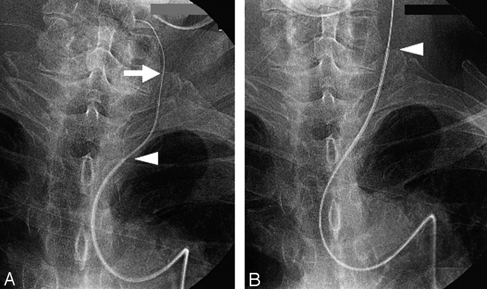
In patients with severe atherosclerosis, the insertion of a catheter, especially a guiding catheter, into the common carotid artery (CCA) can be difficult. Anatomic factors such as a long distance between the origin of the carotid artery and the top of the ascending aorta and sharpness of the angle formed by the CCA and the aorta contribute to this difficulty. Under such conditions, as a first step, a catheter and/or guidewire with a different angle or rigidity must be introduced. Even when the guidewire can be inserted into the CCA, however, its tortuosity may prohibit the introduction of the guiding catheter along the guidewire. In such cases, we manually compress the CCA to stabilize the wire in the artery and then insert the guiding catheter along the wire (Fig 1). When a coaxial catheter system is used, the inner catheter can be compressed for stabilization, and the guiding catheter can then be inserted along the inner catheter. This technique can be used in both conventional angiographic and neurointerventional procedures.

Fig 1.
  • Download figure
  • Open in new tab
  • Download powerpoint
Fig 1.

Carotid-compression technique I. A, After insertion of the guidewire, the CCA is manually compressed (arrow). B, The inner catheter (Glidecath, Terumo, Tokyo, Japan) is first inserted along the guidewire stabilized by manual compression of the CCA. The guiding catheter (Guider, Boston-Scientific, Natick, Mass) is then inserted along the inner catheter. Arrowheads indicate the tip of the guiding catheter.

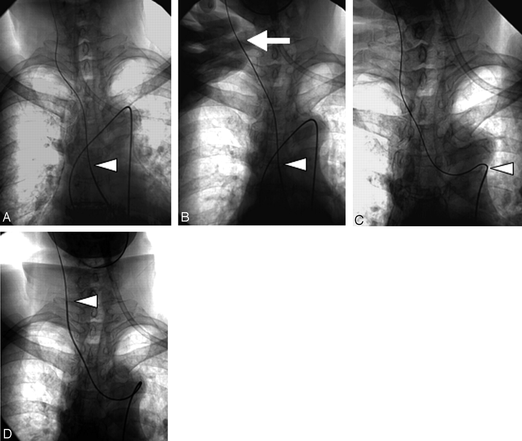
In cases where insertion of the guidewire is impossible, different approaches such as the transbrachial- and transcarotid artery approach can be taken. If the femoral approach is considered optimal, the “turn-over technique” can be used to insert the guidewire into the CCA.1,2 In patients undergoing neurointerventional procedures, the inserted guidewire must be straightened. Otherwise, the inserted guiding catheter moves into the left ventricle over the aortic valve. It is rarely possible to straighten the catheter by simply pulling on the tail of the guidewire, because this maneuver risks withdrawal of the inserted guidewire from the CCA. If the CCA is compressed manually, however, the stabilized guidewire can be straightened with the coaxial catheter by pulling on its tail regardless of the presence or absence of an inner catheter (Fig 2). The catheter can then be inserted into the CCA along the straightened wire and the manual compression is released. The wire and catheter are subsequently advanced as far as necessary in the internal or external carotid artery.

Fig 2.
  • Download figure
  • Open in new tab
  • Download powerpoint
Fig 2.

Carotid-compression technique II. A, The coaxial system (Shuttle, Cook/Glidecath, Terumo, Bloomington, Ind) is inserted into the ascending aorta, and the guidewire and inner catheter are then introduced into the target CCA or ICA by using the turn-over technique. B, The CCA is then compressed manually (arrow) to stabilize the guidewire and inner catheter. C, Withdrawal of the system toward the femoral artery results in its straightening. D, The guiding catheter is then advanced along the inner catheter. Arrowheads indicate the tip of the guiding catheter.

Results and Discussion

Between April 2003 and March 2005, 303 patients with cerebrovascular disease were referred to our institution for endovascular treatment. In 12 of them, regular catheterization was not possible and they (8 men and 4 women, ranging in age from 58 to 89 years; mean age, 74.6 years) underwent our carotid-compression technique. We successfully inserted the guiding catheter in 10 of these 12 patients. In one patient we could not insert the guiding catheter into the CCA because the guidewire slipped down despite sufficient compression of the vessel. In the other patient, the guiding catheter was inserted into the CCA, but it developed a kink because the angle between the origin of the CCA and the aorta was very acute. None of the 12 patients experienced clinical complications, and subsequent procedures were carried out uneventfully.

Our “carotid-compression technique” requires neither special catheters or guidewires nor the puncture of other vessels and is less risky than direct puncture of the carotid artery. Nevertheless, it is not risk-free, because excessive compression of the CCA may result in vessel injury. Therefore, other techniques (eg, distal guidewire insertion, compression of the wire inserted into the occipital artery, the coaxial technique using the microcatheter system, and use of a Simmons-type guide catheter) should be considered before attempting our technique. In addition, before undertaking our procedure, the degree of atherosclerosis in the target carotid artery should be assessed by preoperative MR/CT angiography or by angiography following the injection of contrast material into the origin of the CCA. Braided, nonkinking catheters may yield the best results. Hydrophilic-coated guidewires are sometimes unsuitable because they tend to slip during insertion of the guiding catheter. Our technique can also be used with the transbrachial artery approach.

In patients subjected to neurointerventional procedures, excessive manipulation of the target artery in the course of attempted catheter insertion can result in thromboembolic complications. Thromboemboli after coil embolization3,4 and conventional cerebral angiography have been reported.5,6 Our carotid-compression technique is a simple and relatively safe option in patients who require neurointerventional procedures but whose vascular anatomy is tortuous.

Acknowledgments

We thank Ms. U. Petralia for editing this manuscript.

References

  1. ↵
    Touho H, Karasawa J, Ohnishi H, et al. Transbrachial approach with turn over technique for selective cerebral angiography: technical note. Neurol Med Chir (Tokyo) 1993;33:716–18
    PubMed
  2. ↵
    Touho H, Karasawa J, Ohnishi H, et al. The “turn-over” technique for selective cerebral angiography. Neuroradiology 1994;36:123–24
    PubMed
  3. ↵
    Rordorf G, Bellon RJ, Budzik RE Jr, et al. Silent thromboembolic events associated with the treatment of unruptured cerebral aneurysms by use of Guglielmi detachable coils: prospective study applying diffusion-weighted imaging. AJNR Am J Neuroradiol 2001;22:5–10
    Abstract/FREE Full Text
  4. ↵
    Soeda A, Sakai N, Murao K, et al. Thromboembolic events associated with Guglielmi detachable coil embolization of asymptomatic cerebral aneurysms: evaluation of 66 consecutive cases with use of diffusion-weighted MR imaging. AJNR Am J Neuroradiol 2003;24:127–32
    Abstract/FREE Full Text
  5. ↵
    Heiserman JE. Silent embolism after cerebral angiography: what harm? Lancet 1999;354:1577–78
    PubMed
  6. ↵
    Chuah KC, Stuckey SL, Berman IG. Silent embolism in diagnostic cerebral angiography: detection with diffusion-weighted imaging. Austral Radiol 2004;48:133–38
    CrossRefPubMed
  • Received December 29, 2005.
  • Accepted after revision April 3, 2006.
  • Copyright © American Society of Neuroradiology
View Abstract
PreviousNext
Back to top

In this issue

American Journal of Neuroradiology: 27 (8)
American Journal of Neuroradiology
Vol. 27, Issue 8
September 2006
  • Table of Contents
  • Index by author
Advertisement
Print
Download PDF
Email Article

Thank you for your interest in spreading the word on American Journal of Neuroradiology.

NOTE: We only request your email address so that the person you are recommending the page to knows that you wanted them to see it, and that it is not junk mail. We do not capture any email address.

Enter multiple addresses on separate lines or separate them with commas.
Carotid-compression Technique for the Insertion of Guiding Catheters
(Your Name) has sent you a message from American Journal of Neuroradiology
(Your Name) thought you would like to see the American Journal of Neuroradiology web site.
CAPTCHA
This question is for testing whether or not you are a human visitor and to prevent automated spam submissions.
Cite this article
S. Yoshimura, Y. Enomoto, H. Kitajima, J. Yamada, Y. Kaku, T. Iwama
Carotid-compression Technique for the Insertion of Guiding Catheters
American Journal of Neuroradiology Sep 2006, 27 (8) 1710-1711;

Citation Manager Formats

  • BibTeX
  • Bookends
  • EasyBib
  • EndNote (tagged)
  • EndNote 8 (xml)
  • Medlars
  • Mendeley
  • Papers
  • RefWorks Tagged
  • Ref Manager
  • RIS
  • Zotero
0 Responses
Respond to this article
Share
Bookmark this article
Carotid-compression Technique for the Insertion of Guiding Catheters
S. Yoshimura, Y. Enomoto, H. Kitajima, J. Yamada, Y. Kaku, T. Iwama
American Journal of Neuroradiology Sep 2006, 27 (8) 1710-1711;
del.icio.us logo Twitter logo Facebook logo Mendeley logo
  • Tweet Widget
  • Facebook Like
  • Google Plus One

Jump to section

  • Article
    • Abstract
    • Technical Considerations
    • Results and Discussion
    • Acknowledgments
    • References
  • Figures & Data
  • Info & Metrics
  • Responses
  • References
  • PDF

Related Articles

  • No related articles found.
  • PubMed
  • Google Scholar

Cited By...

  • No citing articles found.
  • Crossref
  • Google Scholar

This article has not yet been cited by articles in journals that are participating in Crossref Cited-by Linking.

More in this TOC Section

  • A Retrospective Study in Tentorial DAVFs
  • Proximal Protection Devices for Carotid Stenting
  • Effect of SARS-CoV2 on Endovascular Thrombectomy
Show more NEUROINTERVENTION

Similar Articles

Advertisement

Indexed Content

  • Current Issue
  • Accepted Manuscripts
  • Article Preview
  • Past Issues
  • Editorials
  • Editor's Choice
  • Fellows' Journal Club
  • Letters to the Editor
  • Video Articles

Cases

  • Case Collection
  • Archive - Case of the Week
  • Archive - Case of the Month
  • Archive - Classic Case

More from AJNR

  • Trainee Corner
  • Imaging Protocols
  • MRI Safety Corner
  • Book Reviews

Multimedia

  • AJNR Podcasts
  • AJNR Scantastics

Resources

  • Turnaround Time
  • Submit a Manuscript
  • Submit a Video Article
  • Submit an eLetter to the Editor/Response
  • Manuscript Submission Guidelines
  • Statistical Tips
  • Fast Publishing of Accepted Manuscripts
  • Graphical Abstract Preparation
  • Imaging Protocol Submission
  • Evidence-Based Medicine Level Guide
  • Publishing Checklists
  • Author Policies
  • Become a Reviewer/Academy of Reviewers
  • News and Updates

About Us

  • About AJNR
  • Editorial Board
  • Editorial Board Alumni
  • Alerts
  • Permissions
  • Not an AJNR Subscriber? Join Now
  • Advertise with Us
  • Librarian Resources
  • Feedback
  • Terms and Conditions
  • AJNR Editorial Board Alumni

American Society of Neuroradiology

  • Not an ASNR Member? Join Now

© 2025 by the American Society of Neuroradiology All rights, including for text and data mining, AI training, and similar technologies, are reserved.
Print ISSN: 0195-6108 Online ISSN: 1936-959X

Powered by HighWire