Advertisement

Main menu

  • Home
  • Content
    • Current Issue
    • Accepted Manuscripts
    • Article Preview
    • Past Issue Archive
    • Video Articles
    • AJNR Case Collection
    • Case of the Week Archive
    • Case of the Month Archive
    • Classic Case Archive
  • Special Collections
    • AJNR Awards
    • Low-Field MRI
    • Alzheimer Disease
    • ASNR Foundation Special Collection
    • Photon-Counting CT
    • View All
  • Multimedia
    • AJNR Podcasts
    • AJNR SCANtastic
    • Trainee Corner
    • MRI Safety Corner
    • Imaging Protocols
  • For Authors
    • Submit a Manuscript
    • Submit a Video Article
    • Submit an eLetter to the Editor/Response
    • Manuscript Submission Guidelines
    • Statistical Tips
    • Fast Publishing of Accepted Manuscripts
    • Graphical Abstract Preparation
    • Imaging Protocol Submission
    • Author Policies
  • About Us
    • About AJNR
    • Editorial Board
    • Editorial Board Alumni
  • More
    • Become a Reviewer/Academy of Reviewers
    • Subscribers
    • Permissions
    • Alerts
    • Feedback
    • Advertisers
    • ASNR Home

User menu

  • Alerts
  • Log in

Search

  • Advanced search
American Journal of Neuroradiology

American Journal of Neuroradiology

ASHNR American Society of Functional Neuroradiology ASHNR American Society of Pediatric Neuroradiology ASSR
  • Alerts
  • Log in

Advanced Search

  • Home
  • Content
    • Current Issue
    • Accepted Manuscripts
    • Article Preview
    • Past Issue Archive
    • Video Articles
    • AJNR Case Collection
    • Case of the Week Archive
    • Case of the Month Archive
    • Classic Case Archive
  • Special Collections
    • AJNR Awards
    • Low-Field MRI
    • Alzheimer Disease
    • ASNR Foundation Special Collection
    • Photon-Counting CT
    • View All
  • Multimedia
    • AJNR Podcasts
    • AJNR SCANtastic
    • Trainee Corner
    • MRI Safety Corner
    • Imaging Protocols
  • For Authors
    • Submit a Manuscript
    • Submit a Video Article
    • Submit an eLetter to the Editor/Response
    • Manuscript Submission Guidelines
    • Statistical Tips
    • Fast Publishing of Accepted Manuscripts
    • Graphical Abstract Preparation
    • Imaging Protocol Submission
    • Author Policies
  • About Us
    • About AJNR
    • Editorial Board
    • Editorial Board Alumni
  • More
    • Become a Reviewer/Academy of Reviewers
    • Subscribers
    • Permissions
    • Alerts
    • Feedback
    • Advertisers
    • ASNR Home
  • Follow AJNR on Twitter
  • Visit AJNR on Facebook
  • Follow AJNR on Instagram
  • Join AJNR on LinkedIn
  • RSS Feeds

AJNR Awards, New Junior Editors, and more. Read the latest AJNR updates

Research ArticleBrain

Underestimation of Cerebral Perfusion on Flow-Sensitive Alternating Inversion Recovery Image: Semiquantitative Evaluation with Time-to-Peak Values

H.S. Kim, S.Y. Kim and J.M. Kim
American Journal of Neuroradiology November 2007, 28 (10) 2008-2013; DOI: https://doi.org/10.3174/ajnr.A0720
H.S. Kim
  • Find this author on Google Scholar
  • Find this author on PubMed
  • Search for this author on this site
S.Y. Kim
  • Find this author on Google Scholar
  • Find this author on PubMed
  • Search for this author on this site
J.M. Kim
  • Find this author on Google Scholar
  • Find this author on PubMed
  • Search for this author on this site
  • Article
  • Figures & Data
  • Info & Metrics
  • Responses
  • References
  • PDF
Loading

Article Figures & Data

Figures

  • Tables
  • Fig 1.
    • Download figure
    • Open in new tab
    • Download powerpoint
    Fig 1.

    The 3 FAIR maps with use of different TIs: (A) 800 ms, (B) 1200 ms, and (C) 1600 ms; and the corresponding TTP map with ROI (D), and a CBF map (E) obtained from the DSC MR image of a 56-year-old man with severe stenosis of the left proximal ICA. The area of decreased CBF (arrows) on the FAIR map with the shortest (800 ms) and intermediate (1200 ms) TIs is larger than those on the FAIR maps with the longest TI (1600 ms).

  • Fig 2.
    • Download figure
    • Open in new tab
    • Download powerpoint
    Fig 2.

    A stem-and-leaf plot of the rCBFs between the ipsilateral lesions and contralateral normal areas on the 3 FAIR images in the area with less prolonged TTP (dTTP <3.2 s) (FAIR, 800 ms; TI, 800 ms; FAIR, 1200 ms; TI, 1200 ms; FAIR, 1600 ms; TI, 1600 ms).
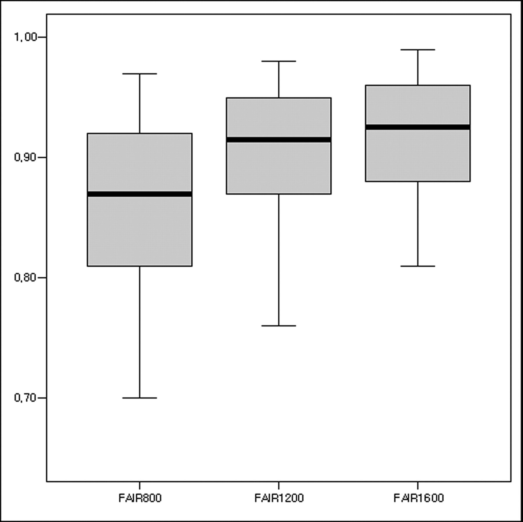
  • Fig 3.
    • Download figure
    • Open in new tab
    • Download powerpoint
    Fig 3.

    A stem-and-leaf plot of the rCBFs between the ipsilateral lesions and contralateral normal areas on the 3 FAIR images in the area with more prolonged TTP (dTTP ≥3.2 s) (FAIR, 800 ms; TI, 800 ms; FAIR, 1200 ms; TI, 1200 ms; FAIR, 1600 ms; TI, 1600 ms).

Tables

  • Figures
    • View popup
    Table 1:

    Width of decreased CBFs among the three FAIR maps with different inversion times according to the TTP difference

    wCBFdTTP <3.2 s (%)dTTP ≥3.2 s (%)
    FAIR 800 < FAIR 12000/25 (0)0/17 (0)
    FAIR 800 = FAIR 120023/25 (92)12/17 (71)
    FAIR 800 > FAIR 12002/25 (8)5/17 (29)
    FAIR 800 < FAIR 16000/25 (0)0/17 (0)
    FAIR 800 = FAIR 160017/25 (68)3/17 (18)
    FAIR 800 > FAIR 16008/25 (32)14/17 (82)
    FAIR 1200 < FAIR 16000/25 (0)0/17 (0)
    FAIR 1200 = FAIR 160022/25 (88)5/17 (29)
    FAIR 1200 > FAIR 16003/25 (12)12/17 (71)
    • Note:—wCBF, width of decreased CBF; dTTP, TTP difference; FAIR 800, TI = 800 ms; FAIR 1200, TI = 1200 ms; FAIR 1600, TI = 1600 ms.

    • View popup
    Table 2:

    Correlation coefficients of rCBF among the three FAIR maps

    GroupsdTTP <3.2 sdTTP ≥3.2 s
    FAIR 800 vs FAIR 1200r = 0.764, P < .01r = 0.712, P < .01
    FAIR 800 vs FAIR 1600r = 0.667, P < .01r = 0.595, P < .01
    FAIR 1200 vs FAIR 1600r = 0.791, P < .01r = 0.612, P < .01
    • Note:—dTTP indicates TTP difference; FAIR 800, TI = 800 ms; FAIR 1200, TI = 1200 ms; FAIR 1600, TI = 1600 ms; r, correlation coefficient.

    • View popup
    Table 3:

    Comparison of rCBF among the three FAIR images in the areas with less prolonged TTP (dTTP <3.2 s)*

    ComparisonMDP Value95% CI
    FAIR 800 vs FAIR 1200−0.031<.05−0.060-−0.003
    FAIR 800 vs FAIR 1600−0.046<.001−0.075-−0.017
    FAIR 1200 vs FAIR 1600−0.015>.05−0.043-−0.014
    • Note:—MD indicates mean difference; CI, confidence interval; FAIR 800, TI = 800 ms; FAIR 1200, TI = 1200 ms; FAIR 1600, TI = 1600 ms.

    • * Statistical analysis was performed with the repeated measures ANOVA and post hoc analysis with the Tukey-Kramer multiple comparisons test.

    • View popup
    Table 4:

    Comparison of rCBF among the three FAIR images in the area with more prolonged TTP (dTTP ≥3.2 s)*

    ComparisonMDP Value95% CI
    FAIR 800 vs FAIR 1200−0.026<.05−0.049-−0.002
    FAIR 800 vs FAIR 1600−0.078<.001−0.101-−0.054
    FAIR 1200 vs FAIR 1600−0.052<.001−0.075-−0.029
    • Note:—MD indicates mean difference; CI, confidence interval; FAIR 800, TI = 800 ms; FAIR 1200, TI = 1200 ms; FAIR 1600, TI = 1600 ms.

    • * Statistical analysis was performed with the repeated measures ANOVA and post hoc analysis with the Tukey-Kramer multiple comparisons test.

PreviousNext
Back to top

In this issue

American Journal of Neuroradiology: 28 (10)
American Journal of Neuroradiology
Vol. 28, Issue 10
November 2007
  • Table of Contents
  • Index by author
Advertisement
Print
Download PDF
Email Article

Thank you for your interest in spreading the word on American Journal of Neuroradiology.

NOTE: We only request your email address so that the person you are recommending the page to knows that you wanted them to see it, and that it is not junk mail. We do not capture any email address.

Enter multiple addresses on separate lines or separate them with commas.
Underestimation of Cerebral Perfusion on Flow-Sensitive Alternating Inversion Recovery Image: Semiquantitative Evaluation with Time-to-Peak Values
(Your Name) has sent you a message from American Journal of Neuroradiology
(Your Name) thought you would like to see the American Journal of Neuroradiology web site.
CAPTCHA
This question is for testing whether or not you are a human visitor and to prevent automated spam submissions.
Cite this article
H.S. Kim, S.Y. Kim, J.M. Kim
Underestimation of Cerebral Perfusion on Flow-Sensitive Alternating Inversion Recovery Image: Semiquantitative Evaluation with Time-to-Peak Values
American Journal of Neuroradiology Nov 2007, 28 (10) 2008-2013; DOI: 10.3174/ajnr.A0720

Citation Manager Formats

  • BibTeX
  • Bookends
  • EasyBib
  • EndNote (tagged)
  • EndNote 8 (xml)
  • Medlars
  • Mendeley
  • Papers
  • RefWorks Tagged
  • Ref Manager
  • RIS
  • Zotero
0 Responses
Respond to this article
Share
Bookmark this article
Underestimation of Cerebral Perfusion on Flow-Sensitive Alternating Inversion Recovery Image: Semiquantitative Evaluation with Time-to-Peak Values
H.S. Kim, S.Y. Kim, J.M. Kim
American Journal of Neuroradiology Nov 2007, 28 (10) 2008-2013; DOI: 10.3174/ajnr.A0720
del.icio.us logo Twitter logo Facebook logo Mendeley logo
  • Tweet Widget
  • Facebook Like
  • Google Plus One
Purchase

Jump to section

  • Article
    • Abstract
    • Materials and Methods
    • Results
    • Discussion
    • Conclusion
    • References
  • Figures & Data
  • Info & Metrics
  • Responses
  • References
  • PDF

Related Articles

  • No related articles found.
  • PubMed
  • Google Scholar

Cited By...

  • Hemodynamic Alterations in Vertebrobasilar Large Artery Disease Assessed by Arterial Spin-Labeling MR Imaging
  • Dynamic contrast-enhanced imaging techniques: CT and MRI
  • Crossref
  • Google Scholar

This article has not yet been cited by articles in journals that are participating in Crossref Cited-by Linking.

More in this TOC Section

  • Usefulness of Quantitative Susceptibility Mapping for the Diagnosis of Parkinson Disease
  • White Matter Alterations in the Brains of Patients with Active, Remitted, and Cured Cushing Syndrome: A DTI Study
  • Qualitative and Quantitative Analysis of MR Imaging Findings in Patients with Middle Cerebral Artery Stroke Implanted with Mesenchymal Stem Cells
Show more Brain

Similar Articles

Advertisement

Indexed Content

  • Current Issue
  • Accepted Manuscripts
  • Article Preview
  • Past Issues
  • Editorials
  • Editor's Choice
  • Fellows' Journal Club
  • Letters to the Editor
  • Video Articles

Cases

  • Case Collection
  • Archive - Case of the Week
  • Archive - Case of the Month
  • Archive - Classic Case

More from AJNR

  • Trainee Corner
  • Imaging Protocols
  • MRI Safety Corner
  • Book Reviews

Multimedia

  • AJNR Podcasts
  • AJNR Scantastics

Resources

  • Turnaround Time
  • Submit a Manuscript
  • Submit a Video Article
  • Submit an eLetter to the Editor/Response
  • Manuscript Submission Guidelines
  • Statistical Tips
  • Fast Publishing of Accepted Manuscripts
  • Graphical Abstract Preparation
  • Imaging Protocol Submission
  • Evidence-Based Medicine Level Guide
  • Publishing Checklists
  • Author Policies
  • Become a Reviewer/Academy of Reviewers
  • News and Updates

About Us

  • About AJNR
  • Editorial Board
  • Editorial Board Alumni
  • Alerts
  • Permissions
  • Not an AJNR Subscriber? Join Now
  • Advertise with Us
  • Librarian Resources
  • Feedback
  • Terms and Conditions
  • AJNR Editorial Board Alumni

American Society of Neuroradiology

  • Not an ASNR Member? Join Now

© 2025 by the American Society of Neuroradiology All rights, including for text and data mining, AI training, and similar technologies, are reserved.
Print ISSN: 0195-6108 Online ISSN: 1936-959X

Powered by HighWire