Advertisement

Main menu

  • Home
  • Content
    • Current Issue
    • Accepted Manuscripts
    • Article Preview
    • Past Issue Archive
    • AJNR Case Collection
    • Case of the Week Archive
    • Classic Case Archive
    • Case of the Month Archive
  • Special Collections
    • Spinal CSF Leak Articles (Jan 2020-June 2024)
    • 2024 AJNR Journal Awards
    • Most Impactful AJNR Articles
  • Multimedia
    • AJNR Podcast
    • AJNR Scantastics
    • Video Articles
  • For Authors
    • Submit a Manuscript
    • Author Policies
    • Fast publishing of Accepted Manuscripts
    • Graphical Abstract Preparation
    • Manuscript Submission Guidelines
    • Imaging Protocol Submission
    • Submit a Case for the Case Collection
  • About Us
    • About AJNR
    • Editorial Board
  • More
    • Become a Reviewer/Academy of Reviewers
    • Subscribers
    • Permissions
    • Alerts
    • Feedback
    • Advertisers
    • ASNR Home
  • Other Publications
    • ajnr

User menu

  • Alerts
  • Log in

Search

  • Advanced search
American Journal of Neuroradiology

American Journal of Neuroradiology

ASHNR American Society of Functional Neuroradiology ASHNR American Society of Pediatric Neuroradiology ASSR
  • Alerts
  • Log in

Advanced Search

  • Home
  • Content
    • Current Issue
    • Accepted Manuscripts
    • Article Preview
    • Past Issue Archive
    • AJNR Case Collection
    • Case of the Week Archive
    • Classic Case Archive
    • Case of the Month Archive
  • Special Collections
    • Spinal CSF Leak Articles (Jan 2020-June 2024)
    • 2024 AJNR Journal Awards
    • Most Impactful AJNR Articles
  • Multimedia
    • AJNR Podcast
    • AJNR Scantastics
    • Video Articles
  • For Authors
    • Submit a Manuscript
    • Author Policies
    • Fast publishing of Accepted Manuscripts
    • Graphical Abstract Preparation
    • Manuscript Submission Guidelines
    • Imaging Protocol Submission
    • Submit a Case for the Case Collection
  • About Us
    • About AJNR
    • Editorial Board
  • More
    • Become a Reviewer/Academy of Reviewers
    • Subscribers
    • Permissions
    • Alerts
    • Feedback
    • Advertisers
    • ASNR Home
  • Follow AJNR on Twitter
  • Visit AJNR on Facebook
  • Follow AJNR on Instagram
  • Join AJNR on LinkedIn
  • RSS Feeds

Welcome to the new AJNR, Updated Hall of Fame, and more. Read the full announcements.


AJNR is seeking candidates for the position of Associate Section Editor, AJNR Case Collection. Read the full announcement.


Getting new auth cookie, if you see this message a lot, tell someone!
LetterLetter

Ectopic Posterior Pituitary Lobe and Cortical Dysplasia

Jill C. Bergson, Vikram K. Garg and Jakwei Chang
American Journal of Neuroradiology February 2007, 28 (2) 198a-199;
Jill C. Bergson
  • Find this author on Google Scholar
  • Find this author on PubMed
  • Search for this author on this site
Vikram K. Garg
  • Find this author on Google Scholar
  • Find this author on PubMed
  • Search for this author on this site
Jakwei Chang
  • Find this author on Google Scholar
  • Find this author on PubMed
  • Search for this author on this site
  • Article
  • Figures & Data
  • Info & Metrics
  • Responses
  • References
  • PDF
Loading

We read with interest the report by Mitchell et al1 of 4 cases of ectopic posterior pituitary lobe and periventricular heterotopia on MR imaging studies. The authors suggested that ectopic posterior pituitary lobe with growth hormone deficiency is part of a spectrum associated with septo-optic dysplasia and concluded that the coexistence of periventricular heterotopia dysplasia implies a common underlying genetic mechanism. Further, in 1 case, they found the presence of a heterozygous HESX1 mutation, a gene associated with ectopic posterior pituitary lobe and septo-optic dysplasia, and suggested that this gene is important in the development of both ectopic posterior pituitary lobe and periventricular heterotopia.

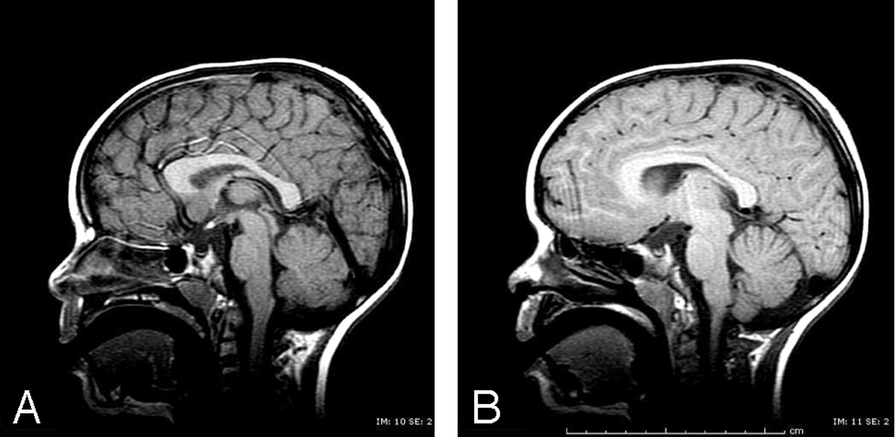
We report a case of a 4-year-old boy with a history of seizures. The patient had an episode of convulsive seizure in March 2006, with a left focal onset. His medical history included panhypopituitarism and optic atrophy of the right eye. Physical examination was significant for a pale right optic disk. MR imaging revealed ectopic posterior pituitary lobe, a hypoplastic pituitary, and a hypoplastic optic chiasm on the T1-weighted sagittal images (Fig 1A, -B). Incidentally, the pituitary stalk could not be visualized. T2-weighted axial and coronal fluid-attenuated inversion recovery (FLAIR) images showed bifrontal cortical dysplasia and a strip of ectopic gray matter extending from the head of the right caudate nuclei to the orbitofrontal lobe (Fig 2A, -B). T2-weighted axial images also revealed small optic nerves, especially on the right side.

The syndrome of septo-optic dysplasia consists of a combination of malformations including hypoplasia or absence of a septum pellucidum or corpus callosum, optic nerve hypoplasia or dysplasia, pituitary-hypothalamic dysfunction, and occasionally other cerebral abnormalities such as schizencephaly or cortical dysplasia. This heterogeneity of features has resulted in some disagreement as to how septo-optic dysplasia is to be defined and categorized. For example, both Barkovich et al2 and Miller et al3 attempted to define and classify septo-optic dysplasia into distinct anatomic subsets. However, Polizzi et al4 suggested that septo-optic dysplasia should be recategorized as a heterogeneous malformation syndrome encompassing multiple brain, endocrine, and systemic anomalies, rather than a single, precisely defined entity. Our patient did not have hypoplasia or absence of the septum pellucidum; however, the presence of an ectopic posterior pituitary lobe, optic nerve hypoplasia, optic chiasm hypoplasia, cortical dysplasia, and ectopic gray matter suggests features of septo-optic dysplasia.

We, therefore, agree with Mitchell et al1 that the presence of ectopic posterior pituitary lobe is often associated with other important cerebral malformations, even in the absence of classic features of septo-optic dysplasia. Like the cases presented by Mitchell et al,1 our patient had an ectopic posterior pituitary lobe. One difference is that the pituitary stalk in our patient could not be well visualized. We are curious as to whether the etiology of this finding had a common mechanism with the other abnormalities or if this was secondary to trauma or some other cause. In addition, our patient presented with bifrontal cortical dysplasia and an ectopic strip of gray matter in the right frontal lobe. These entities are due to abnormalities in neuronal cell migration, as is periventricular heterotopia seen in all 4 of Mitchell’s patients.

Thus, we support the theory that these cerebral malformations may have a common mechanism in the development of ectopic posterior pituitary lobe and suggest that they are part of a complex of features seen in septo-optic dysplasia.

Fig 1.
  • Download figure
  • Open in new tab
  • Download powerpoint
Fig 1.

A, Sagittal T1-weighted image shows a hyperintensity corresponding to an ectopic posterior pituitary lobe located in the hypothalamic area. The pituitary is small.

B, Paramedian sagittal T1-weighted image reveals a thin optic chiasm and a small pituitary.

Fig 2.
  • Download figure
  • Open in new tab
  • Download powerpoint
Fig 2.

A, Axial T2-weighted image reveals thickening of the cortical gray matter at the medial aspect of both frontal lobes, compatible with bifrontal cortical dysplasia (short arrows). Also seen is a strip of ectopic gray matter extending from the head of the right caudate nuclei to the orbitofrontal cortex (long arrow).

B, Coronal FLAIR image shows a strip of ectopic gray matter extending from the head of the right caudate nuclei to the orbitofrontal cortex (long arrow) as well as bifrontal thickening of the cortical gray matter (short arrows).

References

  1. ↵
    Mitchell LA, Thomas PQ, Zacharin MR, et al. Ectopic posterior pituitary lobe and periventricular heterotopia: cerebral malformations with the same underlying mechanism? AJNR Am J Neuroradiol 2002;23:1475–81
    Abstract/FREE Full Text
  2. ↵
    Barkovich AJ, Fram EK, Norman D. Septo-optic dysplaisa: MR imaging. Radiology 1989;171:189–92
    PubMed
  3. ↵
    Miller SP, Shevell MI, Patenaude Y, et al. Septo-optic dysplasia plus: a spectrum of malformations of cortical development. Neurology 2000;54:1701–03
    Abstract/FREE Full Text
  4. ↵
    Polizzi A, Pavone P, Iannetti P, et al. Septo-optic dysplasia complex: a heterogeneous malformation syndrome. Pediatr Neurol 2006;34:66–71
    CrossRefPubMed
  • Copyright © American Society of Neuroradiology
PreviousNext
Back to top

In this issue

American Journal of Neuroradiology: 28 (2)
American Journal of Neuroradiology
Vol. 28, Issue 2
February 2007
  • Table of Contents
  • Index by author
Advertisement
Print
Download PDF
Email Article

Thank you for your interest in spreading the word on American Journal of Neuroradiology.

NOTE: We only request your email address so that the person you are recommending the page to knows that you wanted them to see it, and that it is not junk mail. We do not capture any email address.

Enter multiple addresses on separate lines or separate them with commas.
Ectopic Posterior Pituitary Lobe and Cortical Dysplasia
(Your Name) has sent you a message from American Journal of Neuroradiology
(Your Name) thought you would like to see the American Journal of Neuroradiology web site.
CAPTCHA
This question is for testing whether or not you are a human visitor and to prevent automated spam submissions.
Cite this article
Jill C. Bergson, Vikram K. Garg, Jakwei Chang
Ectopic Posterior Pituitary Lobe and Cortical Dysplasia
American Journal of Neuroradiology Feb 2007, 28 (2) 198a-199;

Citation Manager Formats

  • BibTeX
  • Bookends
  • EasyBib
  • EndNote (tagged)
  • EndNote 8 (xml)
  • Medlars
  • Mendeley
  • Papers
  • RefWorks Tagged
  • Ref Manager
  • RIS
  • Zotero
0 Responses
Respond to this article
Share
Bookmark this article
Ectopic Posterior Pituitary Lobe and Cortical Dysplasia
Jill C. Bergson, Vikram K. Garg, Jakwei Chang
American Journal of Neuroradiology Feb 2007, 28 (2) 198a-199;
del.icio.us logo Twitter logo Facebook logo Mendeley logo
  • Tweet Widget
  • Facebook Like
  • Google Plus One

Jump to section

  • Article
    • References
  • Figures & Data
  • Info & Metrics
  • Responses
  • References
  • PDF

Related Articles

  • No related articles found.
  • PubMed
  • Google Scholar

Cited By...

  • No citing articles found.
  • Crossref
  • Google Scholar

This article has not yet been cited by articles in journals that are participating in Crossref Cited-by Linking.

More in this TOC Section

  • Brain AVM’s Nidus: What if We Hadn’t Understood Anything?
  • Letter to the Editor regarding “Automated Volumetric Software in Dementia: Help or Hindrance to the Neuroradiologist?”
  • Reply:
Show more Letters

Similar Articles

Advertisement

Indexed Content

  • Current Issue
  • Accepted Manuscripts
  • Article Preview
  • Past Issues
  • Editorials
  • Editors Choice
  • Fellow Journal Club
  • Letters to the Editor

Cases

  • Case Collection
  • Archive - Case of the Week
  • Archive - Case of the Month
  • Archive - Classic Case

Special Collections

  • Special Collections

Resources

  • News and Updates
  • Turn around Times
  • Submit a Manuscript
  • Author Policies
  • Manuscript Submission Guidelines
  • Evidence-Based Medicine Level Guide
  • Publishing Checklists
  • Graphical Abstract Preparation
  • Imaging Protocol Submission
  • Submit a Case
  • Become a Reviewer/Academy of Reviewers
  • Get Peer Review Credit from Publons

Multimedia

  • AJNR Podcast
  • AJNR SCANtastic
  • Video Articles

About Us

  • About AJNR
  • Editorial Board
  • Not an AJNR Subscriber? Join Now
  • Alerts
  • Feedback
  • Advertise with us
  • Librarian Resources
  • Permissions
  • Terms and Conditions

American Society of Neuroradiology

  • Not an ASNR Member? Join Now

© 2025 by the American Society of Neuroradiology All rights, including for text and data mining, AI training, and similar technologies, are reserved.
Print ISSN: 0195-6108 Online ISSN: 1936-959X

Powered by HighWire