Advertisement

Main menu

  • Home
  • Content
    • Current Issue
    • Accepted Manuscripts
    • Article Preview
    • Past Issue Archive
    • Video Articles
    • AJNR Case Collection
    • Case of the Week Archive
    • Case of the Month Archive
    • Classic Case Archive
  • Special Collections
    • AJNR Awards
    • Low-Field MRI
    • Alzheimer Disease
    • ASNR Foundation Special Collection
    • Photon-Counting CT
    • View All
  • Multimedia
    • AJNR Podcasts
    • AJNR SCANtastic
    • Trainee Corner
    • MRI Safety Corner
    • Imaging Protocols
  • For Authors
    • Submit a Manuscript
    • Submit a Video Article
    • Submit an eLetter to the Editor/Response
    • Manuscript Submission Guidelines
    • Statistical Tips
    • Fast Publishing of Accepted Manuscripts
    • Graphical Abstract Preparation
    • Imaging Protocol Submission
    • Author Policies
  • About Us
    • About AJNR
    • Editorial Board
    • Editorial Board Alumni
  • More
    • Become a Reviewer/Academy of Reviewers
    • Subscribers
    • Permissions
    • Alerts
    • Feedback
    • Advertisers
    • ASNR Home

User menu

  • Alerts
  • Log in
  • Log out

Search

  • Advanced search
American Journal of Neuroradiology

American Journal of Neuroradiology

ASHNR American Society of Functional Neuroradiology ASHNR American Society of Pediatric Neuroradiology ASSR
  • Alerts
  • Log in
  • Log out

Advanced Search

  • Home
  • Content
    • Current Issue
    • Accepted Manuscripts
    • Article Preview
    • Past Issue Archive
    • Video Articles
    • AJNR Case Collection
    • Case of the Week Archive
    • Case of the Month Archive
    • Classic Case Archive
  • Special Collections
    • AJNR Awards
    • Low-Field MRI
    • Alzheimer Disease
    • ASNR Foundation Special Collection
    • Photon-Counting CT
    • View All
  • Multimedia
    • AJNR Podcasts
    • AJNR SCANtastic
    • Trainee Corner
    • MRI Safety Corner
    • Imaging Protocols
  • For Authors
    • Submit a Manuscript
    • Submit a Video Article
    • Submit an eLetter to the Editor/Response
    • Manuscript Submission Guidelines
    • Statistical Tips
    • Fast Publishing of Accepted Manuscripts
    • Graphical Abstract Preparation
    • Imaging Protocol Submission
    • Author Policies
  • About Us
    • About AJNR
    • Editorial Board
    • Editorial Board Alumni
  • More
    • Become a Reviewer/Academy of Reviewers
    • Subscribers
    • Permissions
    • Alerts
    • Feedback
    • Advertisers
    • ASNR Home
  • Follow AJNR on Twitter
  • Visit AJNR on Facebook
  • Follow AJNR on Instagram
  • Join AJNR on LinkedIn
  • RSS Feeds

AJNR Awards, New Junior Editors, and more. Read the latest AJNR updates

OtherSpine Imaging and Spine Image-Guided Interventions

Spinal Subarachnoid Hematoma Resulting from Lumbar Myelography

M.D. Sather, M.D. Gibson and J.S. Treves
American Journal of Neuroradiology February 2007, 28 (2) 220-221;
M.D. Sather
  • Find this author on Google Scholar
  • Find this author on PubMed
  • Search for this author on this site
M.D. Gibson
  • Find this author on Google Scholar
  • Find this author on PubMed
  • Search for this author on this site
J.S. Treves
  • Find this author on Google Scholar
  • Find this author on PubMed
  • Search for this author on this site
  • Article
  • Figures & Data
  • Info & Metrics
  • Responses
  • References
  • PDF
Loading

Abstract

SUMMARY: We describe a rare complication of myelography. A subarachnoid filling defect was apparent on the postmyelographic CT but not on conventional myelography. MR imaging performed later showed a spinal subarachnoid hematoma (SSAH), extending from L3 to L5. Lumbar puncture may rarely be a cause for SSAH and is more common in patients with coagulopathy. Significant back pain, paresis, radiculopathy, and even altered consciousness or meningeal signs may herald an SSAH.

Myelography carries risks, including infection, spinal hematoma, CSF leak, contrast reaction, radiculopathy, spinal headache, intracranial hemorrhage, and tonsillar herniation. Patients should be monitored closely for these adverse reactions. In particular, severe back pain or new neurologic symptoms following myelography should be investigated further for spinal hematoma, including the rare spinal subarachnoid hematoma (SSAH).

The first surgical description of a hematoma in the spinal subarachnoid space was by Plotkin et al in 1966.1 It was originally described as a type of subarachnoid hemorrhage, and only later was the term “spinal subarachnoid hematoma” used. The first published case of a SSAH resulting from myelography occurred in 1975 and was associated with a subdural component as well.2 Two additional cases of SSAH have been reported in the literature, both from cervical myelography.3,4

Case Report

The patient was a 46-year-old woman who had previously undergone an L5-S1 instrumented fusion. She later developed recurrence of left S1 radiculopathy and underwent repeat lumbar MR imaging, followed by diagnostic myelography.

The patient had no history of coagulopathy, and results of laboratory studies were normal. The procedure was performed with the patient prone on the x-ray table with the dural puncture at the L4–5 level by using a 22-gauge spinal needle. CSF emanated from the hub, and the contrast flowed freely into the subarachnoid space, outlining the nerve roots in the thecal sac. The patient experienced no discomfort during the procedure.

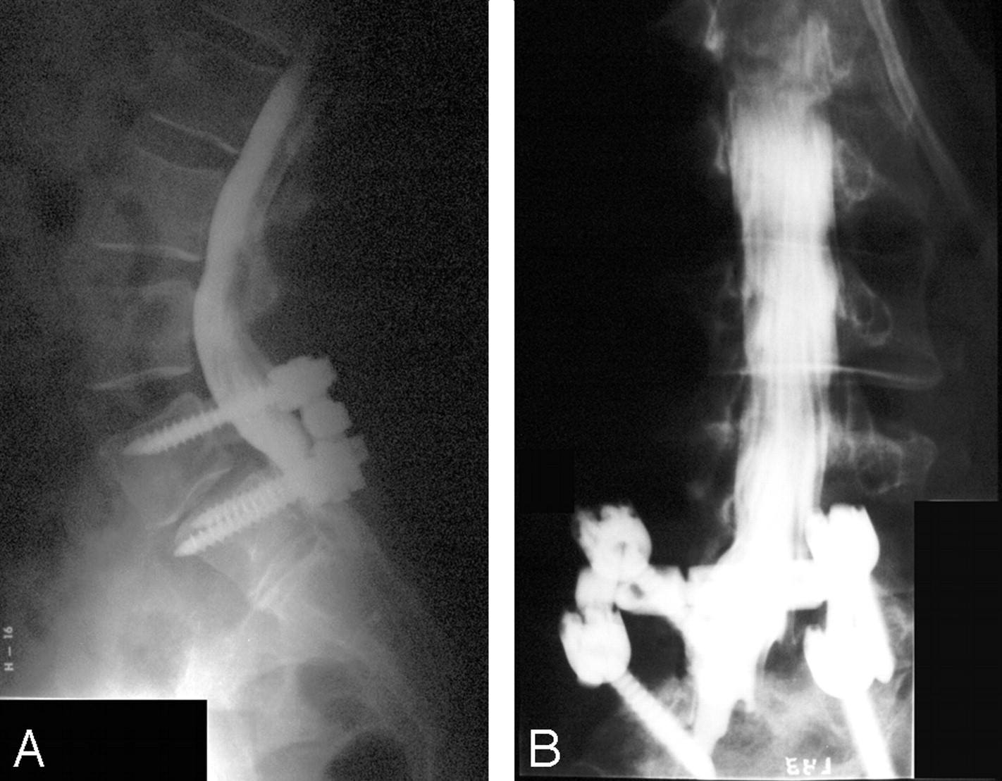
The lateral and oblique myelograms showed only small ventral and dorsal extradural filling defects, but the nerve roots were clearly seen, and there was no subarachnoid abnormality (Fig 1A, -B). CT myelography performed approximately 2 hours after dural puncture showed a subarachnoid filling defect extending from L3 to L5 (Fig 2). The possibility of a neoplasm was suggested, and follow-up MR imaging was recommended.

Fig 1.
  • Download figure
  • Open in new tab
  • Download powerpoint
Fig 1.

Myelograms demonstrate small dorsal and ventral extradural filling defects but normal delineation of nerve roots, indicating no subarachnoid abnormality.

A, Cross-table lateral position.

B, Left-oblique position.

Fig 2.
  • Download figure
  • Open in new tab
  • Download powerpoint
Fig 2.

Representative axial CT at L4–5 with large subarachnoid filling defect displacing the CSF and the nerve roots.

During the next 6 days, the patient developed progressive severe bilateral lower extremity pain and distal L5 and S1 motor weakness. A lumbar MR imaging revealed a subarachnoid hematoma extending from L3 to L5 (Fig 3A, -B). She was taken emergently to the operating room. The dura was dark blue; and once incised, along with the intact arachnoid, the hematoma delivered itself through the opening.

Fig 3.
  • Download figure
  • Open in new tab
  • Download powerpoint
Fig 3.

MR imaging, saggital series, demonstrating spinal subarachnoid hematoma.

A, T1-weighted image showing heterogeneous signal intensity consistent with acute and subacute hematoma from L3 to L5.

B, T2-weighted image showing heterogeneous signal intensity consistent with acute and subacute hematoma.

The discordance between the relatively normal findings on the myelogram and markedly abnormal delayed CT myelogram findings was thought to be secondary to the high attenuation of the contrast material on the myelogram, which might obscure the subarachnoid finding seen on the CT myelogram. Retrospectively, however, the explanation is obvious; the hematoma was not present on the myelogram because the patient hemorrhaged during the 2-hour delay between the myelogram and the CT.

Discussion

A recent article by Sandow and Donnal5 reviewed the potential complications of myelography, including seizures, allergic contrast dye reactions, contrast-induced renal insufficiency in patients on metformin therapy, and rare cases of spinal cord or conus medullaris puncture. However, no specific mention was made of the rare occurrence of SSAH resulting from diagnostic myelography.

Domenicucci et al6 reviewed 69 cases of SSAH and found that 44.9% were iatrogenically caused by lumbar puncture (LP) performed for spinal anesthesia, CSF control, or myelography. Of the patients undergoing LP, approximately 68% had coagulopathy.6 Thus, LP is rarely a cause of SSAH in the absence of coagulopathy, unless the procedure is technically difficult. This finding makes our case quite rare because the myelography was not technically difficult and the patient did not have a coagulopathy.

A literature review performed by Kreppel et al7 of 613 patients with spinal hematomas also found anticoagulant therapy to be a higher risk, representing the second largest causative factor in that series. Anticoagulant therapy alone was not a risk for spontaneous hematoma formation; but in the setting of vascular malformations, tumors, mild trauma, and lumbar puncture, it posed a higher risk.7

These studies by Domenicucci et al6 and Kreppel et al7 suggest the importance of scrutinizing patients for coagulopathy before dural puncture. However, a review of myelography practice patterns revealed that 58% of myelographers studied did not advise discontinuing aspirin or nonsteroidal anti-inflammatory drugs, and 73% did not routinely check prothrombin time (PT) and partial thromboplastin time (PTT).1 Because a much higher proportion of patients with coagulopathy undergoing dural puncture do sustain a complicating SSAH, practice guidelines should favor routinely checking platelet count, PT, and PTT and the discontinuation of antiplatelets and anticoagulants.

Kreppel el al7 also pointed out that although spinal hematomas in general are localized, SSAHs can extend along the entire spine. Spinal hematomas usually present with back pain and developing paresis, yet SSAHs can occasionally be differentiated by their ability to mimic cerebral processes, with altered consciousness, meningeal signs and symptoms, and seizures.7 However, these intracranial symptoms may cause difficulty in distinguishing SSAH from an intracranial hemorrhage, which can occur following myelography. More important, the leading symptom of this complication is postpuncture headache.8

The neuroradiologic characteristics of SSAH are often difficult to separate from those of the more common spinal subdural hematoma. On myelography, the most significant and distinguishing feature is “capping,” described by Frager et al.9 This was not seen in our patient because the hematoma was not apparent until the delayed CT images. However, the CT scans and MR images (Figs 2 and 3) showed a mass that was surrounded by CSF and separated from the dura, which is consistent with subarachnoid location.

Conclusion

SSAH is a rare complication of dural puncture, such as that performed in myelography. It is more predominant in patients with coagulopathy, suggesting the importance of routine laboratory studies and discontinuation of antiplatelet and anticoagulant medications. The myelographer should have a high degree of suspicion for a spinal hematoma and a low threshold for MR imaging of the spine when the patient develops significant back pain, paresis, or radiculopathy. Altered consciousness, meningeal signs, and headache may, more specifically, herald an SSAH or intracranial hemorrhage. CT myelograms are nonspecific. MR imaging is more sensitive to the by-products of acute and subacute hematoma. SSAH should be considered in the differential diagnosis for intradural abnormalities following dural puncture.

Acknowledgments

We thank Debi Smedlund for image preparation.

References

  1. ↵
    Plotkin HB, Ronthal M, Froman C. Spontaneous spinal subarachnoid hemorrhage: report of 3 cases. J Neurosurg 1966;25:443–46
    PubMed
  2. ↵
    Kirkpatrick D, Goodman SJ. Combined subarachnoid and subdural hematoma following spinal puncture. Surg Neurol 1975;3:109–11
    PubMed
  3. ↵
    Abla AA, Rothfu WE, Maroon JC, et al. Delayed spinal subarachnoid hematoma: a rare complication of C1–C2 cervical myelography. AJNR Am J Neuroradiol 1986;7:526–28
    FREE Full Text
  4. ↵
    Aghi M, Couman JV, Brisman JL. Subarachnoid haematoma, hydrocephalus, and aseptic meningitis resulting from a high cervical myelogram. J Spinal Disord Tech 2004;17:348–51
    PubMed
  5. ↵
    Sandow BA, Donnal JF. Myelography complications and current practice patterns. AJR Am J Roentgenol 2005;185:768–71
    PubMed
  6. ↵
    Domenicucci M, Ramieri A, Paolini S, et al. Spinal subarachnoid hematomas: our experience and literature review. Acta Neurochir (Wien) 2005;147:741–50
    PubMed
  7. ↵
    Kreppel D, Antoniadis G, Seeling W. Spinal hematoma: a literature survey with meta-analysis of 613 patients. Neurosurg Rev 2003;26:1–49
    CrossRefPubMed
  8. ↵
    Suess O, Stendel R, Baur S, et al. Intracranial haemorrhage following lumbar myelography: case report and review of literature. Neuroradiology 2000;42:211–14
    CrossRefPubMed
  9. ↵
    Frager D, Zimmerman RD, Wisoff HS, et al. Spinal subarachnoid hematoma. AJNR Am J Neuroradiol 1982;3:77–79
    FREE Full Text
  • Received December 2, 2005.
  • Accepted after revision January 18, 2006.
  • Copyright © American Society of Neuroradiology
View Abstract
PreviousNext
Back to top

In this issue

American Journal of Neuroradiology: 28 (2)
American Journal of Neuroradiology
Vol. 28, Issue 2
February 2007
  • Table of Contents
  • Index by author
Advertisement
Print
Download PDF
Email Article

Thank you for your interest in spreading the word on American Journal of Neuroradiology.

NOTE: We only request your email address so that the person you are recommending the page to knows that you wanted them to see it, and that it is not junk mail. We do not capture any email address.

Enter multiple addresses on separate lines or separate them with commas.
Spinal Subarachnoid Hematoma Resulting from Lumbar Myelography
(Your Name) has sent you a message from American Journal of Neuroradiology
(Your Name) thought you would like to see the American Journal of Neuroradiology web site.
CAPTCHA
This question is for testing whether or not you are a human visitor and to prevent automated spam submissions.
Cite this article
M.D. Sather, M.D. Gibson, J.S. Treves
Spinal Subarachnoid Hematoma Resulting from Lumbar Myelography
American Journal of Neuroradiology Feb 2007, 28 (2) 220-221;

Citation Manager Formats

  • BibTeX
  • Bookends
  • EasyBib
  • EndNote (tagged)
  • EndNote 8 (xml)
  • Medlars
  • Mendeley
  • Papers
  • RefWorks Tagged
  • Ref Manager
  • RIS
  • Zotero
0 Responses
Respond to this article
Share
Bookmark this article
Spinal Subarachnoid Hematoma Resulting from Lumbar Myelography
M.D. Sather, M.D. Gibson, J.S. Treves
American Journal of Neuroradiology Feb 2007, 28 (2) 220-221;
del.icio.us logo Twitter logo Facebook logo Mendeley logo
  • Tweet Widget
  • Facebook Like
  • Google Plus One

Jump to section

  • Article
    • Abstract
    • Case Report
    • Discussion
    • Conclusion
    • Acknowledgments
    • References
  • Figures & Data
  • Info & Metrics
  • Responses
  • References
  • PDF

Related Articles

  • No related articles found.
  • PubMed
  • Google Scholar

Cited By...

  • Aseptic Meningoencephalitis after Iohexol CT Myelography
  • Crossref
  • Google Scholar

This article has not yet been cited by articles in journals that are participating in Crossref Cited-by Linking.

More in this TOC Section

  • Management Outcomes For VO Spine Biopsy
  • Characteristics of SIH Type I Culprit Lesions
  • Advanced Imaging of Type 2 Spinal CSF Leaks
Show more Spine Imaging and Spine Image-Guided Interventions

Similar Articles

Advertisement

Indexed Content

  • Current Issue
  • Accepted Manuscripts
  • Article Preview
  • Past Issues
  • Editorials
  • Editor's Choice
  • Fellows' Journal Club
  • Letters to the Editor
  • Video Articles

Cases

  • Case Collection
  • Archive - Case of the Week
  • Archive - Case of the Month
  • Archive - Classic Case

More from AJNR

  • Trainee Corner
  • Imaging Protocols
  • MRI Safety Corner
  • Book Reviews

Multimedia

  • AJNR Podcasts
  • AJNR Scantastics

Resources

  • Turnaround Time
  • Submit a Manuscript
  • Submit a Video Article
  • Submit an eLetter to the Editor/Response
  • Manuscript Submission Guidelines
  • Statistical Tips
  • Fast Publishing of Accepted Manuscripts
  • Graphical Abstract Preparation
  • Imaging Protocol Submission
  • Evidence-Based Medicine Level Guide
  • Publishing Checklists
  • Author Policies
  • Become a Reviewer/Academy of Reviewers
  • News and Updates

About Us

  • About AJNR
  • Editorial Board
  • Editorial Board Alumni
  • Alerts
  • Permissions
  • Not an AJNR Subscriber? Join Now
  • Advertise with Us
  • Librarian Resources
  • Feedback
  • Terms and Conditions
  • AJNR Editorial Board Alumni

American Society of Neuroradiology

  • Not an ASNR Member? Join Now

© 2025 by the American Society of Neuroradiology All rights, including for text and data mining, AI training, and similar technologies, are reserved.
Print ISSN: 0195-6108 Online ISSN: 1936-959X

Powered by HighWire