Advertisement

Main menu

  • Home
  • Content
    • Current Issue
    • Accepted Manuscripts
    • Article Preview
    • Past Issue Archive
    • AJNR Case Collection
    • Case of the Week Archive
    • Classic Case Archive
    • Case of the Month Archive
  • Special Collections
    • Spinal CSF Leak Articles (Jan 2020-June 2024)
    • 2024 AJNR Journal Awards
    • Most Impactful AJNR Articles
  • Multimedia
    • AJNR Podcast
    • AJNR Scantastics
    • Video Articles
  • For Authors
    • Submit a Manuscript
    • Author Policies
    • Fast publishing of Accepted Manuscripts
    • Graphical Abstract Preparation
    • Manuscript Submission Guidelines
    • Imaging Protocol Submission
    • Submit a Case for the Case Collection
  • About Us
    • About AJNR
    • Editorial Board
  • More
    • Become a Reviewer/Academy of Reviewers
    • Subscribers
    • Permissions
    • Alerts
    • Feedback
    • Advertisers
    • ASNR Home
  • Other Publications
    • ajnr

User menu

  • Alerts
  • Log in

Search

  • Advanced search
American Journal of Neuroradiology

American Journal of Neuroradiology

ASHNR American Society of Functional Neuroradiology ASHNR American Society of Pediatric Neuroradiology ASSR
  • Alerts
  • Log in

Advanced Search

  • Home
  • Content
    • Current Issue
    • Accepted Manuscripts
    • Article Preview
    • Past Issue Archive
    • AJNR Case Collection
    • Case of the Week Archive
    • Classic Case Archive
    • Case of the Month Archive
  • Special Collections
    • Spinal CSF Leak Articles (Jan 2020-June 2024)
    • 2024 AJNR Journal Awards
    • Most Impactful AJNR Articles
  • Multimedia
    • AJNR Podcast
    • AJNR Scantastics
    • Video Articles
  • For Authors
    • Submit a Manuscript
    • Author Policies
    • Fast publishing of Accepted Manuscripts
    • Graphical Abstract Preparation
    • Manuscript Submission Guidelines
    • Imaging Protocol Submission
    • Submit a Case for the Case Collection
  • About Us
    • About AJNR
    • Editorial Board
  • More
    • Become a Reviewer/Academy of Reviewers
    • Subscribers
    • Permissions
    • Alerts
    • Feedback
    • Advertisers
    • ASNR Home
  • Follow AJNR on Twitter
  • Visit AJNR on Facebook
  • Follow AJNR on Instagram
  • Join AJNR on LinkedIn
  • RSS Feeds

Welcome to the new AJNR, Updated Hall of Fame, and more. Read the full announcements.


AJNR is seeking candidates for the position of Associate Section Editor, AJNR Case Collection. Read the full announcement.


OtherBRAIN

Wolfram Syndrome Presenting Marked Brain MR Imaging Abnormalities with Few Neurologic Abnormalities

S. Ito, R. Sakakibara and T. Hattori
American Journal of Neuroradiology February 2007, 28 (2) 305-306;
S. Ito
  • Find this author on Google Scholar
  • Find this author on PubMed
  • Search for this author on this site
R. Sakakibara
  • Find this author on Google Scholar
  • Find this author on PubMed
  • Search for this author on this site
T. Hattori
  • Find this author on Google Scholar
  • Find this author on PubMed
  • Search for this author on this site
  • Article
  • Figures & Data
  • Info & Metrics
  • Responses
  • References
  • PDF
Loading

Abstract

SUMMARY: Wolfram syndrome is a rare autosomal recessive disorder featuring diabetes insipidus, diabetes mellitus, optic atrophy, and deafness; DIDMOAD is a commonly accepted anonym for this disorder. We describe a 35-year-old man with Wolfram syndrome, who had marked atrophy of the brain stem, middle cerebellar peduncle, and cerebellum. Despite these MR imaging findings involving the pontocerebellar tract, the patient had no neurologic abnormalities suggesting dysfunction of the brain stem or cerebellum. Patients with Wolfram syndrome may have discrepancies between neurologic and radiologic findings.

In 1938, Wolfram and Wagener1 described a family in which 4 siblings developed bilateral optic atrophy and diabetes mellitus, followed by deafness, incontinence, and ataxia. The frequent association of diabetes insipidus, diabetes mellitus, optic atrophy, and deafness led to the acronym DIDMOAD, indicating 4 cardinal features.2 DIDMOAD, commonly known as Wolfram syndrome, is a rare autosomal recessive disorder with demonstrable clinical and genetic heterogeneity,2–4 and a gene for Wolfram syndrome (WFS1) has been cloned and mapped to chromosome 4p.5 The onset of Wolfram syndrome is usually juvenile, and most patients are referred to pediatricians or endocrinologists. Therefore, despite the striking neurologic and neuroradiologic features of Wolfram syndrome, the disorder has made little impact on the neurologic and neuroradiologic literature. In the present report, we describe a patient with Wolfram syndrome presenting marked MR imaging abnormalities with few neurologic abnormalities.

Case Report

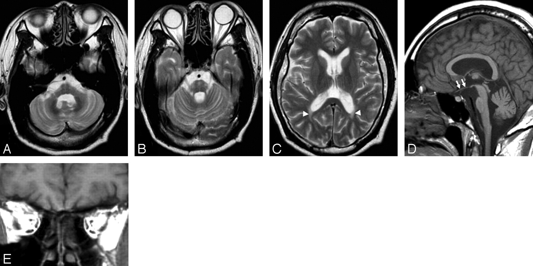
A 35-year-old man had diabetes insipidus, diabetes mellitus, and visual loss due to optic atrophy since he was 3 years of age. He had been diagnosed with Wolfram syndrome on the basis of the typical clinical features and familial history of diabetes mellitus. The man was admitted to our hospital because of consciousness disturbance in June 2005. Laboratory examinations showed diabetic ketoacidosis. He was stuporous and opened his eyes only to painful stimuli. After the patient recovered from diabetic ketoacidosis, neurologic examinations revealed complete visual loss and mild sensory hearing loss, but there were no findings suggesting abnormalities of the brain stem or cerebellum, such as nystagmus, dysarthria, or cerebellar ataxia; however brain MR imaging showed moderate atrophy of the brain stem and middle cerebellar peduncle and mild atrophy of the cerebellum (Fig 1A, -B), in addition to T2 elongation of peritrigonal white matter and absence of the T1 hyperintensity normally recognized in the posterior pituitary lobe (Fig 1C, -D). There was atrophy of intraorbital and intracranial optic nerves and tracts (Fig 1D, -E). The patient was treated by intensive insulin therapy and discharged 3 weeks later. Diabetic mellitus was well controlled, and his neurologic status showed no obvious deterioration in January 2006.

  Fig 1.
  • Download figure
  • Open in new tab
  • Download powerpoint
Fig 1.

Axial T2-weighted MR images show atrophy of the brain stem and middle cerebellar peduncles and areas of elevated signal intensity in the pons (A and B). Cerebellar atrophy is also recognized. Signal-intensity abnormalities in optic radiation on both sides are also seen (C, arrowheads). A sagittal T1-weighted MR image shows intracranial optic nerve atrophy (D, arrows) and absence of T1-hyperinensity normally recognized in the posterior pituitary lobe. A coronal T1-weighted image shows atrophy of both intraorbital optic nerves (E).

Discussion

Our patient had elevated signals in the pontine base and marked atrophy of the brain stem, middle cerebellar peduncle, and cerebellum. These changes involving the pontocerebellar tract resembled MR imaging findings in multiple system atrophy (MSA) and some kind of familial spinocerebellar degeneration (eg, spinocerebellar ataxia [SCA] 1, SCA2, Machado-Joseph disease [MJD or SCA3], or dentatorubral-pallidoluysian atrophy [DRPLA]). Neuroradiologically, MSA features pontocerebellar atrophy and pontine signal-intensity changes widely known as “cross signs.” It is difficult to radiologically differentiate the present case from MSA, but the age at onset of MSA is usually the 5th or 6th decade. The patients with SCA1 have brain stem and cerebellar volume loss and mild supratentorial generalized volume loss.6,7 Patients with SCA2 have more severe olivopontocerebellar atrophy than patients with SCA1 and SCA3, and they also have supratentorial atrophy.7,8 The patients with SCA3 and DRPLA have atrophy of the superior cerebellar peduncle, in addition to pontocerebellar atrophy.7,9–11 In patients with DRPLA, T2-weighted images usually show white matter hyperintensities. In our patient, there was no atrophy of the superior cerebellar peduncle, and white matter hyperintensities were more focal than those in patients with DRPLA.

Although it is difficult to radiologically differentiate the present case from the previously mentioned pontocerebellar disorders, our patient had no neurologic abnormalities suggesting dysfunction of the brain stem or cerebellum. Previous reports of Wolfram syndrome2,12–14 showed nearly the same MR imaging findings as those of our patient, but the previous patients had some neurologic deficits secondary to brain stem and cerebellar atrophy.2,13 For example, a patient described by Scolding et al2 had pendular nystagmus in the primary position and gaze-evoked nystagmus as a vestibulocerebellar dysfunction, and another patient had slurring dysarthria and limb ataxia, which corresponded with pontocerebellar atrophy. A patient reported by Pakdemirli et al13 had dysarthria, and her brain MR imaging showed atrophy of the brain stem and cerebellum. In a pathologically confirmed case of Wolfram syndrome in a patient described by Genís et al,15 the patient had slight dysarthria and gait unsteadiness caused by atrophy of the pons and cerebellar white matter; however, some previous reports of Wolfram syndrome did not contain sufficient description of the neurologic conditions.12,14

In our patient, it is unclear why the neurologic deficits secondary to brain stem and cerebellar atrophy were absent despite apparent MR imaging abnormalities in that region. A previous neuropathologic report of Wolfram syndrome15 showed moderate loss of neurons in pontine nuclei, inferior olives, and dentate nuclei of the cerebellum, and cerebellar white matter that was reduced in size without demyelination. Purkinje cells were reduced and showed axonal ballooning, but the granular cell layer was normal. Similarly, in our patient, the atrophy was more apparent in the brain stem and middle cerebellar peduncle than in the cerebellar cortices. This relative preservation of cerebellar cortical architecture might be one of the causes of the neurologic normality of cerebellar function, which was mismatched with brain stem and cerebellar atrophy on MR imaging. In the previous reports of Wolfram syndrome,2,13 the patients had some neurologic deficits secondary to brain stem and cerebellar atrophy, but these deficits were considered milder than those in patients with multiple system atrophy.

Additionally, our patient had T2 elongation of peritrigonal white matter and absence of T1 hypeintensity normally seen in the posterior pituitary lobe. The former could reflect primary or secondary degeneration of optic radiation, because past microscopic examinations showed atrophy of optic nerves, chiasm, tracts, and optic radiations and neuronal loss in the lateral geniculate nuclei and superior colliculi.15 The latter suggested loss of vasopressin-containing neurons as a cause of diabetes insipidus.

In summary, in patients with Wolfram syndrome, there were marked brain MR imaging abnormalities affecting the pontocerebellar tract, whereas there were few neurologic abnormalities suggesting cerebellar dysfunction. These patients may have discrepancies between neurologic and radiologic findings.

Acknowledgments

We thank Drs. Ichiro Tatsuno and Kenichi Sakurai in the Department of Clinical Cell Biology, Graduate School of Medicine, Chiba University, for their great contributions to diagnosis and treatment of the patients.

References

  1. ↵
    Wolfram DJ, Wagener HP. Diabetes mellitus and simple optic atrophy among siblings: report of four cases. Mayo Clin Proc 1938;9:715–18
  2. ↵
    Scolding NJ, Kellar-Wood HF, Shaw C, et al. Wolfram syndrome: hereditary diabetes mellitus with brainstem and optic atrophy. Ann Neurol 1996;39:352–60
    CrossRefPubMed
  3. Swift RG, Sadler DB, Swift M. Psychiatric findings in Wolfram syndrome homozygotes. Lancet 1990;336:667–69
    CrossRefPubMed
  4. ↵
    Simsek E, Simsek T, Tekgül S, et al. Wolfram (DIDMOAD) syndrome: a multidisciplinary clinical study in nine Turkish patients and review of literature. Acta Pediatr 2003;92:55–61
    PubMed
  5. ↵
    Inoue H, Tanizawa Y, Wasson J, et al. A gene encoding a transmembrane protein is mutated in patients with diabetes mellitus and optic atrophy (Wolfram syndrome). Nat Genet 1998;20:143–48
    CrossRefPubMed
  6. ↵
    Gilman S, Sima AAF, Junck L, et al. Spinocerebellar ataxia type 1 with multiple system degeneration and glial cytoplasmic inclusions. Ann Neurol 1996;39:241–55
    CrossRefPubMed
  7. ↵
    Bürk K, Abele M, Fetter M, et al. Autosomal dominant cerebellar ataxia type I: clinical features and MRI in families with SCA1, SCA2 and SCA3. Brain 1996;119:1497–505
    Abstract/FREE Full Text
  8. ↵
    Giuffrida S, Saponara R, Restivo DA, et al. Supratentorial atrophy in spinocerebellar ataxia type 2: MRI study of 20 patients. J Neurol 1999;246:383–88
    CrossRefPubMed
  9. ↵
    Tokumaru AM, Kamakura K, Maki T, et al. Magnetic resonance imaging findings of Machado-Joseph disease: histopathologic correlation. J Comput Assist Tomogr 2003;27:241–48
    CrossRefPubMed
  10. Uyama E, Kondo I, Uchino M, et al. Dentatorubral-pallidoluysian atrophy (DRPLA): clinical, genetic, and neuroradiologic studies in a family. J Neurol Sci 1995;130:146–53
    CrossRefPubMed
  11. ↵
    Koide R, Onodera O, Ikeuchi T, et al. Atrophy of the cerebellum and brainstem in dentatorubral pallidoluysian atrophy: influence of CAG repeat size on MRI findings. Neurology 1997;49:1605–12
    Abstract/FREE Full Text
  12. ↵
    Hadidy AM, Jarrah NS, Al-Till MI, et al. Radiological findings in Wolfram syndrome. Saudi Med J 2004;25:638–41
    PubMed
  13. ↵
    Pakdemirli E, Karabulut N, Bir LS, et al. Cranial magnetic resonance imaging of Wolfram (DIDMOAD) syndrome. Australas Radiol 2005;49:189–91
    PubMed
  14. ↵
    Yang MS, Chen CC, Chen YY, et al. Imaging characteristics of familial Wolfram syndrome. J Formos Med Assoc 2005;104:129–32
    PubMed
  15. ↵
    Geís D, Dávalos A, Molins A, et al. Wolfram syndrome: a neuropathological study. Acta Neuropathol (Berl) 1997;93:426–29
    CrossRefPubMed
  • Received January 13, 2006.
  • Accepted after revision February 17, 2006.
  • Copyright © American Society of Neuroradiology
View Abstract
PreviousNext
Back to top

In this issue

American Journal of Neuroradiology: 28 (2)
American Journal of Neuroradiology
Vol. 28, Issue 2
February 2007
  • Table of Contents
  • Index by author
Advertisement
Print
Download PDF
Email Article

Thank you for your interest in spreading the word on American Journal of Neuroradiology.

NOTE: We only request your email address so that the person you are recommending the page to knows that you wanted them to see it, and that it is not junk mail. We do not capture any email address.

Enter multiple addresses on separate lines or separate them with commas.
Wolfram Syndrome Presenting Marked Brain MR Imaging Abnormalities with Few Neurologic Abnormalities
(Your Name) has sent you a message from American Journal of Neuroradiology
(Your Name) thought you would like to see the American Journal of Neuroradiology web site.
CAPTCHA
This question is for testing whether or not you are a human visitor and to prevent automated spam submissions.
Cite this article
S. Ito, R. Sakakibara, T. Hattori
Wolfram Syndrome Presenting Marked Brain MR Imaging Abnormalities with Few Neurologic Abnormalities
American Journal of Neuroradiology Feb 2007, 28 (2) 305-306;

Citation Manager Formats

  • BibTeX
  • Bookends
  • EasyBib
  • EndNote (tagged)
  • EndNote 8 (xml)
  • Medlars
  • Mendeley
  • Papers
  • RefWorks Tagged
  • Ref Manager
  • RIS
  • Zotero
0 Responses
Respond to this article
Share
Bookmark this article
Wolfram Syndrome Presenting Marked Brain MR Imaging Abnormalities with Few Neurologic Abnormalities
S. Ito, R. Sakakibara, T. Hattori
American Journal of Neuroradiology Feb 2007, 28 (2) 305-306;
del.icio.us logo Twitter logo Facebook logo Mendeley logo
  • Tweet Widget
  • Facebook Like
  • Google Plus One

Jump to section

  • Article
    • Abstract
    • Case Report
    • Discussion
    • Acknowledgments
    • References
  • Figures & Data
  • Info & Metrics
  • Responses
  • References
  • PDF

Related Articles

  • No related articles found.
  • PubMed
  • Google Scholar

Cited By...

  • Expanding the spectrum of white matter abnormalities in Wolfram syndrome: A retrospective review
  • Longitudinal Assessment of Neuroradiologic Features in Wolfram Syndrome
  • Wolfram Syndrome: A rare cause of progressive cerebellar ataxia
  • Teaching NeuroImages: MRI of brain findings of Wolfram (DIDMOAD) syndrome
  • Teaching NeuroImages: A neuroendocrine rarity: Wolfram syndrome
  • Wolfram syndrome associated with leukoencephalopathy
  • Crossref
  • Google Scholar

This article has not yet been cited by articles in journals that are participating in Crossref Cited-by Linking.

More in this TOC Section

  • Progression of Microstructural Damage in Spinocerebellar Ataxia Type 2: A Longitudinal DTI Study
  • Usefulness of Quantitative Susceptibility Mapping for the Diagnosis of Parkinson Disease
  • Evaluating the Effects of White Matter Multiple Sclerosis Lesions on the Volume Estimation of 6 Brain Tissue Segmentation Methods
Show more Brain

Similar Articles

Advertisement

Indexed Content

  • Current Issue
  • Accepted Manuscripts
  • Article Preview
  • Past Issues
  • Editorials
  • Editors Choice
  • Fellow Journal Club
  • Letters to the Editor

Cases

  • Case Collection
  • Archive - Case of the Week
  • Archive - Case of the Month
  • Archive - Classic Case

Special Collections

  • Special Collections

Resources

  • News and Updates
  • Turn around Times
  • Submit a Manuscript
  • Author Policies
  • Manuscript Submission Guidelines
  • Evidence-Based Medicine Level Guide
  • Publishing Checklists
  • Graphical Abstract Preparation
  • Imaging Protocol Submission
  • Submit a Case
  • Become a Reviewer/Academy of Reviewers
  • Get Peer Review Credit from Publons

Multimedia

  • AJNR Podcast
  • AJNR SCANtastic
  • Video Articles

About Us

  • About AJNR
  • Editorial Board
  • Not an AJNR Subscriber? Join Now
  • Alerts
  • Feedback
  • Advertise with us
  • Librarian Resources
  • Permissions
  • Terms and Conditions

American Society of Neuroradiology

  • Not an ASNR Member? Join Now

© 2025 by the American Society of Neuroradiology All rights, including for text and data mining, AI training, and similar technologies, are reserved.
Print ISSN: 0195-6108 Online ISSN: 1936-959X

Powered by HighWire