
Article Figures & Data
Figures
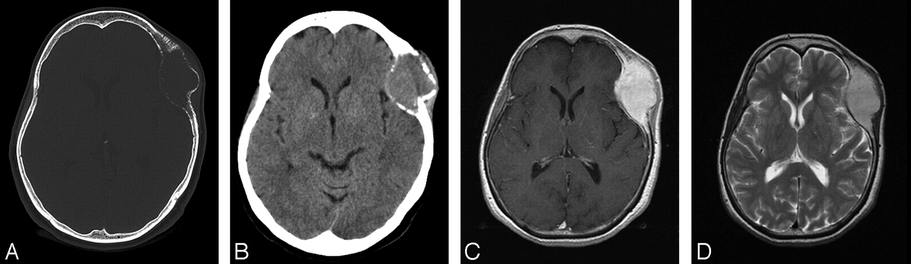
- Fig 1.
A, Noncontrast CT axial projection of a 41-mm minimally hyperattenuated homogeneous lytic mass centered at the coronal suture line with the inner and outer bony tables thinned and partially interrupted. B, Bone window exhibiting an osteolytic lesion with anterior hyperostosis. C, Axial T1-weighted precontrast MR image demonstrating an isointense biconvex mass. D, Axial T1-weighted postcontrast MR image demonstrating intense homogeneous enhancement with a mild dural reaction.
- Fig 2.
Intraoperative image demonstrating an exophytic relatively vascular skull mass.
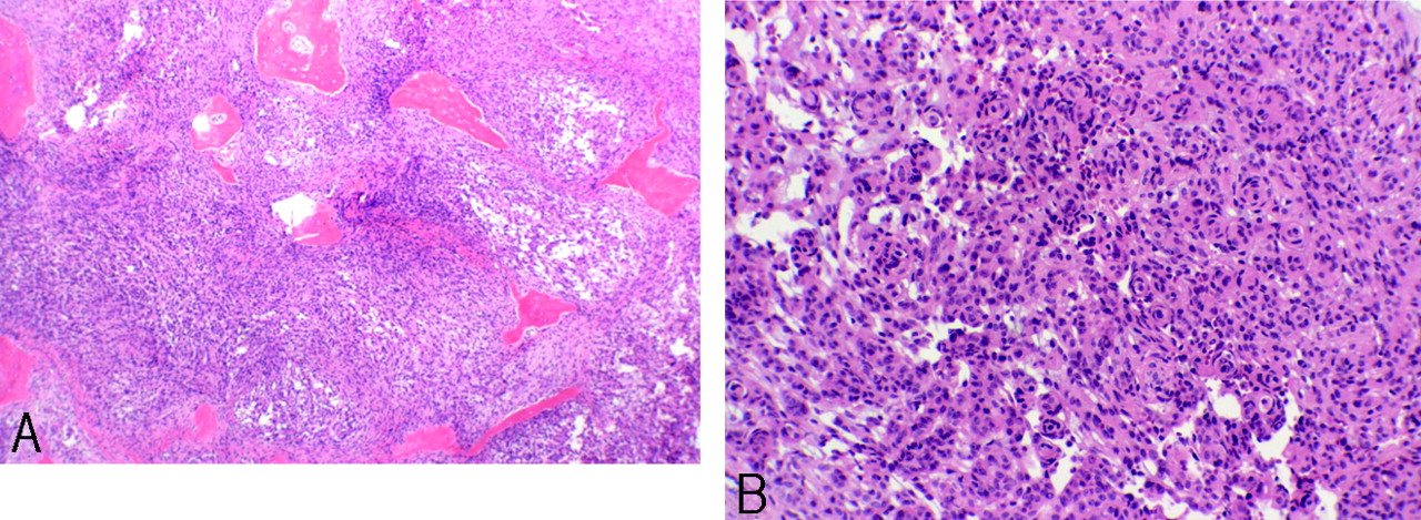
- Fig 3.
A, Light microscopy (H&E; original magnification × 2) of resected tumor involving bone. B, Light microscopy (H&E; original magnification × 40) reveals sheets of small cells with uniform nuclei forming small whorls.


