Advertisement

Main menu

  • Home
  • Content
    • Current Issue
    • Accepted Manuscripts
    • Article Preview
    • Past Issue Archive
    • Video Articles
    • AJNR Case Collection
    • Case of the Week Archive
    • Case of the Month Archive
    • Classic Case Archive
  • Special Collections
    • AJNR Awards
    • Low-Field MRI
    • Alzheimer Disease
    • ASNR Foundation Special Collection
    • Photon-Counting CT
    • View All
  • Multimedia
    • AJNR Podcasts
    • AJNR SCANtastic
    • Trainee Corner
    • MRI Safety Corner
    • Imaging Protocols
  • For Authors
    • Submit a Manuscript
    • Submit a Video Article
    • Submit an eLetter to the Editor/Response
    • Manuscript Submission Guidelines
    • Statistical Tips
    • Fast Publishing of Accepted Manuscripts
    • Graphical Abstract Preparation
    • Imaging Protocol Submission
    • Author Policies
  • About Us
    • About AJNR
    • Editorial Board
    • Editorial Board Alumni
  • More
    • Become a Reviewer/Academy of Reviewers
    • Subscribers
    • Permissions
    • Alerts
    • Feedback
    • Advertisers
    • ASNR Home

User menu

  • Alerts
  • Log in

Search

  • Advanced search
American Journal of Neuroradiology

American Journal of Neuroradiology

ASHNR American Society of Functional Neuroradiology ASHNR American Society of Pediatric Neuroradiology ASSR
  • Alerts
  • Log in

Advanced Search

  • Home
  • Content
    • Current Issue
    • Accepted Manuscripts
    • Article Preview
    • Past Issue Archive
    • Video Articles
    • AJNR Case Collection
    • Case of the Week Archive
    • Case of the Month Archive
    • Classic Case Archive
  • Special Collections
    • AJNR Awards
    • Low-Field MRI
    • Alzheimer Disease
    • ASNR Foundation Special Collection
    • Photon-Counting CT
    • View All
  • Multimedia
    • AJNR Podcasts
    • AJNR SCANtastic
    • Trainee Corner
    • MRI Safety Corner
    • Imaging Protocols
  • For Authors
    • Submit a Manuscript
    • Submit a Video Article
    • Submit an eLetter to the Editor/Response
    • Manuscript Submission Guidelines
    • Statistical Tips
    • Fast Publishing of Accepted Manuscripts
    • Graphical Abstract Preparation
    • Imaging Protocol Submission
    • Author Policies
  • About Us
    • About AJNR
    • Editorial Board
    • Editorial Board Alumni
  • More
    • Become a Reviewer/Academy of Reviewers
    • Subscribers
    • Permissions
    • Alerts
    • Feedback
    • Advertisers
    • ASNR Home
  • Follow AJNR on Twitter
  • Visit AJNR on Facebook
  • Follow AJNR on Instagram
  • Join AJNR on LinkedIn
  • RSS Feeds

AJNR Awards, New Junior Editors, and more. Read the latest AJNR updates

Research ArticleHead and Neck Imaging

Can Pretreatment CT Perfusion Predict Response of Advanced Squamous Cell Carcinoma of the Upper Aerodigestive Tract Treated with Induction Chemotherapy?

A. Zima, R. Carlos, D. Gandhi, I. Case, T. Teknos and S.K. Mukherji
American Journal of Neuroradiology February 2007, 28 (2) 328-334;
A. Zima
  • Find this author on Google Scholar
  • Find this author on PubMed
  • Search for this author on this site
R. Carlos
  • Find this author on Google Scholar
  • Find this author on PubMed
  • Search for this author on this site
D. Gandhi
  • Find this author on Google Scholar
  • Find this author on PubMed
  • Search for this author on this site
I. Case
  • Find this author on Google Scholar
  • Find this author on PubMed
  • Search for this author on this site
T. Teknos
  • Find this author on Google Scholar
  • Find this author on PubMed
  • Search for this author on this site
S.K. Mukherji
  • Find this author on Google Scholar
  • Find this author on PubMed
  • Search for this author on this site
  • Article
  • Figures & Data
  • Info & Metrics
  • Responses
  • References
  • PDF
Loading

Article Figures & Data

Figures

  • Tables
  • Fig 1.
    • Download figure
    • Open in new tab
    • Download powerpoint
    Fig 1.

    Examples of CT perfusion images for a patient with squamous cell carcinoma of the epiglottis.

    A, Regions of interest are drawn within the ipsilateral internal carotid artery (1), internal jugular vein (2), and around the tumor margins (3).

    B, Concentration curves of the previously selected regions of interest are plotted with Hounsfield units on the y-axis and time on the x-axis.

    C, Corresponding BF map of the neck at the level of the epiglottic tumor.

    D, Corresponding BV map of the neck at the level of the epiglottic tumor.
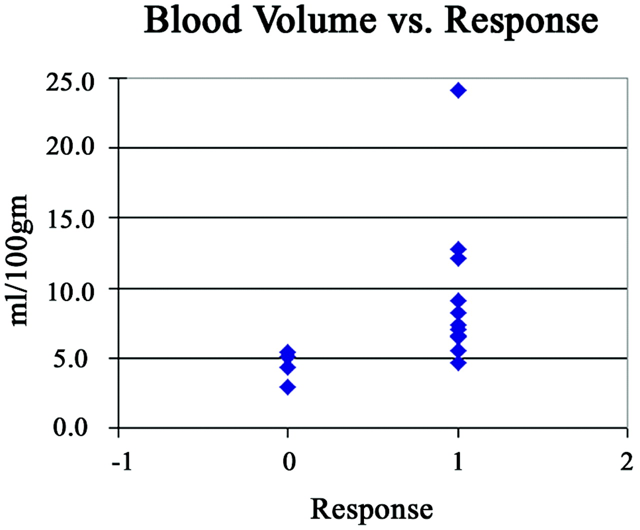
  • Fig 2.
    • Download figure
    • Open in new tab
    • Download powerpoint
    Fig 2.

    The relationship to BV values as a function response, defined as >50% reduction in tumor volume as assessed by endoscopy.

  • Fig 3.
    • Download figure
    • Open in new tab
    • Download powerpoint
    Fig 3.

    The relationship to BF values as a function response, defined as >50% reduction in tumor volume as assessed by endoscopy.

  • Fig 4.
    • Download figure
    • Open in new tab
    • Download powerpoint
    Fig 4.

    The area under the ROC curve for the predictive model.

  • Fig 5.
    • Download figure
    • Open in new tab
    • Download powerpoint
    Fig 5.

    The probability of response to induction chemotherapy as a function of BV.

  • Fig 6.
    • Download figure
    • Open in new tab
    • Download powerpoint
    Fig 6.

    The potential use of the prediction model using the 17 patients in this study in clinical practice.

    A, Actual response. In our patient population, all 17 patients underwent induction chemotherapy. Twelve patients responded (+) and received additional chemotherapy and radiation; 5 patients underwent surgery and radiation.

    B, Using ≥90% pretest probability as a threshold. In this hypothetical application of the prediction rule, 10 patients would be directed immediately to organ preservation, whereas 7 patients would undergo induction chemotherapy. Of those 7 patients, 2 patients would respond and proceed to organ preservation therapy, and the remaining 5 patients would proceed to surgery and radiation.

    C, Using ≥10% pretest probability as a threshold. In this hypothetical application, 2 patients would receive immediate surgery and radiation, whereas 15 would undergo induction chemotherapy. Using this low threshold, 3 patients would not respond to induction chemotherapy with potential delay in more invasive, but appropriate, surgery and radiation therapy.

Tables

  • Figures
    • View popup
    Table 1:

    Location, stage of tumor, and the decrease in tumor volume as assessed by endoscopy

    SexAgeTumor LocationTMNStageEndoscopic Response (%)
    M40Buccal mucosaT3 N0III20
    M66EpiglottisT3 N0III65
    F80Oral tongueT3 N0III70
    F49EpiglottisT3 N0III100
    M53Base of tongueT3 N1III90
    M53Base of tongueT3 N1III40
    M63Base of tongueT1 N2cIV95
    F57TonsilT2 N2aIV70
    M55Base of tongueT2 N2aIV100
    M48TonsilT2 N2bIV90
    M48TonsilT2 N3IV70
    F46EpiglottisT3 N2bIV60
    M46TonsilT3 N2bIV100
    F72Base of tongueT4 N0IV50
    M55TonsilT4 N2bIV50
    F53Base of tongueT4 N2cIV20
    M58Base of tongueT4 N2cIV70
    • Note:—M indicates male; F, female.

    • View popup
    Table 2:

    Means and ranges for the 4 CT perfusion parameters for responders and nonresponders

    BF (ml/100 g)BV (ml/100 g)MTT (seconds)CP (ml/100 g/min)
    Responders
        Mean155.99.46.818.8
        Range39.1–361.04.6–24.22.4–12.69.8–27.1
    Nonresponders
        Mean78.54.65.414.2
        Range54.5–108.53.0–5.44.4–7.010.4–19.1
    • Note:—BF indicates blood flow; BV, blood volume; MTT, mean transit time; CP, capillary permeability

PreviousNext
Back to top

In this issue

American Journal of Neuroradiology: 28 (2)
American Journal of Neuroradiology
Vol. 28, Issue 2
February 2007
  • Table of Contents
  • Index by author
Advertisement
Print
Download PDF
Email Article

Thank you for your interest in spreading the word on American Journal of Neuroradiology.

NOTE: We only request your email address so that the person you are recommending the page to knows that you wanted them to see it, and that it is not junk mail. We do not capture any email address.

Enter multiple addresses on separate lines or separate them with commas.
Can Pretreatment CT Perfusion Predict Response of Advanced Squamous Cell Carcinoma of the Upper Aerodigestive Tract Treated with Induction Chemotherapy?
(Your Name) has sent you a message from American Journal of Neuroradiology
(Your Name) thought you would like to see the American Journal of Neuroradiology web site.
CAPTCHA
This question is for testing whether or not you are a human visitor and to prevent automated spam submissions.
Cite this article
A. Zima, R. Carlos, D. Gandhi, I. Case, T. Teknos, S.K. Mukherji
Can Pretreatment CT Perfusion Predict Response of Advanced Squamous Cell Carcinoma of the Upper Aerodigestive Tract Treated with Induction Chemotherapy?
American Journal of Neuroradiology Feb 2007, 28 (2) 328-334;

Citation Manager Formats

  • BibTeX
  • Bookends
  • EasyBib
  • EndNote (tagged)
  • EndNote 8 (xml)
  • Medlars
  • Mendeley
  • Papers
  • RefWorks Tagged
  • Ref Manager
  • RIS
  • Zotero
0 Responses
Respond to this article
Share
Bookmark this article
Can Pretreatment CT Perfusion Predict Response of Advanced Squamous Cell Carcinoma of the Upper Aerodigestive Tract Treated with Induction Chemotherapy?
A. Zima, R. Carlos, D. Gandhi, I. Case, T. Teknos, S.K. Mukherji
American Journal of Neuroradiology Feb 2007, 28 (2) 328-334;
del.icio.us logo Twitter logo Facebook logo Mendeley logo
  • Tweet Widget
  • Facebook Like
  • Google Plus One

Jump to section

  • Article
    • Abstract
    • Methods
    • Results
    • Discussion
    • Footnotes
    • References
  • Figures & Data
  • Info & Metrics
  • Responses
  • References
  • PDF

Related Articles

  • No related articles found.
  • PubMed
  • Google Scholar

Cited By...

  • CT Texture Analysis Potentially Predicts Local Failure in Head and Neck Squamous Cell Carcinoma Treated with Chemoradiotherapy
  • CT Perfusion Can Predict Overexpression of CXCL8 (Interleukin-8) in Head and Neck Squamous Cell Carcinoma
  • Human Papillomavirus, p16, and Epidermal Growth Factor Receptor Biomarkers and CT Perfusion Values in Head and Neck Squamous Cell Carcinoma
  • Head and Neck Tumors: Assessment of Perfusion-Related Parameters and Diffusion Coefficients Based on the Intravoxel Incoherent Motion Model
  • Neuroradiology Back to the Future: Head and Neck Imaging
  • Biologic Imaging of Head and Neck Cancer: The Present and the Future
  • Prediction of Disease-Free Survival in Patients with Squamous Cell Carcinomas of the Head and Neck Using Dynamic Contrast-Enhanced MR Imaging
  • CT Perfusion of Head and Neck Cancer: Why We Should Care versus Why Should We Care!
  • Perfusion CT in Squamous Cell Carcinoma of the Upper Aerodigestive Tract: Long-Term Predictive Value of Baseline Perfusion CT Measurements
  • Changes in Perfusion CT of Advanced Squamous Cell Carcinoma of the Head and Neck Treated during the Course of Concomitant Chemoradiotherapy
  • Prediction of Response to Chemoradiation Therapy in Squamous Cell Carcinomas of the Head and Neck Using Dynamic Contrast-Enhanced MR Imaging
  • Intra- and Interobserver Agreement and Impact of Arterial Input Selection in Perfusion CT Measurements Performed in Squamous Cell Carcinoma of the Upper Aerodigestive Tract
  • Response and Progression-Free Survival in Oropharynx Squamous Cell Carcinoma Assessed by Pretreatment Perfusion CT: Comparison with Tumor Volume Measurements
  • Diffusion-Weighted Magnetic Resonance Imaging for Predicting and Detecting Early Response to Chemoradiation Therapy of Squamous Cell Carcinomas of the Head and Neck
  • Crossref
  • Google Scholar

This article has not yet been cited by articles in journals that are participating in Crossref Cited-by Linking.

More in this TOC Section

  • Hydrops Herniation into the Semicircular Canals
  • ASL Sensitivity for Head and Neck Paraganglioma
  • Post SRS Peritumoral Hyperintense Signal of VSs
Show more HEAD AND NECK IMAGING

Similar Articles

Advertisement

Indexed Content

  • Current Issue
  • Accepted Manuscripts
  • Article Preview
  • Past Issues
  • Editorials
  • Editor's Choice
  • Fellows' Journal Club
  • Letters to the Editor
  • Video Articles

Cases

  • Case Collection
  • Archive - Case of the Week
  • Archive - Case of the Month
  • Archive - Classic Case

More from AJNR

  • Trainee Corner
  • Imaging Protocols
  • MRI Safety Corner
  • Book Reviews

Multimedia

  • AJNR Podcasts
  • AJNR Scantastics

Resources

  • Turnaround Time
  • Submit a Manuscript
  • Submit a Video Article
  • Submit an eLetter to the Editor/Response
  • Manuscript Submission Guidelines
  • Statistical Tips
  • Fast Publishing of Accepted Manuscripts
  • Graphical Abstract Preparation
  • Imaging Protocol Submission
  • Evidence-Based Medicine Level Guide
  • Publishing Checklists
  • Author Policies
  • Become a Reviewer/Academy of Reviewers
  • News and Updates

About Us

  • About AJNR
  • Editorial Board
  • Editorial Board Alumni
  • Alerts
  • Permissions
  • Not an AJNR Subscriber? Join Now
  • Advertise with Us
  • Librarian Resources
  • Feedback
  • Terms and Conditions
  • AJNR Editorial Board Alumni

American Society of Neuroradiology

  • Not an ASNR Member? Join Now

© 2025 by the American Society of Neuroradiology All rights, including for text and data mining, AI training, and similar technologies, are reserved.
Print ISSN: 0195-6108 Online ISSN: 1936-959X

Powered by HighWire