
Article Figures & Data
Figures
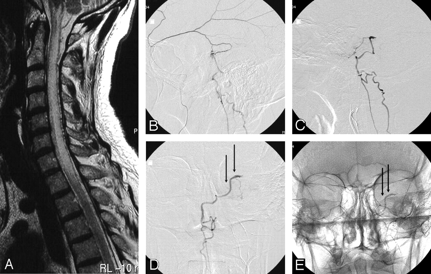
- Fig 1.
A 58-year-old man with progressive cervical cord dysfunction.
A, MR image shows a swollen cervical cord with edema and central myelopathy and dilated perimedullary veins.
B, Lateral view of a selective angiogram of the left middle meningeal artery demonstrates a dural fistula, with drainage to the perimedullary veins.
C and D, Lateral (C) and anteroposterior (D) angiograms of the squamous branch of the middle meningeal artery contributing to the fistula show the beginning of the draining vein (arrows). Glue was injected from this position.
E, Anteroposterior radiograph after glue injection shows glue in the draining vein (arrows).
- Fig 2.
A 65-year-old man with progressive cord dysfunction.
A and B, MR image shows central myelopathy in the cervicothoracic cord, with engorged perimedullary veins.
C and D, Lateral (C) and anteroposterior (D) right external carotid angiograms demonstrate a fistula with drainage to the perimedullary veins (arrows).
E, Anteroposterior projection of selective injection of a branch of the stylomastoid artery supplying the fistula. The proximal part of draining vein is marked with arrows.
F, Radiograph, same as in E, shows glue in the proximal draining vein (arrows).
G, Normal findings on MR image 1 year later.
- Fig 3.
A 72-year-old woman with progressive tetraparesis.
A and B, MR images show a convolute of veins at the level of the foramen magnum, central myelopathy and edema from the medulla oblongata to T6, and dilated posterior medullary veins.
C and D, Lateral (C) and anteroposterior (D) right occipital artery angiograms show a dural fistula at the skull base, draining via a convolute of paramedullary veins into the posterior medullary veins (arrows).
E, Injection via microcatheter shows the proximal draining vein (arrows).
F, Radiograph, same as E, shows glue cast in the proximal draining vein (arrows).


In this issue


Jump to section
Related Articles
Cited By...
- Dural Arteriovenous Fistulas: A Characteristic Pattern of Edema and Enhancement of the Medulla on MRI
- Intracranial Dural Arteriovenous Fistulae
- Unusual case of intracranial dural AV fistula presenting with acute myelopathy
- Cranial Dural Arteriovenous Fistula: Diagnosis and Classification with Time-Resolved MR Angiography at 3T