
Article Figures & Data
Figures
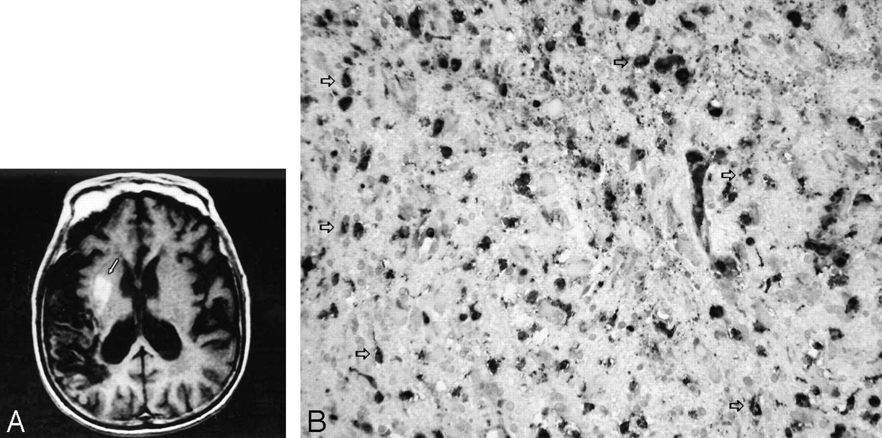
- Fig 1.
A, Axial section of T1-weighted (TR/TE/TI, 550/10/1 ms) MR image at the level of the basal ganglia shows hyperintensities in the right putamen and the external segment of globus pallidus (arrow), along with an infarct in the right temporoparietal cortex. B, Immunohistochemical staining of the brain biopsy specimen for CD68 revealed numerous darkly stained microglia with or without dendritic processes (white arrows) (original magnification ×100).

In this issue


Jump to section
Related Articles
Cited By...
- Transient Hyperintensity in the Subthalamic Nucleus and Globus Pallidus of Newborns on T1-Weighted Images
- Activated microglia in the subthalamic nucleus in hyperglycaemic hemiballism: a case report
- Evolution of Unilateral Perinatal Arterial Ischemic Stroke on Conventional and Diffusion-Weighted MR Imaging