Advertisement

Main menu

  • Home
  • Content
    • Current Issue
    • Accepted Manuscripts
    • Article Preview
    • Past Issue Archive
    • AJNR Case Collection
    • Case of the Week Archive
    • Classic Case Archive
    • Case of the Month Archive
  • Special Collections
    • Spinal CSF Leak Articles (Jan 2020-June 2024)
    • 2024 AJNR Journal Awards
    • Most Impactful AJNR Articles
  • Multimedia
    • AJNR Podcast
    • AJNR Scantastics
    • Video Articles
  • For Authors
    • Submit a Manuscript
    • Author Policies
    • Fast publishing of Accepted Manuscripts
    • Graphical Abstract Preparation
    • Manuscript Submission Guidelines
    • Imaging Protocol Submission
    • Submit a Case for the Case Collection
  • About Us
    • About AJNR
    • Editorial Board
  • More
    • Become a Reviewer/Academy of Reviewers
    • Subscribers
    • Permissions
    • Alerts
    • Feedback
    • Advertisers
    • ASNR Home
  • Other Publications
    • ajnr

User menu

  • Alerts
  • Log in

Search

  • Advanced search
American Journal of Neuroradiology

American Journal of Neuroradiology

ASHNR American Society of Functional Neuroradiology ASHNR American Society of Pediatric Neuroradiology ASSR
  • Alerts
  • Log in

Advanced Search

  • Home
  • Content
    • Current Issue
    • Accepted Manuscripts
    • Article Preview
    • Past Issue Archive
    • AJNR Case Collection
    • Case of the Week Archive
    • Classic Case Archive
    • Case of the Month Archive
  • Special Collections
    • Spinal CSF Leak Articles (Jan 2020-June 2024)
    • 2024 AJNR Journal Awards
    • Most Impactful AJNR Articles
  • Multimedia
    • AJNR Podcast
    • AJNR Scantastics
    • Video Articles
  • For Authors
    • Submit a Manuscript
    • Author Policies
    • Fast publishing of Accepted Manuscripts
    • Graphical Abstract Preparation
    • Manuscript Submission Guidelines
    • Imaging Protocol Submission
    • Submit a Case for the Case Collection
  • About Us
    • About AJNR
    • Editorial Board
  • More
    • Become a Reviewer/Academy of Reviewers
    • Subscribers
    • Permissions
    • Alerts
    • Feedback
    • Advertisers
    • ASNR Home
  • Follow AJNR on Twitter
  • Visit AJNR on Facebook
  • Follow AJNR on Instagram
  • Join AJNR on LinkedIn
  • RSS Feeds

Welcome to the new AJNR, Updated Hall of Fame, and more. Read the full announcements.


AJNR is seeking candidates for the position of Associate Section Editor, AJNR Case Collection. Read the full announcement.


Research ArticleINTERVENTIONAL

Reduction of Radiation Dose for Cerebral Angiography Using Flat Panel Detector of Direct Conversion Type: A Vascular Phantom Study

Y. Hatakeyama, S. Kakeda, N. Ohnari, J. Moriya, N. Oda, K. Nishino, W. Miyamoto and Y. Korogi
American Journal of Neuroradiology April 2007, 28 (4) 645-650;
Y. Hatakeyama
  • Find this author on Google Scholar
  • Find this author on PubMed
  • Search for this author on this site
S. Kakeda
  • Find this author on Google Scholar
  • Find this author on PubMed
  • Search for this author on this site
N. Ohnari
  • Find this author on Google Scholar
  • Find this author on PubMed
  • Search for this author on this site
J. Moriya
  • Find this author on Google Scholar
  • Find this author on PubMed
  • Search for this author on this site
N. Oda
  • Find this author on Google Scholar
  • Find this author on PubMed
  • Search for this author on this site
K. Nishino
  • Find this author on Google Scholar
  • Find this author on PubMed
  • Search for this author on this site
W. Miyamoto
  • Find this author on Google Scholar
  • Find this author on PubMed
  • Search for this author on this site
Y. Korogi
  • Find this author on Google Scholar
  • Find this author on PubMed
  • Search for this author on this site
  • Article
  • Figures & Data
  • Info & Metrics
  • Responses
  • References
  • PDF
Loading

Article Figures & Data

Figures

  • Tables
  • Fig 1.
    • Download figure
    • Open in new tab
    • Download powerpoint
    Fig 1.

    Photograph (A) and schematic drawing (B) of the anthropomorphic vascular phantom used in this study. The phantom was designed to simulate the intracranial arteries with a total of 32 aneurysms. Of 32 aneurysms, 15 had a bleb with diameter of 2 mm.
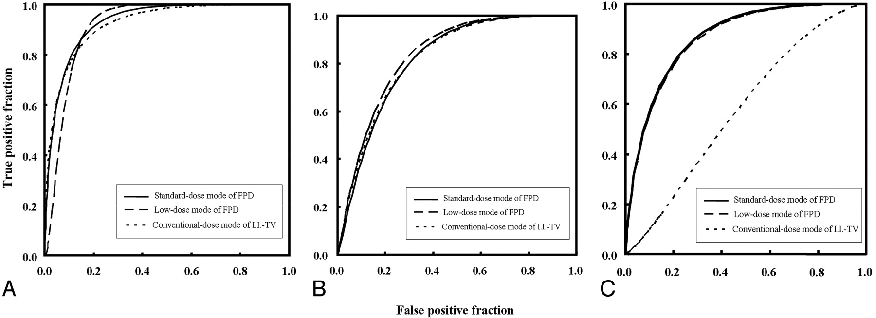
  • Fig 2.
    • Download figure
    • Open in new tab
    • Download powerpoint
    Fig 2.

    ROC curves for the detection of simulated aneurysmal blebs with 3 different contrast material concentrations: 300 mg I/mL (A), 150 mg I/mL (B), and 100 mg I/mL (C). For each contrast material concentration, graph shows the 3 composite ROC curves generated from the pooled data of the 7 observers’ results with the standard- (solid line) and low-dose modes of FPD (dashed line) and the conventional-dose mode of I.I.-TV (dotted line) systems.
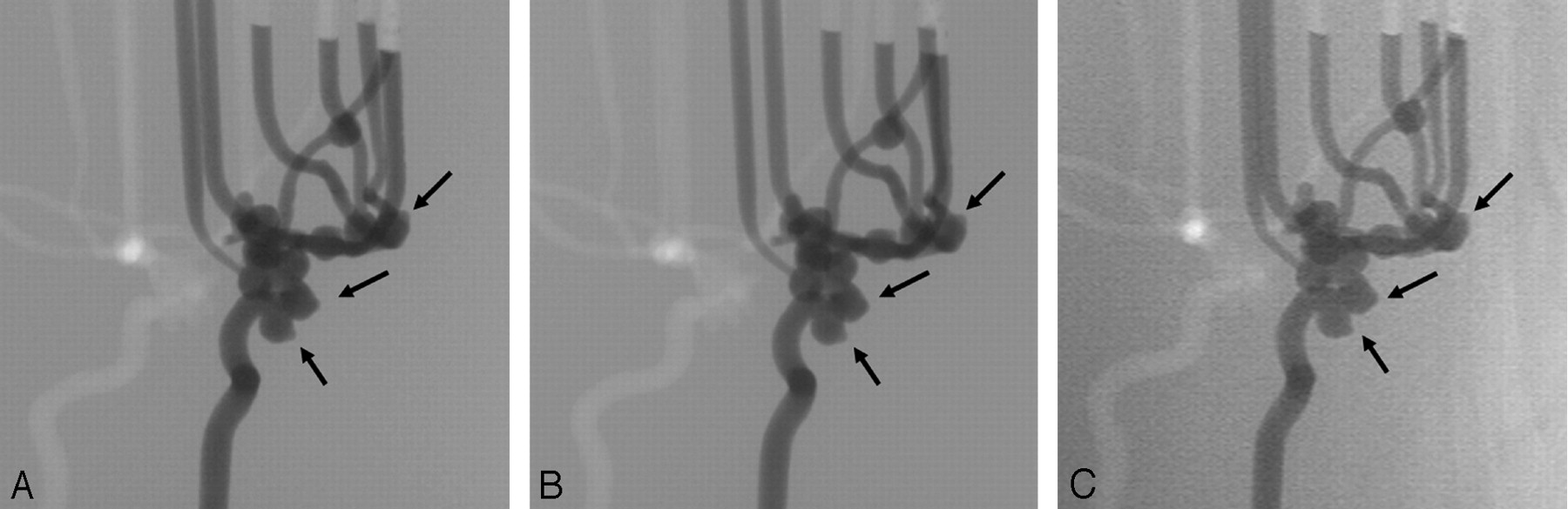
  • Fig 3.
    • Download figure
    • Open in new tab
    • Download powerpoint
    Fig 3.

    2D DSA from the left anterior oblique view at 300 mg I/mL obtained with standard-dose mode of FPD system (A), with low-dose mode of FPD system (B), and with conventional-dose mode of I.I.-TV system shows blebs (arrows) (C). For the depiction of aneurysmal blebs, there is no particular difference among the 3 dose modes.
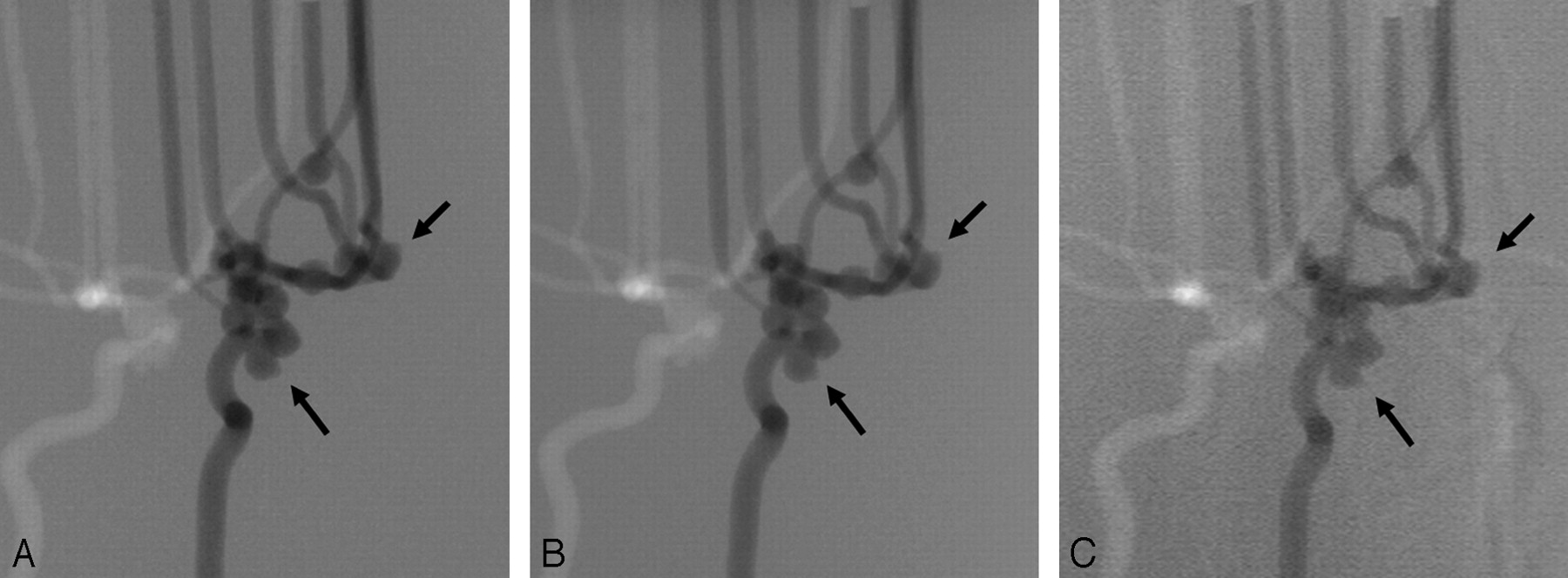
  • Fig 4.
    • Download figure
    • Open in new tab
    • Download powerpoint
    Fig 4.

    2D DSA from the left anterior oblique view at 100 mg I/mL obtained with standard-dose mode of FPD (A), with low-dose mode of FPD (B), and with conventional-dose mode of I.I.-TV system shows blebs (arrows) (C). For the depiction of aneurysmal blebs, the images obtained with both dose modes of FPD system are superior to those obtained with the I.I.-TV system.

Tables

  • Figures
    • View popup
    Table 1:

    Technical features of FPD system and I.I.-TV system

    FeatureFPD systemI.I.-TV system
    Total filtration3 mm of Al + 0.3 mm of Cu3 mm of Al
    Additional filtrationNoneNone
    Frame rate
        Cine mode30 fps30 fps
        Fluoroscopy mode15 fps15 fps
    Image matrix format1440 × 14401024 × 1024
    Field of view9 inches12 inches
    • Note:—FPD indicates flat panel detector; I.I.-TV, image intensifier television; fps, frames per second.

    • View popup
    Table 2:

    Subjective evaluation for the 2D DSA regarding the depiction of simulated aneurysms

    Contrast Material Concentration & Dose ModeScore for Image QualityP
    54321Mean ± SD*
    300 mgI/mL
        Standard-dose mode of FPD system5700005.00 ± 0.00**.038
        Low-dose mode of FPD system5700005.00 ± 0.00**.038
        Conventional-dose mode of I.I.-TV system5232004.88 ± 0.43
    150 mgI/mL
        Standard-dose mode of FPD system5511004.95 ± 0.29**<.001
        Low-dose mode of FPD system5241004.90 ± 0.36**<.001
        Conventional-dose mode of I.I.-TV system41105104.60 ± 0.73
    100 mgI/mL
        Standard-dose mode of FPD system41124004.65 ± 0.61**<.001
        Low-dose mode of FPD system39135004.60 ± 0.65**<.001
        Conventional-dose mode of I.I.-TV system115261323.00 ± 0.85
    • Note:—DSA indicates digital subtraction angiography; FPD, flat panel detector; I.I.-TV, image intensifier television. Data are mean ± SD of scores for image quality evaluated using a 5-point scale (1, clearly visualized; 2, good; 3, reasonably good; 4, poor; 5, very poor)

    • ** Significantly higher to I.I.-TV system (the Wilcoxon signed rank test).

    • View popup
    Table 3:

    Diagnostic performance according of each observer regarding the detection of the simulated aneurysmal blebs

    Contrast Material Concentration & Dose ModeObserverMean Az
    1234567
    300 mgI/mL
        Standard-dose mode of FPD system0.930.910.890.920.910.920.940.92
        Low-dose mode of FPD system0.890.890.910.920.920.940.950.92
        Conventional-dose mode of I.I.-TV system0.920.910.860.980.870.950.900.91
    150 mgI/mL
        Standard-dose mode of FPD system0.810.790.830.830.830.800.820.82
        Low-dose mode of FPD system0.830.790.850.820.830.850.840.83
        Conventional-dose mode of I.I.-TV system0.800.780.850.810.820.840.820.82
    100 mgI/mL
        Standard-dose mode of FPD system0.840.880.870.800.930.870.800.86*
        Low-dose mode of FPD system0.840.880.860.790.930.870.790.85*
        Conventional-dose mode of I.I.-TV system0.670.550.540.570.550.540.520.56
    • Note:—Az indicates areas under the receiver operating characteristic curve; FPD, flat panel detector; I.I.-TV, image intensifier television.

    • * Significantly higher to I.I.-TV system (P < .01).

PreviousNext
Back to top

In this issue

American Journal of Neuroradiology: 28 (4)
American Journal of Neuroradiology
Vol. 28, Issue 4
April 2007
  • Table of Contents
  • Index by author
Advertisement
Print
Download PDF
Email Article

Thank you for your interest in spreading the word on American Journal of Neuroradiology.

NOTE: We only request your email address so that the person you are recommending the page to knows that you wanted them to see it, and that it is not junk mail. We do not capture any email address.

Enter multiple addresses on separate lines or separate them with commas.
Reduction of Radiation Dose for Cerebral Angiography Using Flat Panel Detector of Direct Conversion Type: A Vascular Phantom Study
(Your Name) has sent you a message from American Journal of Neuroradiology
(Your Name) thought you would like to see the American Journal of Neuroradiology web site.
CAPTCHA
This question is for testing whether or not you are a human visitor and to prevent automated spam submissions.
Cite this article
Y. Hatakeyama, S. Kakeda, N. Ohnari, J. Moriya, N. Oda, K. Nishino, W. Miyamoto, Y. Korogi
Reduction of Radiation Dose for Cerebral Angiography Using Flat Panel Detector of Direct Conversion Type: A Vascular Phantom Study
American Journal of Neuroradiology Apr 2007, 28 (4) 645-650;

Citation Manager Formats

  • BibTeX
  • Bookends
  • EasyBib
  • EndNote (tagged)
  • EndNote 8 (xml)
  • Medlars
  • Mendeley
  • Papers
  • RefWorks Tagged
  • Ref Manager
  • RIS
  • Zotero
0 Responses
Respond to this article
Share
Bookmark this article
Reduction of Radiation Dose for Cerebral Angiography Using Flat Panel Detector of Direct Conversion Type: A Vascular Phantom Study
Y. Hatakeyama, S. Kakeda, N. Ohnari, J. Moriya, N. Oda, K. Nishino, W. Miyamoto, Y. Korogi
American Journal of Neuroradiology Apr 2007, 28 (4) 645-650;
del.icio.us logo Twitter logo Facebook logo Mendeley logo
  • Tweet Widget
  • Facebook Like
  • Google Plus One

Jump to section

  • Article
    • Abstract
    • Materials and Methods
    • Results
    • Discussion
    • References
  • Figures & Data
  • Info & Metrics
  • Responses
  • References
  • PDF

Related Articles

  • No related articles found.
  • PubMed
  • Google Scholar

Cited By...

  • No citing articles found.
  • Crossref
  • Google Scholar

This article has not yet been cited by articles in journals that are participating in Crossref Cited-by Linking.

More in this TOC Section

  • SAVE vs. Solumbra Techniques for Thrombectomy
  • CT Perfusion&Reperfusion in Acute Ischemic Stroke
  • Delayed Reperfusion Post-Thrombectomy&Thrombolysis
Show more Interventional

Similar Articles

Advertisement

Indexed Content

  • Current Issue
  • Accepted Manuscripts
  • Article Preview
  • Past Issues
  • Editorials
  • Editors Choice
  • Fellow Journal Club
  • Letters to the Editor

Cases

  • Case Collection
  • Archive - Case of the Week
  • Archive - Case of the Month
  • Archive - Classic Case

Special Collections

  • Special Collections

Resources

  • News and Updates
  • Turn around Times
  • Submit a Manuscript
  • Author Policies
  • Manuscript Submission Guidelines
  • Evidence-Based Medicine Level Guide
  • Publishing Checklists
  • Graphical Abstract Preparation
  • Imaging Protocol Submission
  • Submit a Case
  • Become a Reviewer/Academy of Reviewers
  • Get Peer Review Credit from Publons

Multimedia

  • AJNR Podcast
  • AJNR SCANtastic
  • Video Articles

About Us

  • About AJNR
  • Editorial Board
  • Not an AJNR Subscriber? Join Now
  • Alerts
  • Feedback
  • Advertise with us
  • Librarian Resources
  • Permissions
  • Terms and Conditions

American Society of Neuroradiology

  • Not an ASNR Member? Join Now

© 2025 by the American Society of Neuroradiology All rights, including for text and data mining, AI training, and similar technologies, are reserved.
Print ISSN: 0195-6108 Online ISSN: 1936-959X

Powered by HighWire