
Article Figures & Data
Figures
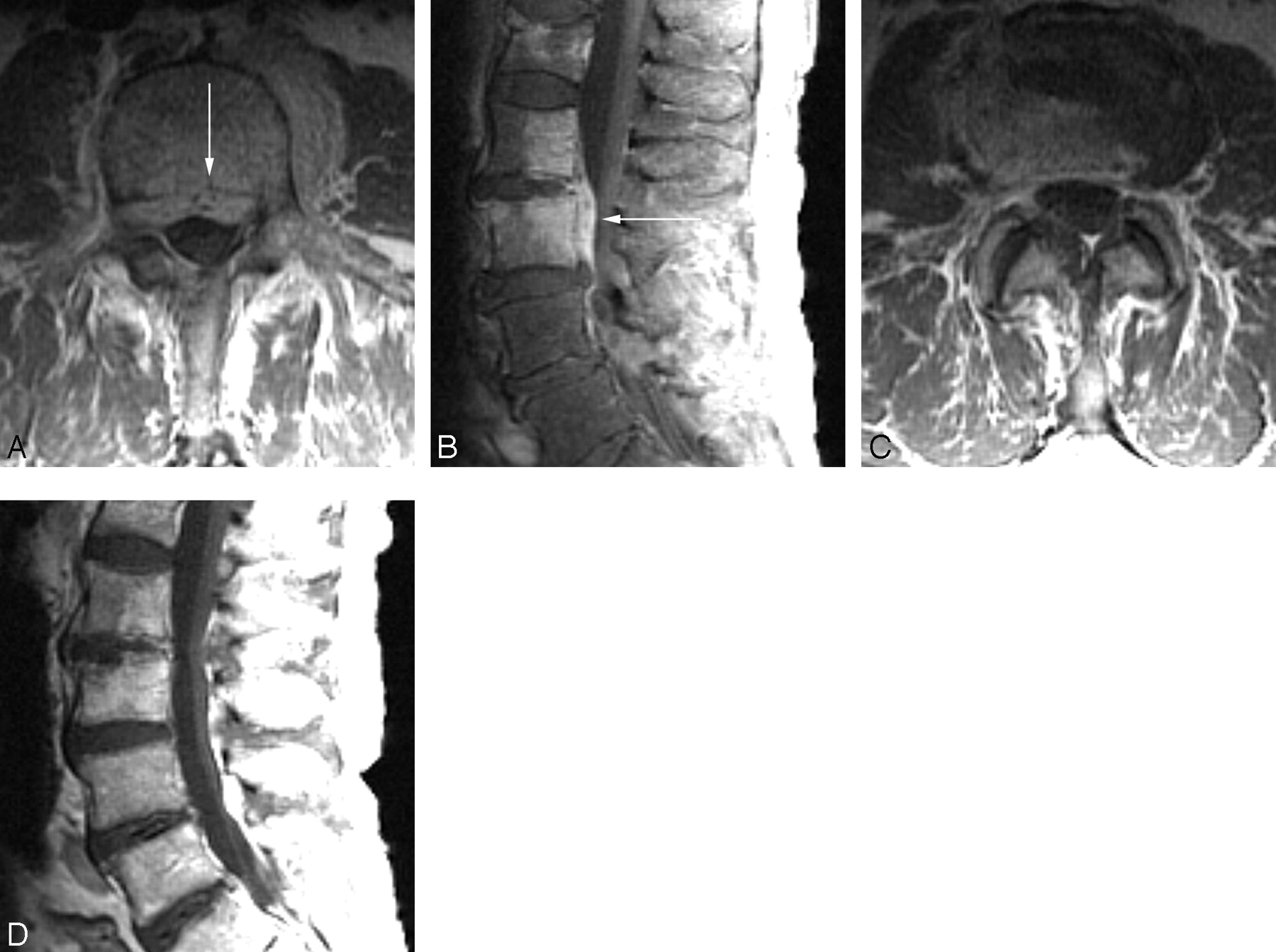
- Fig 1.
Baseline postcontrast axial T1 (A) and sagittal T1 fat-saturated (B) images demonstrate enhancing tissue in the ventral epidural space (arrows) as well as enhancement in the L3 and L4 vertebral bodies. Follow-up axial (C) and sagittal (D) postcontrast T1-weighted images demonstrate resolution of the enhancing epidural tissue. Persistent enhancement of the vertebral bodies and disk space is seen in this patient despite clinical improvement.
- Fig 2.
Baseline postcontrast fat-saturated sagittal (A) T1-weighted imaging demonstrates abnormal enhancement of the paraspinal soft tissues (arrow). Follow-up postcontrast T1-weighted imaging (B) demonstrates persistence of paraspinal enhancement (arrow) and new enhancement in the vertebral bodies and disk space. Despite a worsening appearance on MR imaging, this patient was improving clinically.
- Fig 3.
Baseline postcontrast fat-saturated axial T1-weighted imaging (A) demonstrates a paraspinal phlegmon (arrow). Despite clinical improvement, follow-up postcontrast T1-weighted images demonstrate apparent worsening. The paraspinal phlegmon has increased in size (arrow) and now has a small nonenhancing component suggestive of early abscess formation. There is also increased enhancement in the disk space.


Tables
Characteristic No (%)* (n = 33) Age: median (range) 71 (49–91) Male sex 18 (55) Immunocompromising condition† 2 (6) Systemic comorbidity‡ 6 (18) Presumed source Skin/soft tissue 4 (12) Intravascular device 1 (3) Infection relapse 1 (3) Endovascular 5 (15) Wound infection 4 (12) Septic joint/bursa 2 (6) Unclear 16 (48) Median days of symptoms prior to diagnostic MR imaging (range) 22 (7–162) Microbiologic etiology S aureus 15 (45) Staphylococcus coagulase negative 5 (15) Streptococcus viridans group 5 (15) Gram-negative rod 2 (6) Culture negative 6 (18) Anatomic level involved Cervical 3 (9) Cervicothoracic 1 (3) Cervicothoracic-lumbar 1 (3) Thoracic 6 (18) Thoracolumbar 3 (9) Lumbar 14 (42) Lumbosacral 5 (15) * Unless specified otherwise.
† Corticosteroids, other immunosuppressing medications.
‡ Diabetes mellitus, creatinine >2.0 mg/dL, systemic malignancy, prior radiation therapy, chronic liver disease.
- Table 2:
The presence of specific MR imaging findings at baseline and follow-up MR imaging
MR Imaging Findings Baseline (n = 33) Follow-Up (n = 33)* P† ≥1 level involved 3 (9) 4 (12) 1 Disk space enhancement 22 (67) 25/32 (78) .29 T2 disk space abnormality 29 (88) 24 (73) .13 Epidural enhancement 29 (88) 19/32 (59) .008 Canal abscess 17 (52) 3/32 (9) .001 Canal compromise (%) >25% 19 (58) 10/32 (31) .008 >50% 7 (21) 3/32 (9) .22 Paraspinal abscess 7 (21) 5/32 (16) 1 Paraspinal inflammation None 12 (36) 12/32 (38) Mild 18 (55) 17/32 (53) .75 Moderate 2 (6) 3/32 (9) Severe 1 (3) 0 Vertebral body enhancement None 1 (3) 2 (6) 1%–33% 5 (15) 5 (15) 1 34%–66% 7 (21) 4 (12) >66% 20 (61) 22 (66) Vertebral marrow edema None 1 (3) 1 (3) .80 1%–33% 3 (9) 5 (15) 34%–66% 10 (30) 4 (12) >66% 19 (58) 23 (70) Vertebral body loss of height None 19 (53) 7 (21) 1%–33% 10 (30) 18 (55) <.001 34%–66% 3 (9) 2 (6) >66% 1 (3) 6 (18) Note:—Values represent No. (%) unless otherwise indicated.
* One patient did not receive contrast on the follow-up MR imaging, thereby limiting the evaluation of epidural, paraspinal, and disk space enhancement.
† Baseline MRI vs follow-up MRI.
- Table 3:
Assessment of interval changes of specific MR imaging findings at follow-up MR imaging compared with baseline MR imaging
(n = 33)* Improved Equivocal Worse Findings Absent at Baseline and Follow-Up MRI Disk space enhancement 4 (13) 11 (34) 12 (38) 5 (16) T2 disk space signal 14 (42) 11 (33) 5 (15) 3 (9) Epidural enhancement 21 (66) 5 (16) 3 (9) 3 (9) Epidural canal abscess 15 (47) 1 (3) 1 (3) 15 (47) Paraspinal inflammation 16 (50) 8 (25) 4 (13) 4 (13) Bone marrow edema 3 (9) 22 (67) 7 (21) 1 (3) Vertebral body enhancement 8 (24) 18 (55) 6 (18) 1 (3) Note:—Values represent No. (%).
* One patient did not receive contrast on the follow-up MRI, and therefore was not included in the evaluation of epidural, paraspinal, and disk space enhancement changes.
MR Imaging Findings Clinically Improved (n = 23) Not Clinically Improved* (n = 10) P ≥1 level involved 4 (17) 0 (0) .29 Disk space enhancement 17 (74) 8/9 (89) .64 T2 disk space abnormality 15 (65) 9 (90) .22 Epidural enhancement 13 (57) 6/9 (67) .70 Canal abscess 2 (9) 1 (10) 1 Canal compromise (%) >25% 7 (30) 3/9 (33) 1 >50% 3 (13) 0/9 (0) .54 Paraspinal abscess 3 (13) 2/9 (22) .60 Paraspinal inflammation None 10 (43) 2/9 (22) Mild 11 (48) 6/9 (67) Moderate 2 (9) 1/9 (11) .32 Severe 0 0 Vertebral body enhancement None 2 (9) 0 1%–33% 4 (17) 1 (10) .61 34%–66% 2 (9) 2 (20) >66% 15 (65) 7 (70) Vertebral marrow edema None 1 (4) 0 1%–33% 4 (17) 1 (10) .83 34%–66% 2 (9) 2 (20) >66% 16 (70) 7 (70) Vertebral body loss of height None 7 (30) 0 1%–33% 10 (43) 8 (80) .42 34%–66% 1 (4) 1 (10) >66% 5 (22) 1 (10) Note:—One patient did not receive contrast on the follow-up MR imaging, thereby limiting evaluation of epidural, paraspinal, and disk space enhancement.
* Unless otherwise specified.
MRI Results n Clinical Status at Interval MRI Clinical Impact of Interval MRI Improved Equivocal Worse Prolong Antibiotics Invasive Procedure None Discernible Improved 21 17 4 0 5 2 14 Equivocal 5 3 1 1 3 0 2 Worse 6 5 0 1 2 1 3 Note:—One follow-up MR imaging exam was unable to be given an overall grade because no contrast was administered. Overall grade was based upon the combined assessment of epidural, paravertebral, and T2 disk space changes vs baseline findings.
- Table 6:
Clinical characteristics and patient outcomes by overall follow-up MR imaging results based upon epidural, paravertebral, and T2 disk space changes
Variable Overall Assessment Follow-Up MRI P Improved (n = 21) Unimproved (n = 11) Host Factors Age ≥75 4 (19) 6 (55) .06 Systemic comorbidity* 5 (24) 6 (55) .12 Anatomic site infection† Cervical 3 (14) 1 (9) .87 Thoracic 4 (19) 3 (27) Lumbar 14 (67) 7 (64) BMI >30 11 (52) 5 (45) 1 Microbiologic factors S aureus 11 (52) 5 (36) .47 Culture-negative cases 5 (24) 0 (0) .14 Clinical factors Time from symptom-onset-diagnosis <28 days 11 (52) 4 (36) .47 Multilevel disease 3 (14) 0 (0) .53 Clinical improvement 17 (81) 8 (73) .45 Presence epidural abscess 12 (57) 3 (27) .15 Presence paravertebral/psoas abscess 3 (14) 3 (27) .39 Management factors Surgically managed 3 (14) 1 (9) 1 Total duration of antimicrobials >42 days 6 (29) 6 (55) .25 Use of oral antimicrobials 7 (33) 6 (55) .28 Outcome factors Residual neurologic deficits 2 (10) 2 (18) .59 Residual pain medications 2 (10) 2 (18) .59 Clinical failure 1 (5) 2 (18) .27 Note:—One follow-up MRI exam was unable to be given an overall grade because no contrast was administered.
* Diabetes mellitus, creatinine >2.0 mg/dL, systemic malignancy, prior radiation therapy, chronic liver disease.
† Grouped according to highest level of involvement.
In this issue


Jump to section
Related Articles
Cited By...
- Difficulties in diagnosing vertebral osteomyelitis in a child
- Vertebral osteomyelitis in an immunosuppressed patient with rheumatoid arthritis
- Imaging of spondylodiscitis, inflammatory spondyloarthropathy and pars defects
- Spontaneous abscess of the lumbar spine presenting as subacute back pain
- The toddler refusing to weight-bear: a revised imaging guide from a case series