
Article Figures & Data
Figures
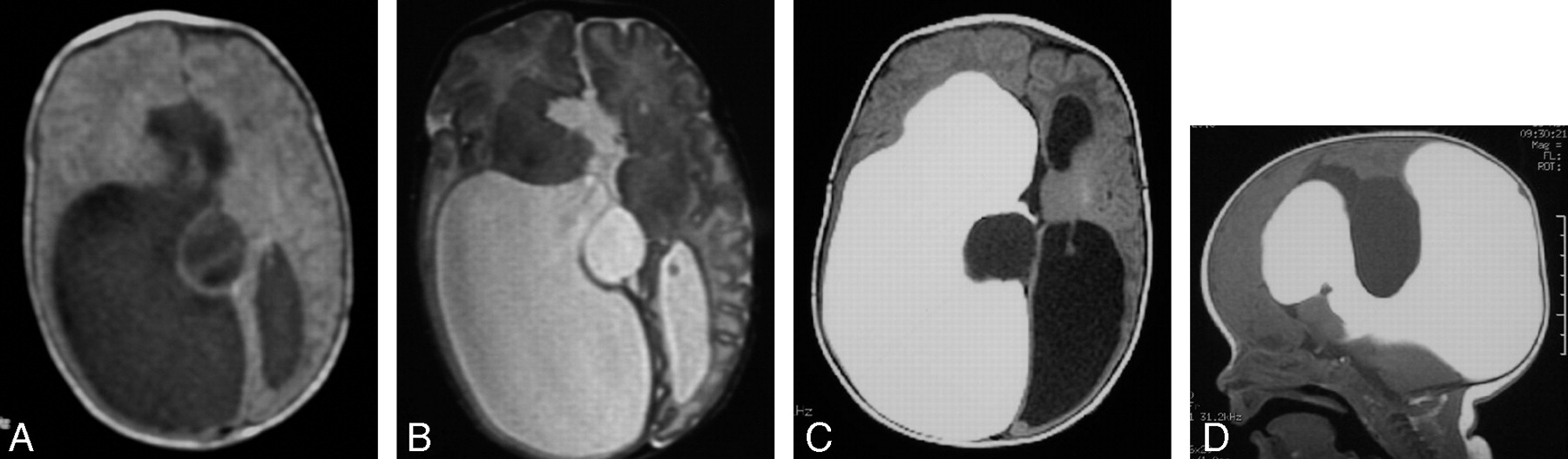
- Fig 1.
Complex hydrocephalus (patient 3). Axial T1-weighted spin-echo (SE) (A) and T2-weighted fast spin-echo (FSE) (B) images showing unilateral ventriculomegaly and midline cyst at the neonate stage and signs of corpus callosum agenesis. Axial (C) and sagittal (D) T1-weighted SE images at 1 month of life, after Gd-DTPA injection via transfontanelle ventriculostomy, show isolated right ventricular enlargement with lack of communication either with the left ventricle or with the midline cyst. Uniportal endoscopic approach was elected for the treatment of both the interhemispheric cyst and the isolated right ventricle.
- Fig 2.
Dysraphic state with hypertelorism, intracranial suspected dermoid tumor, and bifid nose (patient 8). Coronal CT scan through the cribriform plate (A) and sagittal SE T1-weighted MR image through the anterior cranial fossa (B) do not exclude associated encephalocele. Intrathecal Gd-DTPA-enhanced T1-weighted SE fat-saturated images in sagittal (C) and coronal (D) planes show integrity of the anterior cranial fossa and absence of meningoencephalic herniation. Surgery was delayed without the fear of unnoticed sac rupture.
- Fig 3.
Acquired orbital meningoencephalocele in a patient with osteopetrosis (patient 7). Coronal CT scan (A) through the orbits shows intraosseous spheric defect on the roof of the left orbit with intracranial canal connection. Notice additional intraosseous orbital defects in the roof of the right orbit. Coronal FSE T2-weighted image (B) shows an encephalocele contained within the roof of the left orbit and raises questions concerning the contents of the right osseous roof defects. Intrathecal Gd-DTPA-enhanced coronal T1-weighted SE fat-saturated image (C) shows free CSF communication between the brain and left orbital roof defect and an encephalocele contained within the cavity. No additional CSF leaks are seen within the bony defects on the right orbit roof, excluding a bilateral surgical subfrontal approach.
- Fig 4.
Spontaneous otorrhea and dizziness (patient 10). A and B, Axial and coronal CT scans through the left middle ear and osseous labyrinth show absence of osseous cochlear dysplasia (A) and a bulbous appearance of the internal auditory canal (arrow in B), with no middle ear or mastoid cavity filling. C, Intrathecal Gd-DTPA-enhanced axial T1-weighted SE fat-saturated MR image through the upper medulla oblongata shows abnormal filling of the left cochlear structure (arrow). D–F, Coronal T1-weighted SE fat-saturated MR image through the cerebro-pontine angle cisterns. Spot view of the right side (D) shows CSF filling up to the lamina cribrosa. However, further membranous labyrinth structures (semicircular canals) are filled on the left side (arrow in E), as well as the cochlear duct (arrow in F). Additional CSF deposit is seen in the floor of the sphenoidal sinus (arrow in G) (the patient was positioned in a prone head-down position). This patient was eventually diagnosed with juvenile transient osteoporosis because of an upper limb pathologic fracture.


Tables
Demographics, clinical status, and surgical data
Patient Age/Sex Diagnosis Administration Procedure Surgical Implications 1 15 d/F IIIv cystic tumor and asymmetric hydrocephalus TF Ex-vacuo ventriculomegaly and ependymal cyst diagnosed; no surgery 2 1 m/M Multicystic hydrocephalus TF Surgical planning 3 2 m/M Isolated lateral ventricle and interhemispheric cyst TF Agenesis of foramen of Monro diagnosed; surgical planning 4 16 y/M Posttraumatic rhinorrhea LP CSF leakage discarded; avoid surgery 5 11 y/M Progressive paraparesis after medullar tumor resection LP Lumbar arachnoiditis; avoid surgery 6 17 y/M Posttraumatic CSF leakage LP Surgical planning 7 7 y/M Orbital encephalocele; osteopetrosis LP Surgical planning 8 22 m/F Hypertelorism; midline defect LP No associated encephalocele; surgery delayed 9 9 m/M Multicystic hydrocephalus VP shunt Excluded IIIv diagnosed; surgical planning 10 8 y/M Spontaneous multiple CSF fistulae LP Surgical planning; LuP shunt Transient juvenile osteoporosis Avoid open surgery Note:—IIIv indicates 3rd ventricle; TF, transfontanelle; LP, lumbar puncture; VP, ventriculoperitoneal; LuP, lumboperitoneal; d, days; m, month; y, year; F, female; M, male.


