
Article Figures & Data
Figures
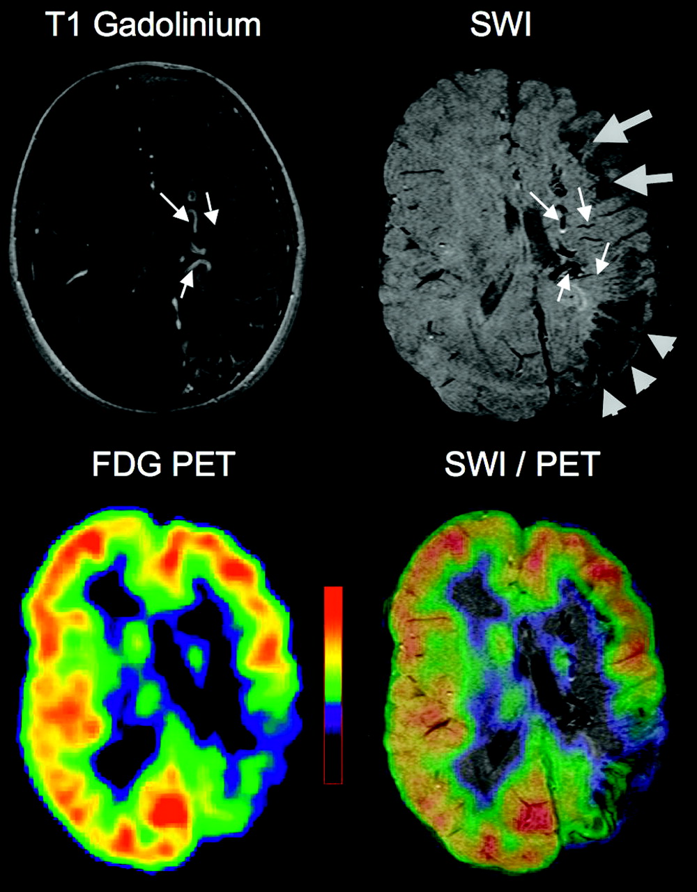
- Fig 1.
Images of a 8.1-year-old girl (patient 12) with Sturge-Weber syndrome affecting the left hemisphere. Postgadolinium T1-weighted MR images show an extensive left hemispheric angioma. SWI shows gyriform cortical signal intensity abnormalities in the left posterior region (arrowheads). A separate area of abnormal SWI is also seen in the left frontal region, located at the gray/white matter junction (thick arrows). Severe hypometabolism on PET is confined to the posterior region, matching well with the cortical SWI changes. In addition to cortical MR imaging abnormalities, abnormal transmedullary deep veins in white matter and periventricular veins are seen on both gadolinium-enhanced T1 and SWI images (thin arrows).
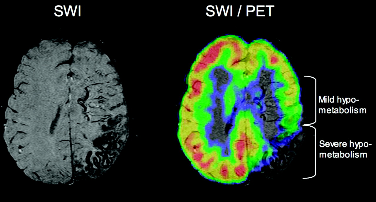
- Fig 2.
Co-registered SWI and PET images of a 6-year-old girl (patient 9) with Sturge-Weber syndrome affecting the left hemisphere. SWI shows gyriform cortical signal intensity abnormalities in the left posterior region in atrophic cortex with severe (>20% decrease) hypometabolism on PET. In front of this area, deep veins are seen in the white matter, connecting mildly hypometabolic (10%–20% decrease) cortex to deep, periventricular veins.
- Fig 3.
Co-registered MR imaging and PET images of a 1.9-year-old boy (patient 2) with Sturge-Weber syndrome and right hemispheric involvement. Contrast-enhanced T1-weighted MR images show a small posterior angioma as well as subtle enhancement in some transmedullary veins. FDG-PET shows no hypometabolism in the right hemisphere. In contrast, SWI demonstrates a number of prominent transmedullary veins in the right frontoparietal region (arrows on phase image); these are joining to a deep periventricular vein (arrowhead). These venous abnormalities are well visualized on the SWI phase images. Magnetic susceptibility effects are also seen in the region of the posterior angioma overlying atrophic right parietal cortex (thick arrow).
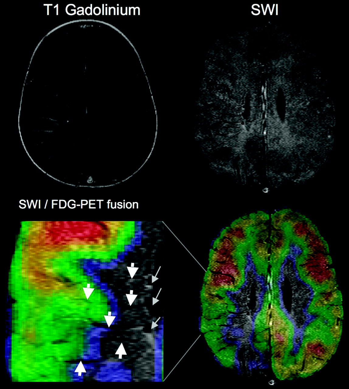
- Fig 4.
T1-weighted and SWI MR imaging fused with PET images of a 2.8 year-old girl (patient 4) with Sturge-Weber syndrome affecting the right hemisphere. T1-weighted images show focal posterior parietal atrophy with leptomeningeal gadolinium enhancement and subtle enhancement in the white matter, consistent with transmedullary veins. The same deep veins are well visualized on SWI (thick arrows), apparently connected to enlarged periventricular veins (thin arrows on the enlarged fused SWI/PET image). FDG-PET shows severe hypometabolism in the area of focal atrophy, while the transmedullary veins reach adjacent cortical regions with mild or no hypometabolism. The SWI/PET fusion image shows well the configuration of multiple draining veins (arrows) originating from the edge of the hypometabolic area.
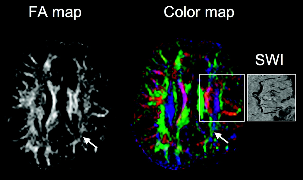
- Fig 5.
Representative DTI fractional anisotropy (FA) and color vector map images of patient 9 with a left parietal angioma. FDG-PET showed severe hypometabolism in the parietal lobe (Fig 2), whereas SWI visualized deep frontocentral transmedullary veins (see affected area in the SWI panel on the right) apparently draining from mildly hypometabolic cortex. FA map demonstrates decreased FA values extending into the frontal lobe. On the color map, fiber directions are color-coded based on the main eigenvector (red, left-right, green, anteroposterior, blue, superior-inferior). Note that in the central white matter region encompassing the deep veins, red and dark purple-coded voxels dominate, indicating an increased number of voxels with transverse (left-right) as well as oblique direction of water diffusion as opposed to the right (normal) side containing more fibers with superior-inferior direction. Both FA and color maps show severe loss and disorganization of white matter in the left parietal area under the angioma (arrows).


Tables
Patient Gender Age at Onset (years) Age at Epilepsy (years) Seizure Frequency Drug IQ 1 F 1.5 1.1 1/month OXC, VPA, PHB 86 2 M 1.9 0.5 Free for 1 year CBZ 70 3 F 2.5 No epilepsy Aspirin 102 4 F 2.8 1.5 Free for 8 months LEV 76 5 F 8.9 0.6 Free for 8 years None 82 6 M 10.3 No epilepsy None 69 7 M 2.7 0.2 2/day OXC 60 8 M 4.3 0.4 A few per year OXC 71 9 F 6.0 0.3 A few per year OXC 60 10 M 6.0 0.5 10–30/day LEV, VPA 35 11 M 6.3 0.8 Free for 2 years CBZ, LEV 55 12 F 8.1 0.3 Free for 3 years CBZ 87 13 F 9.5 0.7 A few per year CBZ 60 Note:—F indicates female; M, male; OXC, oxcarbamazepine; VPA, valproate; PHB, phenobarbital; LEV, levetiracetam; CBZ, carbamazepine.
- Table 2:
Imaging findings of the 13 patients, showing locations of conventional MR imaging, SWI (cortex and white matter), and PET abnormalities; for FDG-PET, areas with severe (>20% decrease) and mild (10–20% decrease) hypometabolism are indicated
Patient Past CT Scan Conventional MRI SWI Abnormality FDG-PET Hypometabolism Cortex White Matter Severe Mild 1 NA L P angioma (small) L F-P L P 2 R calc R P angioma (small), plexus R P, F R O 3 L P calc (very small) L sP angioma (small) 4 Normal (age 1.5 years) R P angioma R pP atrophy R F-P R P R T 5 NA L p plexus L T angioma L T L TO, iF 6 NA R P, pT angioma, atrophy, R p plexus R T R P, pT R T 7 NA R F, P, pT-O angioma R hemisphere atrophy R p plexus R F-P, pT-O L iF [R F*] R sT, sO R P-F R O 8 L hem calc L hemisphere angioma L p plexus L P-O, F, T [L F*] L T-P-O, iF L sF 9 NA L P angioma, plexus L P, T L F, cerebellum L P-T-O L F 10 L P calc L P, pT, iF angioma L P-T-O, F L F L P-T-O L F 11 R P calc R sP, mT angioma R sP-F, mT R mO R aT R pT-O R sP 12 L hem calc L P-T-O, F angioma L posterior plexus L P-T, m-i F L F L T-P-O L T-P-O 13 R F-P calc (age 8 months) R F-P angioma R F, T, P atrophy P p-i F R F-P R med T R T-P-O, F R F R T-P-O Note:—SWI, susceptibility-weighted imaging; FDG, fluorodeoxyglucose; L, left; R, right; T, temporal; F, frontal; P, parietal; O, occipital; i, inferior; p, posterior; a, anterior; s, superior; m, middle; med, medial; WM, white matter; adj, adjacent; calc, calcification, NA, not available; hem, hemisphere.
* Only short streaks in the centrum semiovale.
In this issue


Jump to section
Related Articles
Cited By...
- Alternative Venous Pathways: A Potential Key Imaging Feature for Early Diagnosis of Sturge-Weber Syndrome Type 1
- Study protocol: retrospectively mining multisite clinical data to presymptomatically predict seizure onset for individual patients with Sturge-Weber
- Enlargement of deep medullary veins during the early clinical course of Sturge-Weber syndrome
- Clinical Correlates of White Matter Blood Flow Perfusion Changes in Sturge-Weber Syndrome: A Dynamic MR Perfusion-Weighted Imaging Study
- Attenuation of Cerebral Venous Contrast in Susceptibility-Weighted Imaging of Spontaneously Breathing Pediatric Patients Sedated with Propofol
- Susceptibility-Weighted Imaging: Technical Aspects and Clinical Applications, Part 2
- A Spectrum of Unusual Neuroimaging Findings in Patients with Suspected Sturge-Weber Syndrome