
Article Figures & Data
Figures
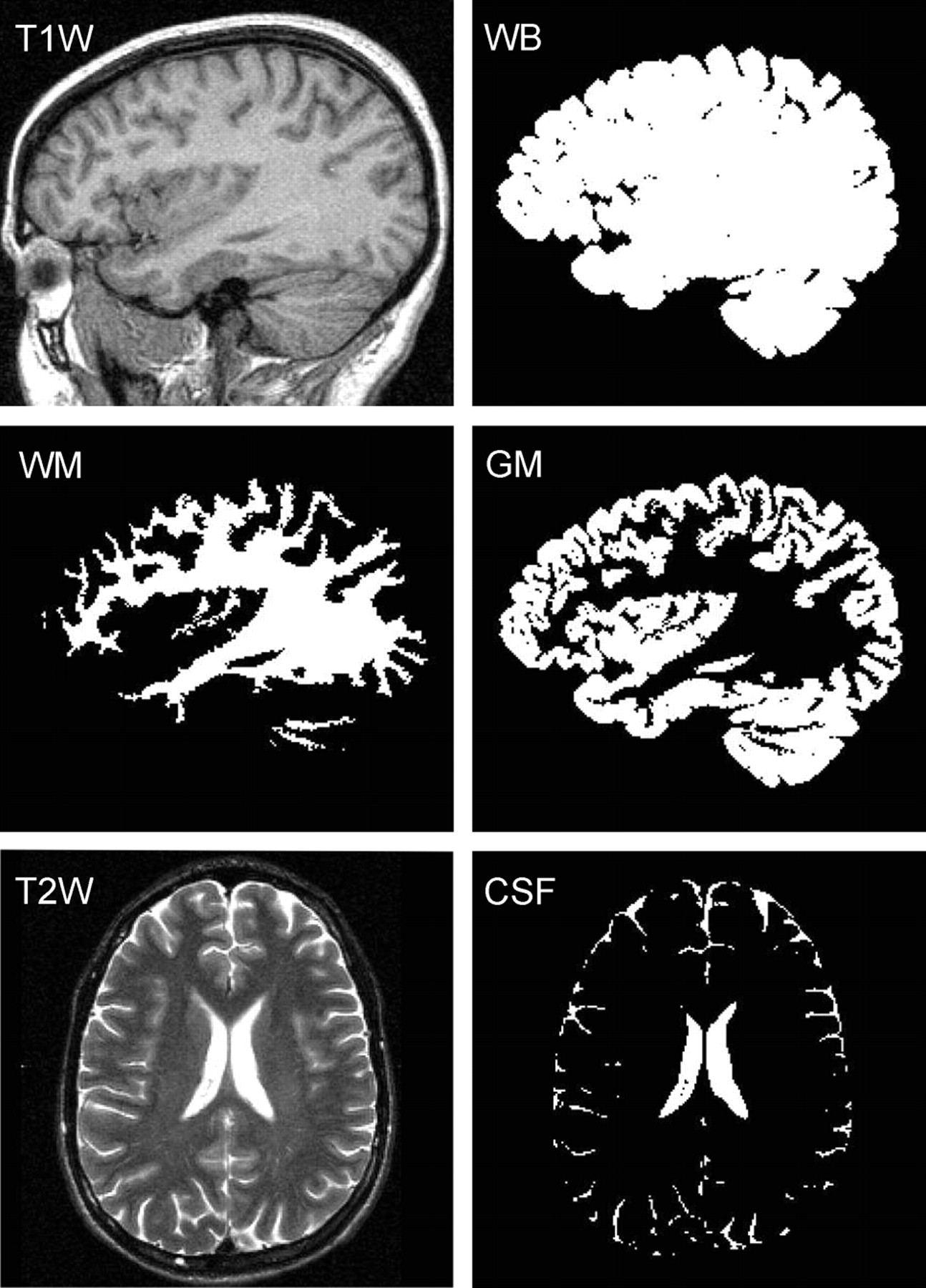
- Fig 1.
Segmentation performance. T1-weighted sagittal images were used to construct a whole-brain (WB) mask, subdivided here into GM and WM masks, using a threshold halfway between their respective signal intensities. Our partial volume technique enables better, subpixel, precision. CSF masks were created from T2-weighted MR imaging.
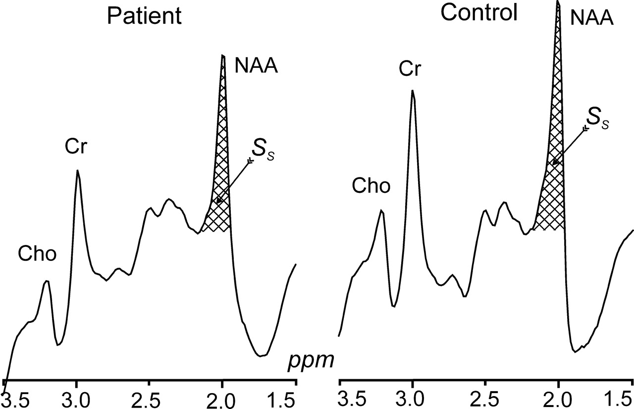
- Fig 2.
Whole-head 1H spectra, left patient 9, right (matched) control subject 5 in Tables 1 and 2. The hatched regions indicate the peak-areas used to obtain QNAA of Eq 1. Note the excellent lipid suppression. Also note that localization relies on knowledge that NAA, unlike the other metabolites (eg, choline, creatine, etc), is exclusive to neuronal cells.
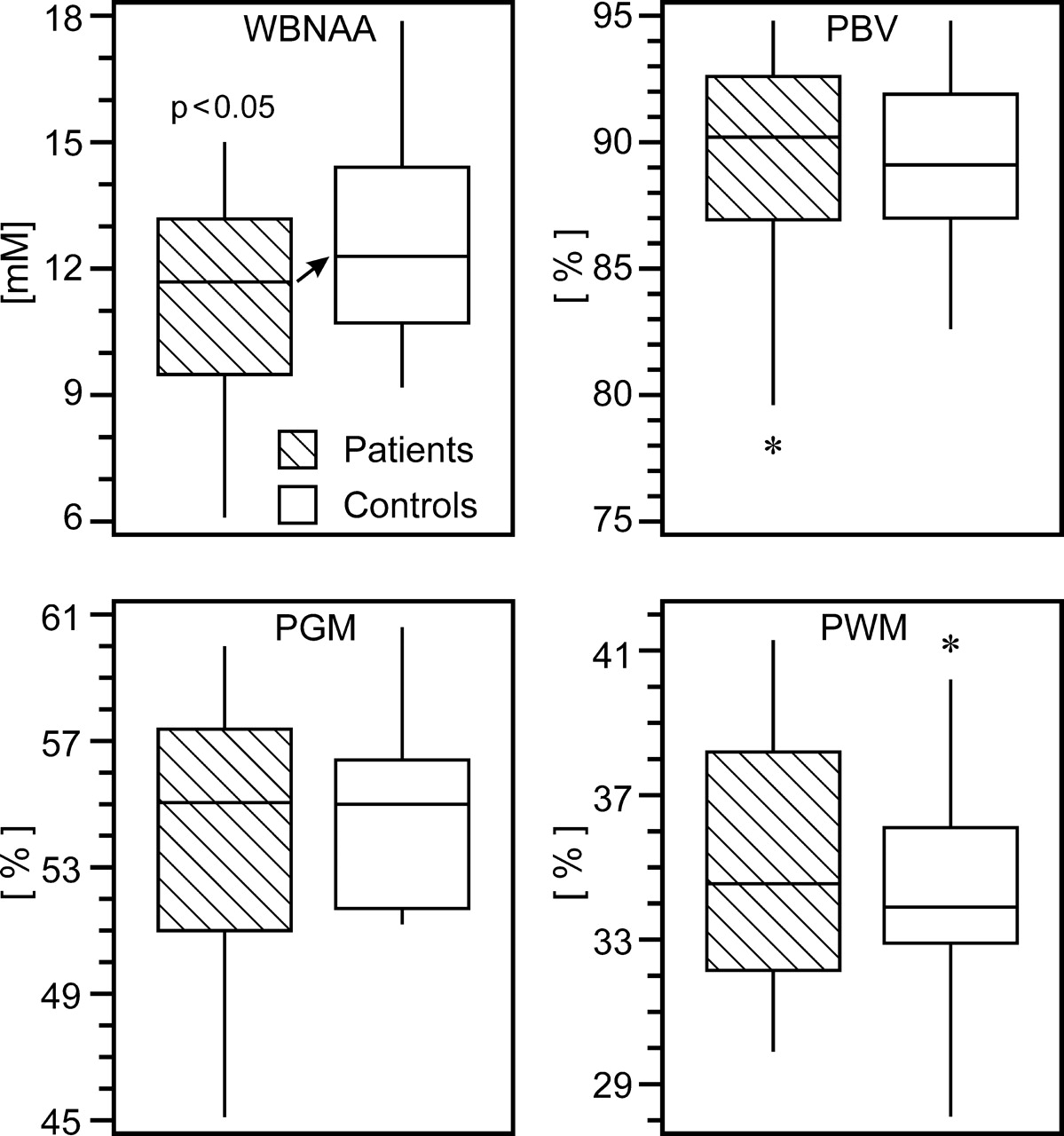
- Fig 3.
Box plots of 1st, 2nd (median), 3rd quartiles (box), ± 95% (whiskers) and outliers (*) for WBNAA, PBV, PGM, and PWM in patients (hatched) and control subjects. Note the significant WBNAA deficit in patients (arrow), reflecting diffuse neuronal injury. Although PBV and PGM did not significantly differ between patients and control subjects, the wide range of the lower half of the patient distributions suggests a subset suffered global and GM atrophy.
- Fig 4.
Scatter plot of WBNAA versus age for mild TBI patients and their matched control subjects. Note that WBNAA deficits increased with age, indicating greater neuronal injury after mild trauma, which may help explain part of their generally worse prognosis.
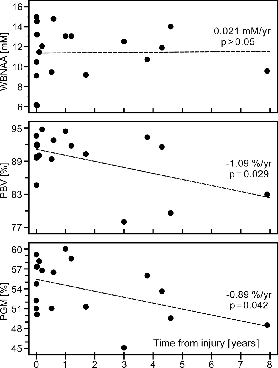
- Fig 5.
Scatter plots of WBNAA, PBV, and PGM versus time from TBI. Although WBNAA remained statistically stable, volumetric measures revealed global atrophy, localized mostly to GM, suggesting that the eventual pathologic outcome of mild TBI is loss of cortical neurons. Stable WBNAA suggests that this loss, measured by QNAA, continued well after the initial traumatic insult but at the same pace as global atrophy.


Tables
- Table 1:
Demographics of patients with mTBI and MR imaging findings suggestive of axonal injury (AI)
No. Age/Sex Injury Elapsed Time* PBV† PGM† PWM† WBNAA (mmol/L) MRI Findings 1 19/F MVA 3.8 years 93.3 56.0 37.4 10.7 Normal 2 21/M Assault 1.2 years 91.8 58.5 33.2 13.1 Punctate AI foci 3 22/M MVA 1 year 94.4 60.0 34.4 13.1 Normal 4 25/M Assault 2.4 months 94.8 56.8 37.9 12.1 Punctate AI foci 5 25/F MVA 8 days 92.0 57.3 34.7 14.6 Normal 6 28/M Assault 1.2 months 90.1 58.2 31.9 11.5 Normal 7 30/F Assault 7 months 92.8 56.5 36.3 14.8 Normal 8 30/M MVA 3 years 78.0 45.1 32.9 12.5 Old contusion, volume loss, AI foci 9 31/F Fall 1 day 93.6 52.2 41.3 6.2 Normal 10 35/F BA 3 days 84.7 54.8 29.9 10.5 Normal 11 35/F MVA 31.5 years 86.1 55.3 30.8 10.4 Old contusion, AI foci 12 36/F Assault 9 days 89.7 50.2 39.5 13.2 Normal 13 37/M BA 3 days 89.6 59.2 30.4 15.0 Normal 14 39/M MVA 6.3 months 89.4 51.0 38.3 9.5 Normal 15 41/M Other† 2 days 89.8 51.0 38.8 9.1 Normal 16 43/M MVA 4.3 years 91.6 53.6 37.9 11.9 Normal 17 47/M Fall 7.9 years 83.0 48.5 34.4 9.6 AI focus 18 47/F MVA 1.7 years 90.3 51.3 39.0 9.2 Normal 19 49/F Fall 4.6 years 79.6 49.6 30.0 14.0 Normal 20 57/M Fall 9 days 91.8 57.4 34.4 6.1 Right frontal hemorrhage Avg ± SD 35 ± 10 89.3 ± 4.7 54.1 ± 4.1 35.2 ± 3.5 11.3 ± 2.6 Note:—mTBI indicates mild traumatic brain injury; PBV, percentage brain volume; PGM, percentage gray matter; PWM, percentage white matter; WBNAA, whole-brain N-acetylaspartate; MVA, motor vehicle crash; BA, bicycle accident. All patients had a GCS score of 15.
* Time from injury to examination.
† Injury involving a power tool.
No. Age/Sex PBV (%) PGM (%) PWM (%) WBNAA (mmol/L) 1 21/M 89.1 55.4 33.7 17.87 2 21/F 87.0 59.0 28.1 12.21 3 23/M 89.4 55.3 34.0 10.60 4 27/M 89.3 51.6 37.8 9.18 5 27/F 89.1 55.5 33.7 16.46 6 27/F 94.6 56.4 38.3 11.49 7 28/M 91.9 51.7 40.2 10.13 8 29/F 87.4 51.4 36.0 9.57 9 31/M 92.4 51.2 41.2 13.87 10 39/F 90.9 55.0 35.9 12.01 11 39/M 85.1 51.2 33.9 13.50 12 39/F 94.8 60.6 34.1 14.40 13 39/M 87.9 56.2 31.6 12.29 14 40/F 92.1 59.0 33.0 10.71 15 41/M 88.5 52.5 36.1 11.86 16 43/M 86.3 53.4 32.9 16.07 17 44/M 87.0 54.5 32.7 16.48 18 50/F 89.6 56.4 33.2 12.60 19 61/M 82.6 52.1 30.5 13.24 Avg ± SD 35 ± 11 89.2 ± 3.1 54.7 ± 2.9 34.6 ± 3.2 12.87 ± 2.49 Note:—PBV indicates percentage brain volume; PGM, percentage gray matter; PWM, percentage white matter; WBNAA, whole-brain N-acetylaspartate.
In this issue


Jump to section
Related Articles
Cited By...
- Value of Advanced MR Imaging Techniques in Mild Traumatic Brain Injury
- Imaging Evidence and Recommendations for Traumatic Brain Injury: Advanced Neuro- and Neurovascular Imaging Techniques
- What is the lowest threshold to make a diagnosis of concussion?
- Whole-Brain N-Acetylaspartate: A Marker of the Severity of Mild Head Trauma