
Article Figures & Data
Figures
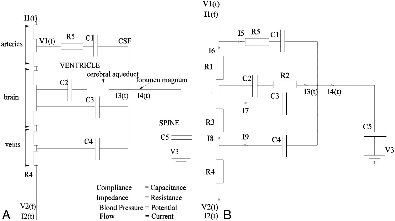
- Fig 1.
The simplified anatomic model on which the electrical equivalence model is based. Boundaries identified by thin lines are considered compliant.
- Fig 2.
A, The electrical equivalence model derived for the anatomic model shown in Fig 1.
B, The simplified equivalent circuit after removal of redundant components.
- Fig 3.
Anatomic (A) and arteriographic (B) scout images showing the location of the measurement planes. Subsequent images show the location of regions of interest for the cerebral aqueduct (1), foramen magnum (2), internal carotid arteries and jugular veins (3), basilar artery (4), and superior sagittal and straight sinuses (5).
- Fig 4.
Measured flow rates for carotid and basilar arteries combined (CAB), superior sagittal sinus (SSS), and combined straight sinus (STS) and SSS venous outflow (Ven) averaged across 16 subjects compared with the predicted venous outflow I2.
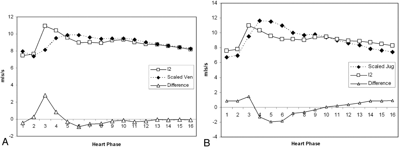
- Fig 5.
A, Comparison of the predicted venous outflow (I2) and the combined STS and SSS venous outflow (Ven). The venous outflow has been scaled to have the same integral as I2. The lower curve shows the difference in the waveforms. Note the prominent systolic peak in the difference.
B, Comparison of the predicted venous outflow (I2) and the jugular venous outflow (JV). The venous outflow has been scaled to have the same integral as I2. The lower curve shows the difference in the waveforms.
- Fig 6.
Mean and 95% confidence intervals for flow at each of 16 heart phases for the predicted venous outflow (I2) and the combined STS and SSS venous outflow (Ven). Asterisks indicate significant differences (∗, P < .05; ∗∗, P < .01).
- Fig 7.
Mean and 95% confidence intervals for flow at each of 16 heart phases for the predicted venous outflow (I2) and the jugular venous outflow. Asterisks indicate significant differences (∗, P < .05).


Tables
- Table 1:
The measured flow volumes and pulsatility indices (PI) from the superior sagittal sinus (SSS), straight sinus (STS), combined venous outflow (SSS + STS = Ven), jugular vein (JV), and arterial inflow (carotid + basilar arteries = CAB), the predicted flow volume for I2 (overall venous outflow in the model), and the predicted indices for I2, I8, and I9
Volume (ml/min) PI (mean ± SD) SSS 305.79 ± 53.59 0.33 ± 0.057 STS 87.23 ± 16.61 0.28 ± 0.068 Ven 392 ± 61.4 0.31 ± 0.041 JV 312 ± 138 0.51 ± 0.045 CAB 536.21 ± 73.61 0.81 ± 0.21 I2 536.21 ± 71.14 0.45 ± 0.16 I8 0.0037 ± 0.0013 I9 1.91 ± 0.53 - Table 2:
The estimated relative values for parameters R1–R5 and C1–C4 (Fig. 2) together with their approximate physiologic characteristics
Parameter Physical Interpretation Value SE R1 Impedance of arterial capillaries 1.0 0.0025 R2 Impedance of cerebral aqueduct ≈0.0 R3 Impedance of venous capillaries ≈0.0 R5 Impedance of arteries 0.01 8 × 1.8−3 C1 Elastic capacitance of arteries ≈Large C2 Elastic capacitance of ventricles 4.11 0.11 C3 Elastic capacitance of capillaries ≈0.0 5 × 10⁻³ C4 Elastic capacitance of veins 271.017 18


